Vitamin D deficiency in mice impairs colonic antibacterial activity and predisposes to colitis
- PMID: 20392825
- PMCID: PMC2875827
- DOI: 10.1210/en.2010-0089
Vitamin D deficiency in mice impairs colonic antibacterial activity and predisposes to colitis
Abstract
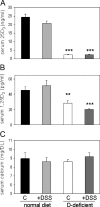
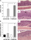
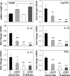
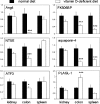
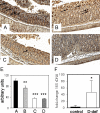
Vitamin D insufficiency is a global health issue. Although classically associated with rickets, low vitamin D levels have also been linked to aberrant immune function and associated health problems such as inflammatory bowel disease (IBD). To test the hypothesis that impaired vitamin D status predisposes to IBD, 8-wk-old C57BL/6 mice were raised from weaning on vitamin D-deficient or vitamin D-sufficient diets and then treated with dextran sodium sulphate (DSS) to induce colitis. Vitamin D-deficient mice showed decreased serum levels of precursor 25-hydroxyvitamin D(3) (2.5 +/- 0.1 vs. 24.4 +/- 1.8 ng/ml) and active 1,25-dihydroxyvitamin D(3) (28.8 +/- 3.1 vs. 45.6 +/- 4.2 pg/ml), greater DSS-induced weight loss (9 vs. 5%), increased colitis (4.71 +/- 0.85 vs. 1.57 +/- 0.18), and splenomegaly relative to mice on vitamin D-sufficient chow. DNA array analysis of colon tissue (n = 4 mice) identified 27 genes consistently (P < 0.05) up-regulated or down-regulated more than 2-fold in vitamin D-deficient vs. vitamin D-sufficient mice, in the absence of DSS-induced colitis. This included angiogenin-4, an antimicrobial protein involved in host containment of enteric bacteria. Immunohistochemistry confirmed that colonic angiogenin-4 protein was significantly decreased in vitamin D-deficient mice even in the absence of colitis. Moreover, the same animals showed elevated levels (50-fold) of bacteria in colonic tissue. These data show for the first time that simple vitamin D deficiency predisposes mice to colitis via dysregulated colonic antimicrobial activity and impaired homeostasis of enteric bacteria. This may be a pivotal mechanism linking vitamin D status with IBD in humans.
Figures


Similar articles
-
Free versus total serum 25-hydroxyvitamin D in a murine model of colitis.J Steroid Biochem Mol Biol. 2019 May;189:204-209. doi: 10.1016/j.jsbmb.2019.01.015. Epub 2019 Jan 30. J Steroid Biochem Mol Biol. 2019. PMID: 30710745 Free PMC article.
-
Vitamin D deficiency predisposes to adherent-invasive Escherichia coli-induced barrier dysfunction and experimental colonic injury.Inflamm Bowel Dis. 2015 Feb;21(2):297-306. doi: 10.1097/MIB.0000000000000282. Inflamm Bowel Dis. 2015. PMID: 25590952
-
Effect of Chronic Vitamin D Deficiency on the Development and Severity of DSS-Induced Colon Cancer in Smad3-/- Mice.Comp Med. 2020 Apr 1;70(2):120-130. doi: 10.30802/AALAS-CM-19-000021. Epub 2020 Feb 3. Comp Med. 2020. PMID: 32014085 Free PMC article.
-
Dietary vitamin D3 deficiency alters intestinal mucosal defense and increases susceptibility to Citrobacter rodentium-induced colitis.Am J Physiol Gastrointest Liver Physiol. 2015 Nov 1;309(9):G730-42. doi: 10.1152/ajpgi.00006.2015. Epub 2015 Sep 3. Am J Physiol Gastrointest Liver Physiol. 2015. PMID: 26336925 Free PMC article.
-
Protective role of 1,25(OH)2 vitamin D3 in the mucosal injury and epithelial barrier disruption in DSS-induced acute colitis in mice.BMC Gastroenterol. 2012 May 30;12:57. doi: 10.1186/1471-230X-12-57. BMC Gastroenterol. 2012. PMID: 22647055 Free PMC article.
Cited by
-
Vitamin D and colorectal cancer: molecular, epidemiological and clinical evidence.Br J Nutr. 2016 May;115(9):1643-60. doi: 10.1017/S0007114516000696. Br J Nutr. 2016. PMID: 27245104 Free PMC article. Review.
-
Vitamin D Supplementation in Laboratory-Bred Mice: An In Vivo Assay on Gut Microbiome and Body Weight.Microbiol Insights. 2020 Jul 28;13:1178636120945294. doi: 10.1177/1178636120945294. eCollection 2020. Microbiol Insights. 2020. PMID: 32782431 Free PMC article.
-
Influence of Foods and Nutrition on the Gut Microbiome and Implications for Intestinal Health.Int J Mol Sci. 2022 Aug 24;23(17):9588. doi: 10.3390/ijms23179588. Int J Mol Sci. 2022. PMID: 36076980 Free PMC article. Review.
-
Role of diet in development of non-communicable diseases: focus on gut microbiome.Cent Eur J Public Health. 2024 Sep;32(3):200-204. doi: 10.21101/cejph.a8138. Cent Eur J Public Health. 2024. PMID: 39352096 Review.
-
Vitamin D and Phenylbutyrate Supplementation Does Not Modulate Gut Derived Immune Activation in HIV-1.Nutrients. 2019 Jul 21;11(7):1675. doi: 10.3390/nu11071675. Nutrients. 2019. PMID: 31330899 Free PMC article. Clinical Trial.
References
-
- Vagianos K, Bector S, McConnell J, Bernstein CN 2007 Nutrition assessment of patients with inflammatory bowel disease. J Parenter Enteral Nutr 31:311–319 - PubMed
-
- Pappa HM, Grand RJ, Gordon CM 2006 Report on the vitamin D status of adult and pediatric patients with inflammatory bowel disease and its significance for bone health and disease. Inflamm Bowel Dis 12:1162–1174 - PubMed
-
- Jahnsen J, Falch JA, Mowinckel P, Aadland E 2002 Vitamin D status, parathyroid hormone and bone mineral density in patients with inflammatory bowel disease. Scand J Gastroenterol 37:192–199 - PubMed
Publication types
MeSH terms
Substances
Grants and funding
LinkOut - more resources
Full Text Sources
Other Literature Sources
Medical
Molecular Biology Databases

