. 2010 Apr;22(4):1299-312.
doi: 10.1105/tpc.109.071431. Epub 2010 Apr 13.

Arrangement of photosystem II and ATP synthase in chloroplast membranes of spinach and pea

Affiliations

Arrangement of photosystem II and ATP synthase in chloroplast membranes of spinach and pea

Bertram Daum et al. Plant Cell. 2010 Apr.

Abstract

We used cryoelectron tomography to reveal the arrangements of photosystem II (PSII) and ATP synthase in vitreous sections of intact chloroplasts and plunge-frozen suspensions of isolated thylakoid membranes. We found that stroma and grana thylakoids are connected at the grana margins by staggered lamellar membrane protrusions. The stacking repeat of grana membranes in frozen-hydrated chloroplasts is 15.7 nm, with a 4.5-nm lumenal space and a 3.2-nm distance between the flat stromal surfaces. The chloroplast ATP synthase is confined to minimally curved regions at the grana end membranes and stroma lamellae, where it covers 20% of the surface area. In total, 85% of the ATP synthases are monomers and the remainder form random assemblies of two or more copies. Supercomplexes of PSII and light-harvesting complex II (LHCII) occasionally form ordered arrays in appressed grana thylakoids, whereas this order is lost in destacked membranes. In the ordered arrays, each membrane on either side of the stromal gap contains a two-dimensional crystal of supercomplexes, with the two lattices arranged such that PSII cores, LHCII trimers, and minor LHCs each face a complex of the same kind in the opposite membrane. Grana formation is likely to result from electrostatic interactions between these complexes across the stromal gap.


Figures
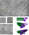
Figure 1.
Figure 1.
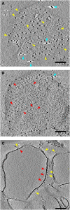
Electron Tomography of Vitreous Spinach Chloroplast Sections. (A) Tomographic slice through the 3D reconstruction of a vitreous chloroplast section. Stacked grana and unstacked stroma thylakoid membranes are easily distinguished. ATP synthase molecules (yellow arrowheads) protrude from the flat regions of grana end membranes and unstacked stromal thylakoids into the stroma. Rows of PSII complexes with a regular repeat distance are visible within some grana membranes (red arrowheads). Bar = 100 nm. (B) and (C) The accurate stacking repeat of grana thylakoids in a subvolume (B) is shown by its power spectrum (C). Zero, first, and second orders indicate a repeat distance of 15.7 nm. (D) Stroma thylakoids either are continuous with a grana thylakoid (green arrowheads) or bifurcate to merge with two adjacent grana thylakoids (blue arrowheads). (E) to (G) Surface representation of connections between grana (green) and stroma (purple) thylakoids reveals their 3D organization at the grana margin. The stroma lamellae are often tilted with respect to the plane of the grana membranes. (E) Subvolume of a grana stack with two stroma thylakoids. (F) and (G) Two different views of the grana stack shown in (E). For simplicity, each thylakoid is depicted as a solid volume rather than a membrane pair.
Figure 2.
Figure 2.
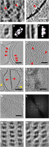
Electron Tomography of Plunge-Frozen, Isolated Thylakoid Membranes. Three tomographic slices at different z heights and angles through the same tomogram of a plunge-frozen, unstacked pea thylakoid membrane prepared by osmotic shock. The tangential slices in (A) and (B) run parallel to the thylakoid membrane, ∼9 nm above (A) or ∼3 nm below (B) the membrane surface. In the slice along the stromal side of the membrane (A), the cF1 heads of the ATP synthase are seen as dark, uniform 12-nm globular protein densities (yellow arrowheads); several dense ∼6-nm gold fiducial markers, used for tomographic reconstruction of the tilt series, are also seen (blue arrowheads, white shadows). The slice in (B) on the lumenal side of the membrane shows dimers of the OEC (red arrowheads) as well as numerous smaller protein densities that cannot be assigned. The tomographic slice in (C) cuts at right angles through the thylakoid membrane, so that the OECs (red arrowheads) and ATP synthase (yellow arrowheads) are seen on opposite sides of the membrane. Bars = 100 nm.
Figure 3.
Figure 3.
Organization of PSII in Grana Thylakoids. Detailed views of tomograms from isolated thylakoid membranes. Membranes isolated from pea chloroplasts by osmotic shock ([A] and [B]) or from spinach by mild digitonin treatment ([C], [E], and [G] to [N]). (A) OECs (red arrowheads) protruding from the membrane surface in a tomographic slice parallel to the membrane. (B) Cross section of PSII dimers with OECs (red arrowhead) in the thylakoid membrane. Bars = 20 nm in (A) and (B). (C) to (L) Top (C) and side view (E) of averaged OEC volumes, compared with the top (D) and side view (F) of the PSII/LHCII supercomplex (Nield and Barber, 2006) drawn at 3-nm resolution. PSII complexes (red arrowheads) are closely but randomly packed in destacked grana thylakoids, as seen in cross (G) and tangential sections (H) of tomographic volumes. In stacked grana thylakoids, PSII complexes occasionally form pairs of crystalline arrays, as seen in cross (I) or oblique sections (J) of a pair of thylakoid vesicles that contain both stacked and unstacked membranes. Yellow arrowheads point to ATP synthases in the unstacked membrane regions that are, in effect, grana end membranes. A tomographic slice of such a PSII array cut parallel to the membrane plane (K) and its power spectrum (L) clearly shows the crystallinity. Bars = 100 nm in (G) to (K). (M) and (N) Slices through each of the two crystalline PSII arrays indicate that the two lattices of the membrane pair are in register across the stromal gap, forming together a 2D crystal of p222 symmetry.
Figure 4.
Figure 4.
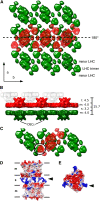
Interaction of PSII/LHCII Supercomplexes in Appressed Grana Thylakoids. (A) A 3D map of the PSII/LHCII supercomplex (Nield and Barber, 2006) at 3-nm resolution fitted manually to the averaged 2D lattices shown in Figures 3M and 3N, respectively. The lower layer is shown in green and the upper layer in red. The OECs in the upper layer face the viewer. The two layers are related by an in-plane twofold axis (dashed line). The flat stromal surfaces of the supercomplexes are in contact across the stromal gap, while the OECs project into the lumenal space. In this arrangement, the PSII dimers, LHCII trimers, and minor LHCs each interact with another complex of the same kind in the opposite membrane. Black arrows indicate the crystal axes a and b of the 2D array. (B) Side view of (A). The bar on the right indicates how the lumenal gap (l), the two membranes (m), and the stromal gap (s) add up to the stacking repeat distance of 15.7 nm (all distances in nm). The narrow lumenal gap means that the OECs of PSII complexes in one grana thylakoid (red and gray) interdigitate. (C) View of four supercomplexes as in (A), showing how a single supercomplex (red) connects to three others (green) in the opposite membrane. (D) Electrostatic surfaces calculated from the x-ray structure (Standfuss et al., 2005) of two LHCII trimers at pH 8.0 in the arrangement entailed by the interacting PSII arrays (black circle in [C]). In this arrangement, the modeled, positively charged N termini (blue) interact with the negatively charged stromal surface (red) of the opposite trimer. Black lines delineate the membranes (m) and the stromal gap (s). (E) Charge distribution on the stromal surface of one LHCII trimer. Black arrowheads in (D) and (E) indicate the positively charged N termini.
Figure 5.
Figure 5.
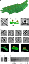
Organization of the Chloroplast ATP Synthase in Thylakoid Membranes. (A) Segmented subvolume of a grana stack with connected stroma lamellae (green) from a vitreous spinach chloroplast section. Individual ATP synthase molecules are indicated by yellow 12-nm spheres. The ATP synthases are randomly distributed over the minimally curved grana end membranes and stromal lamellae but are absent from the appressed regions within the grana and from the highly curved grana margins. (B) Subvolume of a single ATP synthase within an isolated, plunge-frozen pea thylakoid. The cF1 part, the central stalk, and the cFo part in the membrane can be distinguished. Bar = 15 nm. (C) Isosurface representation of two ATP synthase molecules (yellow) in an isolated pea thylakoid membrane (green). lu, lumenal side; st, stromal side. (D) Single-particle average of 50 ATP synthase volumes obtained from isolated spinach thylakoids. Shape and dimensions are consistent with the chloroplast ATP synthase (Mellwig and Böttcher, 2003). (E) Gallery of tomographic slices (top two rows) and surface renderings of segmented subvolumes (bottom row) of multiple copies of ATP synthase in isolated pea thylakoid membranes. Most are monomers, some are pairs, and a small number forms random groups of three to six complexes. Bars = 20 nm. (F) and (G) Clear-native PAGE (F) and in-gel ATPase assay (G) of digitonin-solubilized thylakoid membranes. No ATP synthase oligomers are detected. (H) Second dimension SDS-PAGE of the active band in (G) reveals all subunits of the chloroplast ATP synthase.

Similar articles

Cited by

References

    1. Al-Amoudi A., Studer D., Dubochet J. (2005). Cutting artefacts and cutting process in vitreous sections for cryo-electron microscopy. J. Struct. Biol. 150: 109–121 - PubMed
    1. Albertsson P.-A. (1982). Interaction between the lumenal sides of the thylakoid membrane. FEBS Lett. 149: 186–190
    1. Allen J.F. (1992). Protein phosphorylation in regulation of photosynthesis. Biochim. Biophys. Acta 1098: 275–335 - PubMed
    1. Anderson J.M., Chow W.S., De Las Rivas J. (2008). Dynamic flexibility in the structure and function of photosystem II in higher plant thylakoid membranes: the grana enigma. Photosynth. Res. 98: 575–587 - PubMed
    1. Andersson B., Anderson J.M. (1980). Lateral heterogeneity in the distribution of chlorophyll-protein complexes of the thylakoid membranes of spinach chloroplasts. Biochim. Biophys. Acta 593: 427–440 - PubMed

Publication types

MeSH terms

Substances