Glutaminase 2, a novel p53 target gene regulating energy metabolism and antioxidant function
- PMID: 20378837
- PMCID: PMC2867677
- DOI: 10.1073/pnas.1001006107
Glutaminase 2, a novel p53 target gene regulating energy metabolism and antioxidant function
Abstract
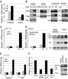
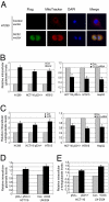
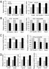
Whereas cell cycle arrest, apoptosis, and senescence are traditionally thought of as the major functions of the tumor suppressor p53, recent studies revealed two unique functions for this protein: p53 regulates cellular energy metabolism and antioxidant defense mechanisms. Here, we identify glutaminase 2 (GLS2) as a previously uncharacterized p53 target gene to mediate these two functions of the p53 protein. GLS2 encodes a mitochondrial glutaminase catalyzing the hydrolysis of glutamine to glutamate. p53 increases the GLS2 expression under both nonstressed and stressed conditions. GLS2 regulates cellular energy metabolism by increasing production of glutamate and alpha-ketoglutarate, which in turn results in enhanced mitochondrial respiration and ATP generation. Furthermore, GLS2 regulates antioxidant defense function in cells by increasing reduced glutathione (GSH) levels and decreasing ROS levels, which in turn protects cells from oxidative stress (e.g., H(2)O(2))-induced apoptosis. Consistent with these functions of GLS2, the activation of p53 increases the levels of glutamate and alpha-ketoglutarate, mitochondrial respiration rate, and GSH levels and decreases reactive oxygen species (ROS) levels in cells. Furthermore, GLS2 expression is lost or greatly decreased in hepatocellular carcinomas and the overexpression of GLS2 greatly reduced tumor cell colony formation. These results demonstrated that as a unique p53 target gene, GLS2 is a mediator of p53's role in energy metabolism and antioxidant defense, which can contribute to its role in tumor suppression.
Conflict of interest statement
The authors declare no conflict of interest.
Figures


Comment in
-
Alternative fuel--another role for p53 in the regulation of metabolism.Proc Natl Acad Sci U S A. 2010 Apr 20;107(16):7117-8. doi: 10.1073/pnas.1002656107. Epub 2010 Apr 14. Proc Natl Acad Sci U S A. 2010. PMID: 20393124 Free PMC article. No abstract available.
-
Journal club. A cancer biologist weighs up p53, metabolism and cancer.Nature. 2010 Aug 19;466(7309):905. doi: 10.1038/466905d. Nature. 2010. PMID: 20725003 No abstract available.
Similar articles
-
Phosphate-activated glutaminase (GLS2), a p53-inducible regulator of glutamine metabolism and reactive oxygen species.Proc Natl Acad Sci U S A. 2010 Apr 20;107(16):7461-6. doi: 10.1073/pnas.1002459107. Epub 2010 Mar 29. Proc Natl Acad Sci U S A. 2010. PMID: 20351271 Free PMC article.
-
Knock-down of glutaminase 2 expression decreases glutathione, NADH, and sensitizes cervical cancer to ionizing radiation.Biochim Biophys Acta. 2013 Dec;1833(12):2996-3005. doi: 10.1016/j.bbamcr.2013.08.003. Epub 2013 Aug 13. Biochim Biophys Acta. 2013. PMID: 23954443
-
Both GLS silencing and GLS2 overexpression synergize with oxidative stress against proliferation of glioma cells.J Mol Med (Berl). 2014 Mar;92(3):277-90. doi: 10.1007/s00109-013-1105-2. Epub 2013 Nov 26. J Mol Med (Berl). 2014. PMID: 24276018 Free PMC article.
-
A systematic review of p53 regulation of oxidative stress in skeletal muscle.Redox Rep. 2018 Dec;23(1):100-117. doi: 10.1080/13510002.2017.1416773. Epub 2018 Jan 3. Redox Rep. 2018. PMID: 29298131 Free PMC article. Review.
-
Glutaminase isoenzymes as key regulators in metabolic and oxidative stress against cancer.Curr Mol Med. 2013 May;13(4):514-34. doi: 10.2174/1566524011313040005. Curr Mol Med. 2013. PMID: 22934847 Review.
Cited by
-
The Mechanism of Ferroptosis and Applications in Tumor Treatment.Front Pharmacol. 2020 Jul 22;11:1061. doi: 10.3389/fphar.2020.01061. eCollection 2020. Front Pharmacol. 2020. PMID: 32774303 Free PMC article. Review.
-
Ferroptosis: Emerging mechanisms, biological function, and therapeutic potential in cancer and inflammation.Cell Death Discov. 2024 Jan 24;10(1):45. doi: 10.1038/s41420-024-01825-7. Cell Death Discov. 2024. PMID: 38267442 Free PMC article. Review.
-
Unraveling Cell Death Pathways during Malaria Infection: What Do We Know So Far?Cells. 2021 Feb 23;10(2):479. doi: 10.3390/cells10020479. Cells. 2021. PMID: 33672278 Free PMC article. Review.
-
Activation of SAT1 engages polyamine metabolism with p53-mediated ferroptotic responses.Proc Natl Acad Sci U S A. 2016 Nov 1;113(44):E6806-E6812. doi: 10.1073/pnas.1607152113. Epub 2016 Oct 3. Proc Natl Acad Sci U S A. 2016. PMID: 27698118 Free PMC article.
-
Tumor suppression in the absence of p53-mediated cell-cycle arrest, apoptosis, and senescence.Cell. 2012 Jun 8;149(6):1269-83. doi: 10.1016/j.cell.2012.04.026. Cell. 2012. PMID: 22682249 Free PMC article.
References
-
- Vousden KH, Prives C. Blinded by the light: The growing complexity of p53. Cell. 2009;137:413–431. - PubMed
-
- Levine AJ, Hu W, Feng Z. The P53 pathway: What questions remain to be explored? Cell Death Differ. 2006;13:1027–1036. - PubMed
-
- Matoba S, et al. p53 regulates mitochondrial respiration. Science. 2006;312:1650–1653. - PubMed
-
- Bensaad K, et al. TIGAR, a p53-inducible regulator of glycolysis and apoptosis. Cell. 2006;126:107–120. - PubMed
Publication types
MeSH terms
Substances
Grants and funding
LinkOut - more resources
Full Text Sources
Other Literature Sources
Medical
Molecular Biology Databases
Research Materials
Miscellaneous

