. 2010 Jul 15;182(2):207-19.
doi: 10.1164/rccm.200909-1414OC. Epub 2010 Apr 8.

Epithelial stress and apoptosis underlie Hermansky-Pudlak syndrome-associated interstitial pneumonia

Affiliations

Epithelial stress and apoptosis underlie Hermansky-Pudlak syndrome-associated interstitial pneumonia

Poornima Mahavadi et al. Am J Respir Crit Care Med. .

Abstract

Rationale: The molecular mechanisms underlying Hermansky-Pudlak syndrome-associated interstitial pneumonia (HPSIP) are poorly understood but, as in idiopathic pulmonary fibrosis, may be linked to chronic alveolar epithelial type II cell (AECII) injury.

Objectives: We studied the development of fibrosis and the role of AECII injury in various murine models of HPS.

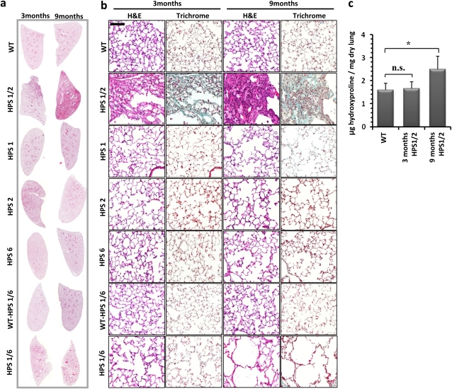
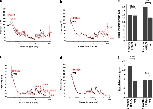
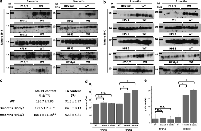
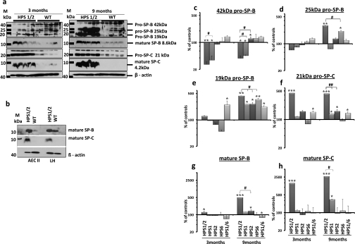
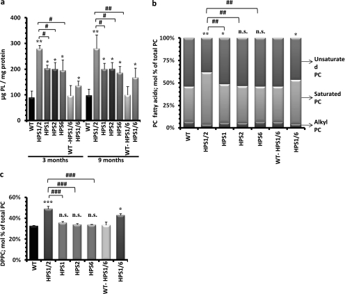
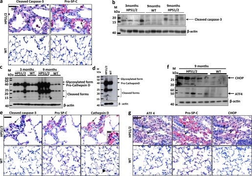
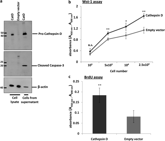
Methods: HPS1, HPS2, and HPS6 monomutant mice, and HPS1/2 and HPS1/6 double-mutant and genetic background mice, were killed at 3 and 9 months of age. Quantitative morphometry was undertaken in lung sections stained with hemalaun-eosin. The extent of lung fibrosis was assessed by trichrome staining and hydroxyproline measurement. Surfactant lipids were analyzed by electrospray ionization mass spectrometry. Surfactant proteins, apoptosis, and lysosomal and endoplasmic reticulum stress markers were studied by Western blotting and immunohistochemistry. Cell proliferation was measured by water-soluble tetrazolium salt-1 and bromodeoxyuridine assays.

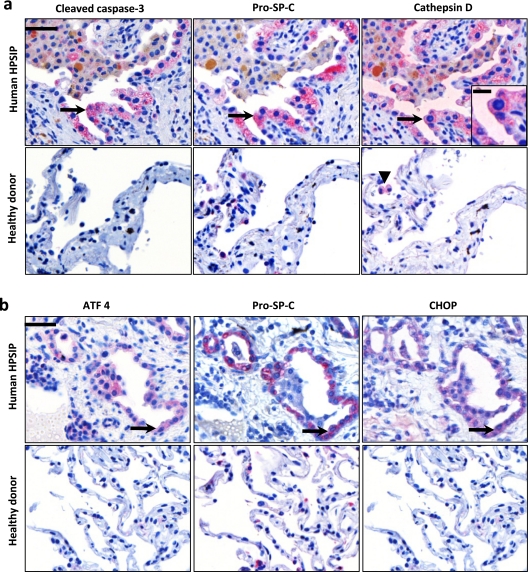
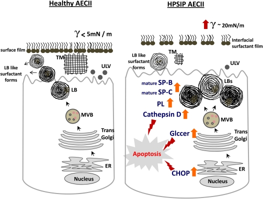
Measurements and main results: Spontaneous and slowly progressive HPSIP was observed in HPS1/2 double mutants, but not in other HPS mutants, with subpleural onset at 3 months and full-blown fibrosis at 9 months. In these mice, extensive surfactant abnormalities were encountered in AECII and were paralleled by early lysosomal stress (cathepsin D induction), late endoplasmic reticulum stress (activating transcription factor-4 [ATF4], C/EBP homologous protein [CHOP] induction), and marked apoptosis. These findings were fully corroborated in human HPSIP. In addition, cathepsin D overexpression resulted in apoptosis of MLE-12 cells and increased proliferation of NIH 3T3 fibroblasts incubated with conditioned medium of the transfected cells.

Conclusions: Extensively impaired surfactant trafficking and secretion underlie lysosomal and endoplasmic reticulum stress with apoptosis of AECII in HPSIP, thereby causing the development of HPSIP.


Figures

Figure 1.
Figure 1.
Development of pulmonary fibrosis only in HPS1/2 double-mutant mice: (a) Representative hematoxylin–eosin (H&E) stainings of complete right lungs of HPS monomutant mice, double-mutant mice, and background wild-type (WT) control mice at the ages of 3 and 9 months. (b) Higher magnification pictures of H&E- and trichrome-stained lungs of all the HPS mutants and WT controls analyzed in this study at the ages of 3 and 9 months. Original magnification, ×200 (scale bar, 100 μm). (c) Quantitative determination of lung hydroxyproline concentration in HPS1/2 double-mutant mice in comparison with WT control mice. *P < 0.05, n = 5 mice per group. n.s. = not significant.
Figure 2.
Figure 2.
Increase in mean linear intercept in Hermansky-Pudlak syndrome (HPS) mice: Graphic representation of the length distribution of airspaces in (a) HPS1/2 mice and (b) HPS1/6 mice as well as the length distribution of septal thickness of (c) HPS1/2 mice and (d) HPS1/6 mice in comparison with respective wild-type (WT) controls. Twenty to 30 regions, covering the entire periphery of one digitally scanned lung slide stained with hematoxylin–eosin, were introduced with grids, comprising a total of 40,000 chords in the horizontal and vertical directions, with a distance of 40 μm. Each caption was analyzed by automated line-to-line intercept analysis, according to the criteria mentioned in Methods. Five 9-month-old mice per group were analyzed and the data are represented as the frequency distribution of either (a and b) alveolar diameter or (c and d) septal thickness. In addition, the conventional parameters “mean linear intercept” and septal thickness are indicated in (e) and (f), respectively. ***P < 0.001,**P < 0.01, *P < 0.05; n.s. = not significant.
Figure 3.
Figure 3.
Loss of surfactant in bronchoalveolar lavage fluid (BALF) of HPS1/2 mice: Western blot analysis of (a) mature surfactant protein (SP)-B and (b) mature SP-C in BALF from Hermansky-Pudlak syndrome (HPS) mice along with their respective wild-type (WT) control mice at the ages of 3 and 9 months. An equal volume (20 μl per sample) of BALF was used for each analysis. (c) Data set showing phospholipid (PL) concentration and the relative amount of large surfactant aggregates (LA content, given as a percentage of total PL) in BALF of HPS1/2 and WT control mice. (d and e) Surface activity of LA from HPS1/2, HPS1/6, and WT control mice was characterized at a PL concentration of 2 mg/ml, employing a pulsating bubble surfactometer. (d) γads (surface tension after 12 s of film adsorption) and (e) γmin (minimal surface tension after 5 min of film oscillation) are given as millinewtons per meter (mN/m). *P < 0.05, **P < 0.01; n = 5 mice per group.
Figure 4.
Figure 4.
Accumulation of hydrophobic surfactant proteins in HPS1/2 lung tissues: (a) Western blot analysis of lung homogenates (LH; equal protein load) of (left) 3-month-old and (right) 9-month-old HPS1/2 double-mutant and wild-type (WT) controls for pro–surfactant protein (SP)-B, mature SP-B, pro–SP-C, and mature SP-C. (b) Western blot analysis of isolated alveolar epithelial type II cells (AECII; equal protein load) and LH for mature SP-B and mature SP-C from 3-month-old HPS1/2 mice and WT controls. [Please note that the blots in (a) and (b) have been developed to show adequate staining for HPS1/2 mouse samples; longer development forwarded staining for mature SP-C also in AECII and LH of WT mice.] Representative blots are from n = 5 mice per group, and three independent experiments are shown. (ch) Densitometry was performed from all Western blot analyses of all HPS mouse lung homogenates. The target protein/β-actin ratio was calculated and is given as a percentage of the respective WT controls. ***P < 0.001,**/##P < 0.01, */#P < 0.05; n = 5 mice per group.
Figure 5.
Figure 5.
Accumulation of surfactant phospholipids in HPS1/2 mouse lungs: (a) Total phospholipids were extracted from lung tissue of all 3- and 9-month-old Hermansky-Pudlak syndrome (HPS) and wild-type (WT) control mice. The resulting phospholipid (PL) concentrations were normalized against their respective protein concentrations and values are expressed as micrograms of phospholipids per milligram of protein. (b and c) Lipidomic study was performed for 9-month-old HPS mice as compared with the respective WT control mice. Columns depicted here represent fatty acid distribution of (b) the phosphatidylcholine (PC) fraction and (c) the relative content of dipalmitoylated phosphatidylcholine (DPPC) within the PC fraction. Values are expressed as the molar percentage (mol %) of the respective lipid class, related to total PC. */#P < 0.05, **/##P < 0.01, ***/###P < 0.001; n.s. = not significant; n = 5 mice per group.
Figure 6.
Figure 6.
Alveolar epithelial type II cell (AECII) stress and apoptosis in HPS1/2 mice: (a) Serial paraffin-embedded lung tissue sections from (top) 9-month-old HPS1/2 mice and (bottom) wild-type (WT) controls were immunostained either for (left) cleaved caspase-3 or (right) pro–surfactant protein (SP)-C. Arrows indicate the same AECII stained for both proteins. (b) Western blot analysis of cleaved caspase-3 in lung homogenates of 3- and 9-month-old HPS1/2 mice in comparison with that of 9-month-old WT controls. (c and d) Western blot analysis of cathepsin D in (c) lung homogenates of HPS1/2 mice (3 and 9 mo of age) and in (d) isolated AECII of HPS1/2 and WT controls (3 mo of age). (e) Immunohistochemistry on serial sections of (top) HPS1/2 and (bottom) WT mouse lungs for (left) cleaved caspase-3, (middle) pro–SP-C, and (right) cathepsin D. Arrows indicate positive staining of AECII for all three proteins. Inset: High-magnification image, illustrating diffuse cytoplasmic staining of cathepsin D within the AECII of HPS1/2 mice. Arrowhead indicates cathepsin D staining in macrophages in a WT mouse lung tissue section. (f) Western blot analysis for activating transcription factor-4 (ATF4) and C/EBP homologous protein (CHOP) in lung homogenates of HPS1/2 mice and age-matched WT controls. (g) Serial sections were stained for either (left) ATF4, (middle) pro–SP-C, or (right) CHOP in 9-month-old HPS1/2 and WT controls. Arrows indicate the same AECII stained positive for all three proteins. Original magnification, ×400 (scale bar, 50 μm). Original magnification of inset, ×800 (scale bar, 20 μm). Representative blots and stainings from each group are shown, from n = 5 mice per group.
Figure 7.
Figure 7.
Apoptosis of alveolar epithelial type II cells (AECII) due to lysosomal and endoplasmic reticulum stress in human Hermansky-Pudlak syndrome–associated interstitial pneumonia (HPSIP): (a) Representative immunohistochemistry performed on serial paraffin-embedded lung tissue sections from (top) human patients with HPS and (bottom) healthy donors for (left) cleaved caspase-3, (middle) pro–surfactant protein (SP)-C, and (right) cathepsin D. Arrows indicate the same AECII stained for all three proteins. Inset: High-magnification image, illustrating diffuse cytoplasmic staining of cathepsin D within the AECII of HPSIP. Arrowhead indicates cathepsin D staining in macrophages in a lung tissue section of a healthy donor. (b) Serial lung tissue sections from (top) human patients with HPS and (bottom) healthy donors were stained for (left) activating transcription factor-4 (ATF4), (middle) pro–SP-C, or (right) C/EBP homologous protein (CHOP). Arrows indicate positive staining of an AECII for all three proteins. Representative sections from patients with HPS1 and four healthy donors are shown. Original magnification, ×400 (scale bar, 50 μm). Original magnification of inset, ×800 (scale bar, 20 μm).
Figure 8.
Figure 8.
Overexpression of cathepsin D (CatD) drives apoptosis of an alveolar epithelial cell line and proliferation of fibroblasts: (a) Total protein extracts prepared from cathepsin D– and empty vector–transfected mouse lung epithelial (MLE)-12 cells as well as cells collected from supernatant after cathepsin D transfection (after 24 h) were subjected to Western blotting for (top) cathepsin D and for (middle) cleaved caspase-3, with equal protein loading (β-actin, bottom). Supernatant from cells transfected with empty vector did not contain enough cells (protein) to be included. (b and c) Conditioned medium from either cathepsin D– or empty vector–transfected MLE-12 cells was applied to an equal number of NIH 3T3 fibroblasts and their viability was assessed by (b) water-soluble tetrazolium salt (WST)-1 assay at various cell numbers or their proliferation was measured by (c) bromodeoxyuridine (BrdU) incorporation. *P < 0.05, **P < 0.01; n.s. = not significant.
Figure 9.
Figure 9.
Proposed model for induction of apoptosis of alveolar epithelial cells type II (AECII) in Hermansky-Pudlak syndrome–associated interstitial pneumonia (HPSIP) lungs: On the left, a healthy AECII is shown, with regular surfactant processing, transport, and secretion. On the right, our data on HPSIP are summarized, in which giant lamellar bodies, defective surfactant transport, and impaired secretion of surfactant are shown. Alveolar surface tension in HPSIP lungs is increased, indicating the reduced surface activity of the alveolar surfactant pool in these lungs. Within the AECII, an increase in cathepsin D, glucosylceramides, and CHOP-induced apoptosis of the cell may result in the development of lung fibrosis. CHOP = C/EBP homologous protein; ER = endoplasmic reticulum; Glccer = glucosylceramides; LBs = lamellar bodies; MVB = multivesicular bodies; PL = phospholipids; SP = surfactant protein; TM = tubular myelin; ULV = unilamellar vesicles.

Similar articles

Cited by

References

    1. Hermansky F, Pudlak P. Albinism associated with hemorrhagic diathesis and unusual pigmented reticular cells in the bone marrow: report of two cases with histochemical studies. Blood 1959;14:162–169. - PubMed
    1. Li W, Rusiniak ME, Chintala S, Gautam R, Novak EK, Swank RT. Murine Hermansky-Pudlak syndrome genes: regulators of lysosome-related organelles. Bioessays 2004;26:616–628. - PubMed
    1. Huizing M, Gahl WA. Disorders of vesicles of lysosomal lineage: the Hermansky-Pudlak syndromes. Curr Mol Med 2002;2:451–467. - PubMed
    1. Anderson PD, Huizing M, Claassen DA, White J, Gahl WA. Hermansky-Pudlak syndrome type 4 (HPS-4): clinical and molecular characteristics. Hum Genet 2003;113:10–17. - PubMed
    1. Wei ML. Hermansky-Pudlak syndrome: a disease of protein trafficking and organelle function. Pigment Cell Res 2006;19:19–42. - PubMed

Publication types

MeSH terms

Substances