. 2010 Jun;30(12):2918-32.
doi: 10.1128/MCB.00131-10. Epub 2010 Apr 5.

Genetic demonstration of a redundant role of extracellular signal-regulated kinase 1 (ERK1) and ERK2 mitogen-activated protein kinases in promoting fibroblast proliferation

Affiliations

Genetic demonstration of a redundant role of extracellular signal-regulated kinase 1 (ERK1) and ERK2 mitogen-activated protein kinases in promoting fibroblast proliferation

Laure Voisin et al. Mol Cell Biol. 2010 Jun.

Abstract

The extracellular signal-regulated kinase 1 and 2 (ERK1/2) mitogen-activated protein (MAP) kinase signaling pathway plays an important role in the proliferative response of mammalian cells to mitogens. However, the individual contribution of the isoforms ERK1 and ERK2 to cell proliferation control is unclear. The two ERK isoforms have similar biochemical properties and recognize the same primary sequence determinants on substrates. On the other hand, analysis of mice lacking individual ERK genes suggests that ERK1 and ERK2 may have evolved unique functions. In this study, we used a robust genetic approach to analyze the individual functions of ERK1 and ERK2 in cell proliferation using genetically matched primary embryonic fibroblasts. We show that individual loss of either ERK1 or ERK2 slows down the proliferation rate of fibroblasts to an extent reflecting the expression level of the kinase. Moreover, RNA interference-mediated silencing of ERK1 or ERK2 expression in cells genetically disrupted for the other isoform similarly reduces cell proliferation. We generated fibroblasts genetically deficient in both Erk1 and Erk2. Combined loss of ERK1 and ERK2 resulted in a complete arrest of cell proliferation associated with G(1) arrest and premature replicative senescence. Together, our findings provide compelling genetic evidence for a redundant role of ERK1 and ERK2 in promoting cell proliferation.


Figures
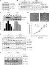
FIG. 1.
FIG. 1.
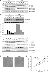
Disruption of Erk1 gene attenuates the proliferation rate of primary MEFs in a CD-1 background. (A) Relative expression of ERK1 and ERK2 isoforms in CD-1 MEFs. Total lysates from exponentially proliferating CD-1 MEFs (n = 4) were analyzed by immunoblotting using antibodies specific for phosphorylated (P) ERK1/ERK2, total ERK1/ERK2, and α-tubulin. (B) MEFs derived from Erk1/ embryos (ERK1−/−) or wild-type control littermates (WT) were made quiescent and then stimulated with 10% NBCS for the indicated times. Total lysates were analyzed by immunoblotting. (C) Kinetics of ERK2 activity. ERK1−/− or WT MEFs were treated as in panel B. Cell lysates were prepared, and the phosphotransferase activity of endogenous ERK2 was measured by immune complex kinase assay using MBP and [γ-32P]ATP as substrates. The top panel shows an autoradiogram and Coomassie staining of MBP. The lower panel is a bar histogram showing the quantification of 32P incorporation into MBP. (D) Morphology of ERK1−/− and WT MEFs. (E) ERK1−/− or WT MEFs were treated as in panel B. The expression and phosphorylation of Mnk1 and p38 were analyzed by immunoblotting. (F) ERK1−/− or WT MEFs were made quiescent, pretreated with SB203580 (10 μM) or DMSO (0.1%) for 30 min, and then stimulated with 10% NBCS for 15 min. Total lysates were analyzed by immunoblotting using antibodies specific for phosphorylated MNK1, phosphorylated MK2, and α-tubulin. (G) Proliferation rates of P3 MEFs prepared from Erk1/ or wild-type littermate embryos were measured by the MTT assay. Values are expressed as fold increase in cell number and correspond to the mean ± standard error of the mean of five independent MEF preparations. The data are representative of three different experiments. *, P < 0.05.
FIG. 2.
FIG. 2.
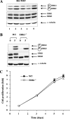
Cell proliferation analysis of Erk1/ primary MEFs in C57BL/6J background. (A) Relative expression of ERK1 and ERK2 isoforms in C57BL/6J MEFs. (B) MEFs derived from ERK1−/− or wild-type (WT) control littermate embryos were made quiescent and then stimulated with 10% NBCS for 5 min. The expression and phosphorylation of ERK1/ERK2 were analyzed by immunoblotting. (C) Proliferation curves of MEFs prepared from Erk1/ or wild-type littermate embryos in a C57BL/6J background. Values correspond to the mean ± standard error of the mean of six independent MEF preparations. The data are representative of three different experiments.
FIG. 3.
FIG. 3.
Rescue of Erk2/ mice from early embryonic lethality by tetraploid aggregation. Each eight-cell-stage embryo isolated from intercrosses of CD-1 Erk2+/ mice was aggregated with two tetraploid four-cell-stage wild-type embryos in vitro. After development to the blastocyst stage, chimeric embryos were transferred into the uteri of pseudopregnant females. (A) Macroscopic views of wild-type and Erk2/ embryos at E13.5 (upper panels) and contribution of wild-type GFP-positive cells to the placenta and yolk sac endoderm (bottom panels). (B) Genotyping analysis of embryos obtained from tetraploid aggregation experiments. (C) Number and genotype of embryos obtained by tetraploid aggregation rescue experiments at E13.5.
FIG. 4.
FIG. 4.
Disruption of Erk2 slows down the proliferation of primary MEFs in a CD-1 background. MEFs derived from ERK2−/− or wild-type (WT) control littermates were made quiescent and then stimulated with 10% NBCS for the indicated times. (A) The expression and phosphorylation of ERK1/ERK2 were analyzed by immunoblotting. (B) Kinetics of ERK1 activity. The phosphotransferase activity of endogenous ERK1 was measured by immune complex kinase assay as described in the legend of Fig. 1. (C) The expression and phosphorylation of Mnk1 and p38 were analyzed by immunoblotting. (D) Morphology of ERK2−/− and wild-type MEFs. (E) Proliferation curves of P3 MEFs prepared from Erk2/ or wild-type littermate embryos obtained by tetraploid aggregation experiments. Values correspond to the mean ± standard error of the mean of two independent MEF preparations. The data are representative of three different experiments. *, P < 0.05.
FIG. 5.
FIG. 5.
RNAi silencing of ERK1 or ERK2 restrains the proliferation of MEFs expressing a single ERK isoform. Erk1/ or Erk2/ MEFs were infected with lentiviruses encoding shRNAs to the ERK2 or ERK1 gene, respectively, and populations of transduced cells were selected with puromycin. After 72 h, the cells were replated at low density (day 0) to measure the rate of cell proliferation. A nontarget shRNA (NT) was used as a control. (A) The expression of ERK1/ERK2 and phospho-ERK1/2 was analyzed by immunoblotting at day 3. (B) Cell proliferation was measured by the MTT assay. Values are expressed as fold increase in cell number and correspond to the mean ± standard error of the mean of triplicate determinations. The data are representative of three different experiments.
FIG. 6.
FIG. 6.
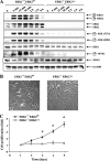
Genetic inactivation of Erk1 and Erk2 genes arrests the proliferation of MEFs. (A) Erk1+/+; Erk2flox/flox and Erk1/; Erk2Δ/Δ MEFs were made quiescent and then stimulated with 10% NBCS for the indicated times. Total lysates were analyzed by immunoblotting using antibodies specific for phospho-ERK1/ERK2, total ERK1/ERK2, phospho-RSK (Thr573), phospho-RSK (Ser380), total RSK1, phospho-Mnk1, total Mnk1, and GAPDH. (B) Morphology of Erk1+/+; Erk2flox/flox and Erk1/; Erk2Δ/Δ MEFs. (C) Proliferation rates of Erk1+/+; Erk2flox/flox and Erk1/; Erk2Δ/Δ MEFs at P5 were measured by the MTT assay. Values are expressed as fold increase in cell number and correspond to the mean ± standard error of the mean of three independent experiments. *, P < 0.05.
FIG. 7.
FIG. 7.
Immortalization of Erk1/ and Erk2/ MEFs. MEF preparations derived from individual embryos were cultured according to a 3T3 protocol. The cells were counted, and the cumulative number of total population doublings was plotted at each passage.
FIG. 8.
FIG. 8.
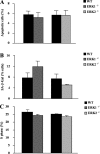
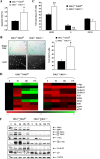
Impact of the loss of ERK1 or ERK2 on apoptosis, replicative senescence, and cell cycle progression of primary MEFs. Proliferating MEFs (P3) derived from CD-1 Erk1/ or Erk2/ mice or from their respective wild-type control littermate embryos were harvested at day 3 after plating. (A) Apoptosis was evaluated by two-color annexin V staining. Results are expressed as the mean ± standard error of the mean (n = 3). (B) Analysis of replicative senescence. Proliferating MEFs were fixed and stained for SA-β-Gal activity. Results are presented as the mean percentage ± standard error of the mean of SA-β-Gal-positive cells for wild-type (n = 4), ERK1−/− (n = 5), and ERK2−/− (n = 2) MEFs. (C) Cell cycle analysis. Asynchronously proliferating MEFs were pulsed with BrdU for 1 h prior to harvesting at day 3 after seeding. The cells were fixed and stained with PI, and the percentage of cells in S phase was determined by FACS analysis. Results are expressed as the mean ± standard error of the mean (n = 3).
FIG. 9.
FIG. 9.
Combined loss of ERK1 and ERK2 impairs cell cycle progression and promotes replicative senescence. Proliferating Erk1+/+; Erk2flox/flox and Erk1/; Erk2Δ/Δ MEFs (P5) were harvested at day 3 after plating. (A) Apoptosis was measured by two-color annexin V staining. Results are expressed as the mean ± standard error of the mean (n = 3). (B) Replicative senescence was assayed by staining for SA-β-Gal activity. Bright-field and fluorescence (DAPI) photographs of representative fields are shown. The graph at right shows the quantification of SA-β-Gal activity. Results are presented as the mean percentage ± standard error of the mean of SA-β-Gal-positive cells (n = 3). (C) Cell cycle analysis. Asynchronously proliferating MEFs were pulsed with BrdU for 1 h prior to harvesting. The percentage of cells in S phase was determined by FACS analysis. Results are expressed as the mean ± standard error of the mean (n = 3). (D) Expression of cell cycle-regulatory genes. Erk1+/+; Erk2flox/flox and Erk1/; Erk2Δ/Δ MEFs were made quiescent and then stimulated with 10% NBCS for the indicated times. The expression of cell cycle genes was analyzed by quantitative PCR and is displayed in a heat map. Results represent the mean of two experiments. (E) Relative expression levels of the genes named in panel D. Gene expression levels were plotted as fold change over the value observed in Erk1+/+; Erk2flox/flox cells at time zero, which was arbitrarily set to 1. (F) Immunoblot analysis of cell cycle-regulatory genes. Results are representative of two experiments.
FIG. 9.
FIG. 9.
Combined loss of ERK1 and ERK2 impairs cell cycle progression and promotes replicative senescence. Proliferating Erk1+/+; Erk2flox/flox and Erk1/; Erk2Δ/Δ MEFs (P5) were harvested at day 3 after plating. (A) Apoptosis was measured by two-color annexin V staining. Results are expressed as the mean ± standard error of the mean (n = 3). (B) Replicative senescence was assayed by staining for SA-β-Gal activity. Bright-field and fluorescence (DAPI) photographs of representative fields are shown. The graph at right shows the quantification of SA-β-Gal activity. Results are presented as the mean percentage ± standard error of the mean of SA-β-Gal-positive cells (n = 3). (C) Cell cycle analysis. Asynchronously proliferating MEFs were pulsed with BrdU for 1 h prior to harvesting. The percentage of cells in S phase was determined by FACS analysis. Results are expressed as the mean ± standard error of the mean (n = 3). (D) Expression of cell cycle-regulatory genes. Erk1+/+; Erk2flox/flox and Erk1/; Erk2Δ/Δ MEFs were made quiescent and then stimulated with 10% NBCS for the indicated times. The expression of cell cycle genes was analyzed by quantitative PCR and is displayed in a heat map. Results represent the mean of two experiments. (E) Relative expression levels of the genes named in panel D. Gene expression levels were plotted as fold change over the value observed in Erk1+/+; Erk2flox/flox cells at time zero, which was arbitrarily set to 1. (F) Immunoblot analysis of cell cycle-regulatory genes. Results are representative of two experiments.
FIG. 10.
FIG. 10.
The rate of cell proliferation is tightly correlated with the level of phosphorylated ERK1/ERK2 in MEFs. The extent of phospho-ERK1/2 staining was quantified at 5 min after NBCS stimulation and is expressed as a percentage of the level observed in control littermate MEFs (n ≥ 2). The rate of cell proliferation was estimated from the MTT assay at day 5 or 6 (last day of the experiment) and is expressed as a percentage of the proliferation measured in control MEFs (n = 3).

Similar articles

Cited by

References

    1. Anjum, R., and J. Blenis. 2008. The RSK family of kinases: emerging roles in cellular signalling. Nat. Rev. Mol. Cell Biol. 9:747-758. - PubMed
    1. Balmanno, K., S. D. Chell, A. S. Gillings, S. Hayat, and S. J. Cook. 2009. Intrinsic resistance to the MEK1/2 inhibitor AZD6244 (ARRY-142886) is associated with weak ERK1/2 signalling and/or strong PI3K signalling in colorectal cancer cell lines. Int. J. Cancer. 125:2332-2341. - PubMed
    1. Bost, F., M. Aouadi, L. Caron, P. Even, N. Belmonte, M. Prot, C. Dani, P. Hofman, G. Pages, J. Pouyssegur, Y. Le Marchand-Brustel, and B. Binetruy. 2005. The extracellular signal-regulated kinase isoform ERK1 is specifically required for in vitro and in vivo adipogenesis. Diabetes 54:402-411. - PubMed
    1. Boulton, T. G., and M. H. Cobb. 1991. Identification of multiple extracellular signal-regulated kinases (ERKs) with antipeptide antibodies. Cell Regul. 2:357-371. - PMC - PubMed
    1. Boulton, T. G., S. H. Nye, D. J. Robbins, N. Y. Ip, E. Radziejewska, S. D. Morgenbesser, R. A. DePinho, N. Panayotatos, M. H. Cobb, and G. D. Yancopoulos. 1991. ERKs: a family of protein-serine/threonine kinases that are activated and tyrosine phosphorylated in response to insulin and NGF. Cell 65:663-675. - PubMed

Publication types

MeSH terms

Substances

LinkOut - more resources