. 2010 May;120(5):1762-73.
doi: 10.1172/JCI40891. Epub 2010 Apr 1.

IL-17 is essential for host defense against cutaneous Staphylococcus aureus infection in mice

Affiliations

IL-17 is essential for host defense against cutaneous Staphylococcus aureus infection in mice

John S Cho et al. J Clin Invest. 2010 May.

Abstract

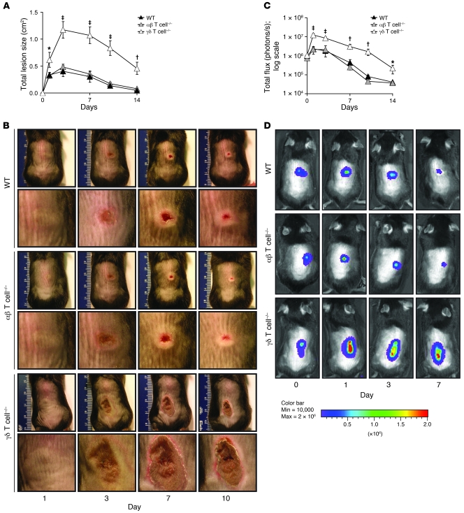
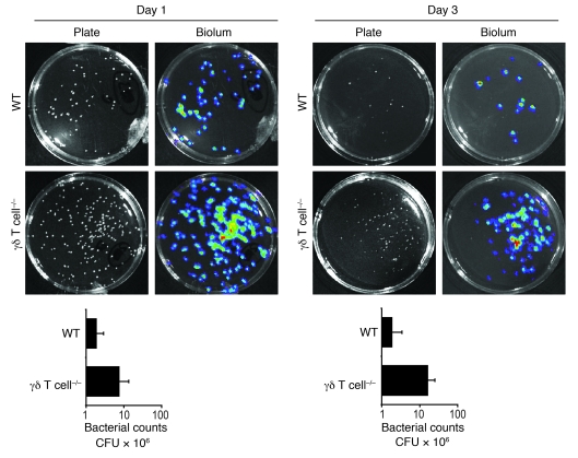
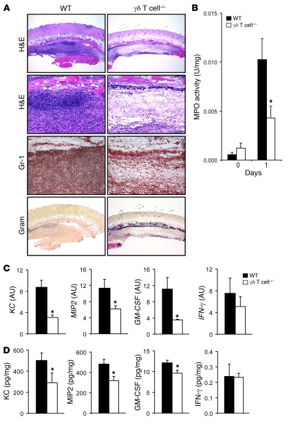
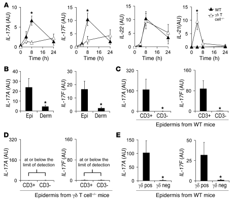
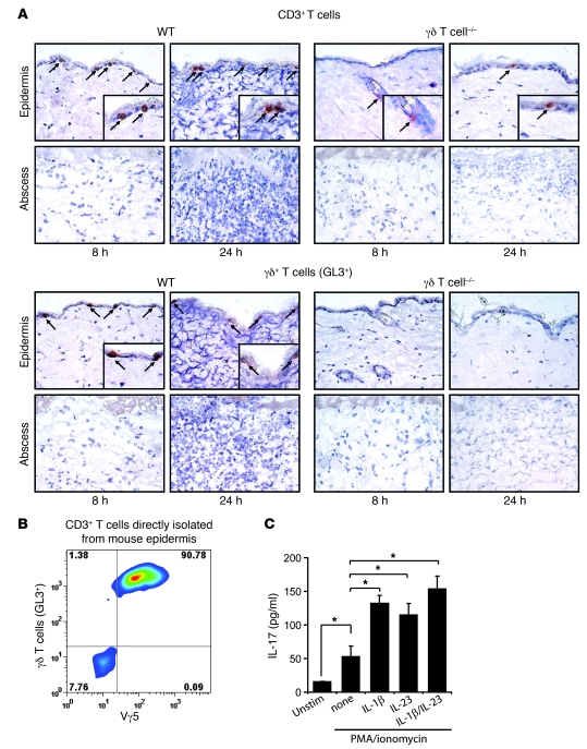
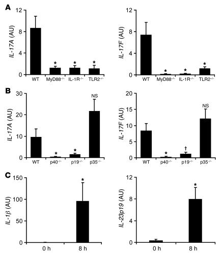
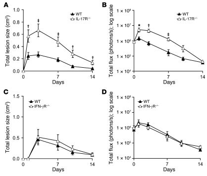
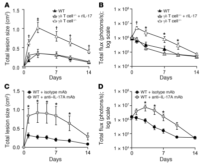
Staphylococcus aureus is the most common cause of skin and soft tissue infections, and rapidly emerging antibiotic-resistant strains are creating a serious public health concern. If immune-based therapies are to be an alternative to antibiotics, greater understanding is needed of the protective immune response against S. aureus infection in the skin. Although neutrophil recruitment is required for immunity against S. aureus, a role for T cells has been suggested. Here, we used a mouse model of S. aureus cutaneous infection to investigate the contribution of T cells to host defense. We found that mice deficient in gammadelta but not alphabeta T cells had substantially larger skin lesions with higher bacterial counts and impaired neutrophil recruitment compared with WT mice. This neutrophil recruitment was dependent upon epidermal Vgamma5+ gammadelta T cell production of IL-17, but not IL-21 and IL-22. Furthermore, IL-17 induction required IL-1, TLR2, and IL-23 and was critical for host defense, since IL-17R-deficient mice had a phenotype similar to that of gammadelta T cell-deficient mice. Importantly, gammadelta T cell-deficient mice inoculated with S. aureus and treated with a single dose of recombinant IL-17 had lesion sizes and bacterial counts resembling those of WT mice, demonstrating that IL-17 could restore the impaired immunity in these mice. Our study defines what we believe to be a novel role for IL-17-producing epidermal gammadelta T cells in innate immunity against S. aureus cutaneous infection.


Figures

Figure 1
Figure 1. γδ but not αβ T cell–deficient mice develop markedly larger skin lesions compared with WT mice in response to cutaneous challenge with S. aureus.
γδ T cell–deficient, αβ T cell–deficient, and WT mice were inoculated intradermally with S. aureus SH1000 strain. (A) Mean total lesion size (cm2) ± SEM. (B) Representative lesions for each mouse strain. Shown are entire dorsal backs (top, mm ruler shown for scale) and close-ups of lesions (bottom). (C) Mean total flux (photons/s) ± SEM. (D) Representative in vivo bioluminescence. Data are from 2 experiments with at least 6 mice/group per experiment. *P < 0.05, P < 0.01, P < 0.001, γδ T cell–deficient versus WT (Student’s t test).
Figure 2
Figure 2. γδ but not αβ T cell–deficient mice develop higher bacterial counts compared with WT mice in response to cutaneous challenge with S. aureus.
Representative bacterial culture plates after overnight culture with or without bioluminescence, and CFUs of S. aureus recovered from 8-mm lesional punch biopsies on days 1 and 3 (n = 5 per group).
Figure 3
Figure 3. γδ T cell–deficient mice have markedly impaired neutrophil recruitment and production of neutrophil chemokines and granulopoiesis factors in response to cutaneous challenge with S. aureus.
γδ T cell–deficient and WT mice were inoculated intradermally with S. aureus. (A) Representative photomicrographs of sections labeled with H&E stain and anti–Gr-1 mAb (immunoperoxidase method) and Gram stain of lesional skin at 1 day after inoculation. Original magnification, ×40 (H&E, top, and Gram); ×200 (H&E, bottom, and Gr-1). (B) Mean MPO activity (U/mg tissue weight) ± SEM of lesional skin. (C) Mean level of mRNA (AU) ± SEM of KC, MIP2, GM-CSF, and IFN-γ from lesional skin homogenates of skin biopsies performed by Q-PCR 8 hours after S. aureus inoculation. (D) Mean protein levels (pg/mg tissue weight) ± SEM of KC, MIP2, GM-CSF, and IFN-γ from lesional skin homogenates of skin biopsies performed at 8 hours after S. aureus inoculation. Data are from 2 experiments with at least 5 mice/group per experiment. *P < 0.05, γδ T cell–deficient versus WT (Student’s t test).
Figure 4
Figure 4. Impaired induction of IL-17, but not IL-22 or IL-21, in γδ T cell–deficient mice after S. aureus cutaneous challenge.
Mice were inoculated intradermally with S. aureus, and the mean level of mRNA (AU) ± SEM of IL-17A and IL-17F by Q-PCR were determined from (A) lesional skin homogenates of skin biopsies from γδ T cell–deficient and WT mice performed 0, 4, 8, and 24 hours after inoculation (levels of IL-22 and IL-21 also evaluated); (B) epidermal and dermal split specimens of skin biopsies from WT mice performed 8 hours after inoculation; (C and D) positively selected cells (CD3+) and negative fractions (CD3) of epidermal cell suspensions after enrichment for CD3+ cells (MACS) from skin biopsies taken from WT (C) or γδ T cell–deficient (D) mice performed 8 hours after inoculation; and (E) positively selected cells (γδ pos) and negative fractions (γδ neg) of epidermal cell suspensions after enrichment for γδ T cells (MACS) from skin biopsies taken from WT mice 8 hours after inoculation. Values in D were at or below the limit of detection. Data are from 2 experiments with at least 5 mice/group per experiment. *P < 0.05 between experimental groups (Student’s t test).
Figure 5
Figure 5. γδ T cells expressing the Vγ5 chain are present in the epidermis of WT mice after skin challenge with S. aureus and produce IL-17 after in vitro stimulation.
(A) Representative photomicrographs labeled with specific mAbs for CD3+ (arrows) and GL3+ (i.e., γδ T) cells (arrows; immunoperoxidase method) of histologic sections from skin biopsies from γδ T cell–deficient and WT mice performed 8 and 24 hours after skin inoculation with S. aureus. Insets show positively labeled cells. Isotype controls are shown in Supplemental Figure 2. Original magnification, ×400; ×800 (insets). (B) Epidermal cell suspensions of skin biopsies from normal uninfected WT mouse skin were labeled with specific mAbs for CD3, γδ T cells (GL3+), and Vγ5, and expression was analyzed by flow cytometry. Cells were gated on CD3+ cells, and the percentage of cells in each quadrant is indicated. (C) Purified GL3+ epidermal γδ T cells (106 cells/ml) from uninfected C57BL/6 mice were left unstimulated or were activated with PMA/ionomycin and cultured in the presence or absence of 20 ng/ml IL-1β, 20 ng/ml IL-23, or 20 ng/ml of both IL-1β and IL-23. Supernatants were collected after 24 hours for analysis of IL-17 protein levels (pg/ml) by ELISA. Data are from 2 experiments with at least 4 mice/group per experiment. *P < 0.05 (Student’s t test).
Figure 6
Figure 6. IL-17A and IL-17F mRNA production in response to S. aureus skin challenge is dependent upon IL-1R, TLR2, and IL-23.
Mice were inoculated intradermally with S. aureus, and the mean level of mRNA (AU) ± SEM of IL-17A and IL-17F by Q-PCR were determined from lesional skin homogenates of skin biopsies from (A) MyD88-deficient, IL-1R–deficient, TLR2-deficient, and WT mice and (B) IL-12/23p40–deficient, IL-23p19–deficient, IL-12p35–deficient, and WT mice, performed 8 hours after inoculation. (C) Mean mRNA level (AU) ± SEM of IL-1β and IL-23p19 from lesional skin homogenates of skin biopsies performed 8 hours after inoculation by Q-PCR. Data are from 2 experiments with at least 5 mice/group per experiment. *P < 0.05, P < 0.01 versus appropriate WT or 0-hour control (Student’s t test).
Figure 7
Figure 7. IL-17R–deficient mice, but not IFN-γR–deficient mice, develop increased lesion sizes and bacterial counts resembling those of γδ T cell–deficient mice in response to S. aureus cutaneous challenge.
IL-17R–deficient, IFN-γR–deficient, and WT mice were inoculated intradermally with S. aureus. (A and C) Mean total lesion size (cm2) ± SEM. (B and D) Mean total flux (photons/s) ± SEM. Data are from 2 experiments with at least 6 mice/group per experiment. *P < 0.05, P < 0.01, P < 0.001, IL-17R–deficient versus WT (Student’s t test).
Figure 8
Figure 8. Administration of exogenous rIL-17 with the S. aureus inoculum restores an effective immune response against S. aureus in γδ T cell–deficient mice.
(A and B) γδ T cell–deficient and WT mice were inoculated intradermally with S. aureus along with administration of a single dose of rIL-17 (1,000 ng/100 μl) or vehicle alone to γδ T cell–deficient mice. (A) Mean total lesion size (cm2) ± SEM. (B) Mean total flux (photons/s) ± SEM. Data are from 2 experiments with at least 6 mice/group per experiment. (C and D) WT mice (n = 7 mice/group) were inoculated intradermally with S. aureus along with an anti–IL-17A neutralizing mAb or an isotype control mAb. (C) Mean total lesion size (cm2) ± SEM. (D) Mean total flux (photons/s) ± SEM. *P < 0.05, P < 0.01, P < 0.001 versus all other groups (Student’s t test).

Similar articles

Cited by

References

    1. McCaig LF, McDonald LC, Mandal S, Jernigan DB. Staphylococcus aureus-associated skin and soft tissue infections in ambulatory care. . Emerg Infect Dis. 2006;12(11):1715–1723. - PMC - PubMed
    1. Moran GJ, et al. Methicillin-resistant S. aureus infections among patients in the emergency department. . N Engl J Med. 2006;355(7):666–674. doi: 10.1056/NEJMoa055356. - DOI - PubMed
    1. Klevens RM, et al. Invasive methicillin-resistant Staphylococcus aureus infections in the United States. . JAMA. 2007;298(15):1763–1771. doi: 10.1001/jama.298.15.1763. - DOI - PubMed
    1. Deleo FR, Chambers HF. Reemergence of antibiotic-resistant Staphylococcus aureus in the genomics era. . J Clin Invest. 2009;119(9):2464–2474. doi: 10.1172/JCI38226. - DOI - PMC - PubMed
    1. Boucher HW, Corey GR. Epidemiology of methicillin-resistant Staphylococcus aureus. . Clin Infect Dis. 2008;46(Suppl 5):S344–S349. doi: 10.1086/533590. - DOI - PubMed

Publication types