. 2010 May 1;184(9):4898-906.
doi: 10.4049/jimmunol.1000142. Epub 2010 Mar 29.

IL-17 enhancement of the IL-6 signaling cascade in astrocytes

Affiliations

IL-17 enhancement of the IL-6 signaling cascade in astrocytes

Xiangyu Ma et al. J Immunol. .

Abstract

Astrocytes have important physiological roles in CNS homeostasis and serve as a bridge between the CNS and immune system. IL-17 and IL-6 are important in many CNS disorders characterized by neuroinflammation. We examined the role of IL-17 on the IL-6 signaling cascade in primary astrocytes. IL-17 functioned in a synergistic manner with IL-6 to induce IL-6 expression in astrocytes. The synergistic effect involved numerous signaling pathways including NF-kappaB, JNK MAPK, and p38 MAPK. The NF-kappaB pathway inhibitor BAY-11, JNK inhibitor JNKi II, and p38 inhibitor SB203580 suppressed the synergistic effect of IL-6 and IL-17 on IL-6 expression. IL-17 synergized with IL-6 to enhance the recruitment of activated NF-kappaB p65, c-Fos, c-Jun, and the histone acetyltransferases CREB-binding protein and p300 to the IL-6 promoter in vivo to induce IL-6 transcription. This was accompanied by enhanced acetylation of histones H3 and H4 on the IL-6 promoter. Moreover, we elucidated an important role for suppressor of cytokine signaling (SOCS) 3 in IL-17 enhancement of IL-6 signaling in astrocytes. SOCS3 small interfering RNA knockdown and SOCS3 deletion in astrocytes augmented the synergistic effect of IL-6 and IL-17 due to an enhancement of activation of the NF-kappaB and MAPK pathways. These results indicate that astrocytes can serve as a target of Th17 cells and IL-17 in the CNS, and SOCS3 participates in IL-17 functions in the CNS as a negative feedback regulator.


Figures
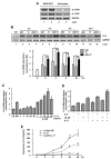
Figure 1
Figure 1. IL-17 Enhances IL-6/sIL-6R-mediated IL-6 Expression in Primary Astrocytes
A, IL-17RA and IL-17RC mRNA expression was determined by RT-PCR in RAW264.7 cells (positive control), and primary astrocytes, in duplicate. B, Primary astrocytes were treated with medium (UN), IL-6/R (IL-6, 10 ng/ml and sIL-6R, 25 ng/ml), IL-17 (25 ng/ml) or IL-6/R plus IL-17 for up to 24 h, and levels of IL-6 and GAPDH mRNA expression were determined by RT-PCR and QRT-PCR. C, Primary astrocytes were treated with medium (UN), IL-6/R, different concentrations of IL-17 (1-50 ng/ml) or IL-6/R + IL-17 (1-50 ng/ml) for 4 h, and levels of IL-6 mRNA expression were determined by QRT-PCR. D, Primary astrocytes were treated with medium (UN), IL-6, sIL-6R or IL-17 in various combinations for 4 h, and levels of IL-6 mRNA expression were determined by QRT-PCR. E, Primary astrocytes were treated with medium (UN), IL-6/R, IL-17 or IL-6/R plus IL-17 for 2 - 48 h, and supernatants were analyzed for IL-6 protein using ELISA. Experiments shown are representative of at least three experiments. All data are the mean ± SD of three experiments. *, p ≤0.05; **, p ≤0.01.
Figure 2
Figure 2. IL-17 Synergizes with IL-6/R for Activation of the NF-κB and MAPK Pathways
A, Astrocytes were incubated with medium (UN), IL-6/R, IL-17 or IL-6/R plus IL-17 for 15 and 30 min, and then cell lysates were immunoblotted with antibodies against IκBα and GAPDH. The basal level of the untreated sample was set at 100, and the percentage change of IκBα upon IL-6/R, IL-17 or IL-6/R + IL-17 treatment compared with the basal value. B, Astrocytes were incubated with medium (UN), IL-6/R, IL-17 or IL-6/R plus IL-17 for 30 and 60 min, and then cell lysates were immunoblotted with antibodies against phospho-p65 Ser 536, p65 and GAPDH. C, Astrocytes were incubated with medium (UN), IL-6/R, IL-17 or IL-6/R plus IL-17 for 15 and 30 min, and then cell lysates were immunoblotted with antibodies against phospho-ERK1/2, ERK1/2 and GAPDH. D, Astrocytes were incubated with medium (UN), IL-6/R, IL-17 or IL-6/R and IL-17 for 30 min and 60 min, and then cell lysates were immunoblotted with antibodies against phospho-p38, p38 and GAPDH. E, Astrocytes were incubated with medium (UN), IL-6/R, IL-17 or IL-6/R plus IL-17 for 30 and 60 min, and then cell lysates were immunoblotted with antibodies against phospho-JNK, JNK and GAPDH. The basal level of the untreated sample was set at 1.0 and fold activation upon IL-6/R, IL-17 or IL-6/R + IL-17 treatment was compared with that value (B, C, D and E). Representative of at least three experiments.
Figure 3
Figure 3. IL-17 and IL-6/R Induction of IL-6 Depends on NF-κB, p38 and JNK MAPK Activity
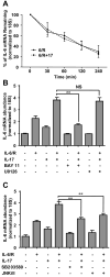
A, Astrocytes were cultured in absence or presence of IL-17 followed by IL-6/R treatment for 4 h. Actinomycin D (5 ng/ml) was then added, and cells harvested at 0, 30, 60, 120, and 240 min after addition. The abundance of IL-6 mRNA was determined by QRT-PCR. B, C, DMSO vehicle, BAY 11 (5 μM), U0126 (10 μM), SB203580 (10 μM) or JNKi II (10 μM) were added to cultures 1 h before cytokine addition, and then astrocytes were incubated with medium, IL-6/R, IL-17 or IL-6/R plus IL-17 for 4 h. Levels of IL-6 mRNA expression were determined by QRT-PCR. All data are the mean ± SD of three experiments. **, p ≤ 0.01; NS = not significant.
Figure 4
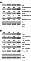
Figure 4. IL-6/R plus IL-17 Enhances Recruitment of p65, P-p65, c-Jun, c-Fos, CBP, p300, and RNA Pol II to the IL-6 Promoter
A, Primary astrocytes were treated with medium (UN), IL-6/R, IL-17 or IL-6/R plus IL-17 for 90 min, and then cells were cross-linked with formaldehyde. Soluble chromatin was subjected to immunoprecipitation with Abs against p65, P-p65, c-Fos, c-Jun, or normal rabbit IgG. PCR analysis of the positive control (input) indicates that soluble chromatin samples obtained from each time point had equal amounts of chromatin fragments containing the IL-6 promoter. B, Primary astrocytes were treated as above. Soluble chromatin was subjected to immunoprecipitation with Abs against histone acetylation (Ac-H3 and Ac-H4), p300, CBP, RNA Pol II or normal rabbit IgG. The basal level of the untreated sample was set at 1.0 and fold activation upon IL-6/R, IL-17 or IL-6/R plus IL-17 treatment was compared with that value. Representative of at least three experiments.
Figure 5
Figure 5. IL-17 Enhances IL-6-mediated SOCS3 Expression in Primary Astrocytes, and SOCS3 is a Negative Regulator of the Synergistic Effect of IL-6 and IL-17
A, Astrocytes were treated with medium (UN), IL-6/R, IL-17 or IL-6/R plus IL-17 for 2 - 8 h, and levels of SOCS3 mRNA expression were determined by QRT-PCR. B and C, Astrocytes were transfected with SOCS3 siRNA (100 nM) or siRNA control (100 nM) for 48 h. Transfected cells were then treated with medium (UN) or IL-6/R plus IL-17 for 4 h. Levels of SOCS3 (B) and IL-6 (C) mRNA expression was determined by QRT-PCR. D, Astrocytes were transfected with SOCS3 siRNA or siRNA control for 48 h. Transfected cells were then treated with medium (UN) or IL-6/R plus IL-17 for 24 h, and supernatants analyzed for IL-6 protein by ELISA. E, SOCS3 floxed astrocytes were infected with GFP as control or GFP-Cre for deletion. After 48 h in culture, cells were harvested. Digestion of genomic DNA distinguishes the full-length (fl) and excised alleles (Δ). F, SOCS3 floxed astrocytes were infected with GFP as control or GFP-Cre for deletion. After 48 h in culture, the cells were treated with medium (UN), IL-6/R, IL-17 or IL-6/R plus IL-17 for 4 h, and levels of IL-6 mRNA expression determined by QRT-PCR. G, SOCS3 floxed astrocytes were infected with GFP as control or GFP-Cre for deletion. After 48 h in culture, cells were incubated with medium (UN), IL-6/R, IL-17 or IL-6/R plus IL-17 for 16 or 24 h, and supernatants analyzed for IL-6 protein by ELISA. All data are the mean ± SD of three experiments. *, p ≤ 0.05;**, p ≤ 0.01.
Figure 6
Figure 6. Enhanced Activation of NF-κB and MAPK Pathways in SOCS3 Deficient Astrocytes
To evaluate NF-κB and MAPK activation in the absence or presence of SOCS3, SOCS3 floxed astrocytes were infected with GFP as control or GFP-Cre for deletion. After 48 h in culture, cells were treated with medium (UN) or IL-6/R plus IL-17 for 15, 30, 60 or 120 min, and then cell lysates were immunoblotted with antibodies against phospho-p65 Ser 536, p65, phospho-p38, p38, phospho-JNK, JNK or GAPDH. The basal level of the untreated GFP infected sample was set at 1.0 and fold activation upon IL-6/R plus IL-17 treatment compared with that value. Representative of at least three experiments.
Figure 7
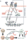
Figure 7. Proposed Model of IL-17 Enhancement of the IL-6 Signaling Cascade in Astrocytes
A, IL-6/R and IL-17 activate the NF-κB and MAPK pathway, which then induce IL-6 gene expression. The synergistic effect of these two mediators depends on NF-κB, p38 and JNK MAPK activity. Activated NF-κB p65, c-Fos and c-Jun bind to the IL-6 promoter. Concurrent with NF-κB and MAPK recruitment, IL-6/R and IL-17 leads to the recruitment of coactivators CBP and p300, modifications in AcH3 and AcH4, and recruitment of RNA Pol II to the IL-6 promoter, which results in transcriptional activation of the IL-6 gene. IL-17 can also enhance IL-6/R induced SOCS3 expression, and SOCS3 inhibits IL-6/R plus IL-17-induced NF-κB and MAPK activation, which results in a reduction of IL-6 gene expression in astrocytes. B, Naive CD4+ T cells, after activation by signaling through the T cell receptor and co-stimulatory molecules, can differentiate into Th17 cells in the presence of IL-6, TGF-β, IL-1 and IL-23. IL-17 together with IL-6/R triggers a positive-feedforward loop of IL-6 expression in astrocytes, which may also influence Th17 cell differentiation. SOCS3 participates in these processes as a negative feedback regulator. See text for details.

Similar articles

Cited by

References

    1. Iadecola C, Nedergaard M. Glial regulation of the cerebral microvasculature. Nat Neurosci. 2007;10:1369–1376. - PubMed
    1. Dong Y, Benveniste EN. Immune function of astrocytes. Glia. 2001;36:180–190. - PubMed
    1. Sofroniew MV. Molecular dissection of reactive astrogliosis and glial scar formation. Trends Neurosci. 2009;32:638–647. - PMC - PubMed
    1. Williams A, Piaton G, Lubetzki C. Astrocytes--friends or foes in multiple sclerosis? Glia. 2007;55:1300–1312. - PubMed
    1. Allen NJ, Barres BA. Neuroscience: Glia - more than just brain glue. Nature. 2009;457:675–677. - PubMed

Publication types

MeSH terms