. 2010 May;137(9):1441-50.
doi: 10.1242/dev.047654. Epub 2010 Mar 24.

The C. elegans homolog of nucleoporin Nup98 is required for the integrity and function of germline P granules

Affiliations

The C. elegans homolog of nucleoporin Nup98 is required for the integrity and function of germline P granules

Ekaterina Voronina et al. Development. 2010 May.

Abstract

C. elegans P granules are conserved cytoplasmic ribonucleoprotein complexes that are unique to the germline and essential for fertility. During most of germline development, P granules are perinuclear and associate with clusters of nuclear pores. In an RNAi screen against nucleoporins, we have identified a specific nucleoporin essential for P granule integrity and function. The C. elegans homolog of vertebrate Nup98 (CeNup98) is enriched in P granules and associates with the translationally repressed, P granule-enriched mRNA nos-2 (nanos homolog). Loss of CeNup98 causes P granules to disperse in the cytoplasm and to release nos-2 mRNA. Embryos depleted for CeNup98 express a nos-2 3'UTR reporter prematurely. In the mouse, Nup98 immunoprecipitates with the germ granule component MVH. Our findings suggest that, in germ cells, the function of Nup98 extends beyond transport at the nuclear pore to include mRNA regulation in the cytoplasm.


Figures

Fig. 1.
Fig. 1.
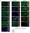
A subset of C. elegans nucleoporins is required for P granule integrity. (A-H) Single-plane confocal sections of 13- to 28-cell stage embryos derived from mothers treated with the indicated RNAi, and co-stained with antibodies against nuclear pore proteins (mAb414, red) and PGL-1 (K76, green). Embryos are oriented with anterior to the left and posterior to the right in this and all subsequent figures. P granules are perinuclear in control (empty vector), npp-1(RNAi), npp-19(RNAi) and lmn-1(RNAi), but mislocalized after npp-7(RNAi), npp-8(RNAi), npp-9(RNAi) and npp-10(RNAi). (I) Comparison of the percentage of embryos expressing the zygotic transgene pes-10::GFP and those with wild-type P granules following the indicated RNAi treatments (error bars are s.e.m.). *, P<0.01 (Student's t-test): a statistically significant reduction in the number of pes-10::GFP-positive embryos in RNAi versus control. The number of embryos scored (n) is shown below each bar.
Fig. 2.
Fig. 2.
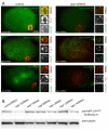
GFP::NPP-10 is enriched in P granules and this localization depends on npp-7, npp-8 and npp-9. (A) Single confocal plane images of 25- to 30-cell stage C. elegans embryos expressing the indicated GFP fusions and double stained with anti-GFP (green) and anti-PGL-1 (red) antibodies. A non-transgenic wild-type (N2) embryo is shown for comparison. (B) Same as A, but with the indicated RNAi treatments. Note that GFP::NPP-10 disappears from P granules under all RNAi treatments, whereas GFP::NPP-8 remains associated with dispersed P granules after npp-7(RNAi) and 10(RNAi). A summary of the results is shown at the bottom.
Fig. 3.
Fig. 3.
Endogenous CeNup98 is enriched in P granules in embryos. (A) Confocal sections of C. elegans embryos co-stained with the indicated anti-nucleoporin antibodies (green) and anti-PGL-1 (red), with and without npp-10(RNAi). npp-10(RNAi) eliminates all anti-nucleoporin staining. (B) Western blot with anti-CeNup98 antibody 1 and with anti-tubulin antibody of extracts from wild-type worms treated with the indicated RNAi. Fifty worms were loaded per lane. Full blots are shown in Fig. S5 in the supplementary material.
Fig. 4.
Fig. 4.
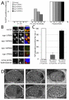
CeNup98 is required for P granule integrity in larvae. (A) Western blot of wild-type and ok467 C. elegans larvae extracts probed with anti-CeNup98, anti-CeNup96 and anti-tubulin (loading control) antibodies. Black arrowhead points to full-length CeNup98 and gray arrowhead points to the CeNup98 truncation product. Low levels of full-length CeNup98 are detected in the ok467 extract, which is likely to be maternal CeNup98. The CeNup96 antibody recognizes a doublet; both bands are eliminated by npp-10(RNAi) (Galy et al., 2003). (B) Fluorescence micrographs of an ok467/qC1 L1 larva (expressing lag-2::GFP in several somatic cells) and an ok467/ok467 L1 larva (lacking the lag-2::GFP-marked qC1 balancer). Both show the wild-type PGL-1::GFP pattern in the two primordial germ cells (arrow). (C) Fluorescence micrographs of L2 gonads of the indicated genotypes expressing PGL-1::GFP, or stained with anti-GFP antibody (green; denoting the presence of the qC1 balancer) and OIC1D4 antibody (red; P granules). (D) Fluorescence micrographs of live L2-stage gonads expressing GFP::PGL-1 or GFP::Histone H2B (to highlight nuclei). RNAi treatments were applied by feeding starting at the L1 stage. Note that only npp-10(RNAi) disrupts GFP::PGL-1.
Fig. 5.
Fig. 5.
The P granule-associated mRNA nos-2 is prematurely translated and released from P granules in npp-7(RNAi) and npp-10(RNAi) embryos. (A) C. elegans JH1999 hermaphrodites (GFP::Histone H2B::nos-2 3′UTR) were treated with the indicated RNAi for 22 hours and their embryos co-stained for PGL-1, GFP and DNA. The percentage of embryos positive for GFP is reported for each indicated stage (n is shown below each bar). We observed two GFP-positive cells in 30% of 12- to 28-cell stage embryos following npp-8(RNAi) and a single GFP-positive cell in the remaining positive embryos at the 12- to 28-cell stage. Excess positive cells were observed in 100% of post-28-cell embryos after npp-7(RNAi), npp-8(RNAi) and npp-10(RNAi), and in ~30% of embryos after npp-19(RNAi). (B) Confocal sections of P3 blastomeres hybridized with nos-2 probe (green) and immunostained with anti-PGL-1 (red). Co-localization of green and red foci is lost in npp-10(RNAi) and npp-7(RNAi) embryos. (C) The average percentage of P granules positive for nos-2 mRNA from ten embryos for each RNAi treatment. Bar colors match those in A. Confocal sections of P3 blastomeres co-stained as in B were scanned for P granules. Each P granule was scored as nos-2 mRNA-positive if the pixel intensity of the green fluorescence (nos-2 probe) overlapping the granule was above background. Error bars are s.e.m. (D) Wild-type embryos treated with the indicated RNAi and hybridized with nos-2 antisense or sense probes (dark coloration indicates hybridization). Under conditions that do not disrupt P granules [vector, npp-1(RNAi), npp-19(RNAi)], the nos-2 signal is concentrated around the nucleus of one cell (the P blastomere). In npp-7(RNAi) and npp-10(RNAi) embryos, nos-2 RNA is diffuse in the cytoplasm and in two cells (presumably the P blastomere and its somatic sister).
Fig. 6.
Fig. 6.
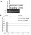
CeNup98 is in a complex with nos-2 mRNA. (A) nos-2 and control mRNAs were amplified by RT-PCR in GFP::NPP-10Nup98 and control GFP::mCherry immunoprecipitates. nos-2 mRNA is only detected in the GFP::NPP-10Nup98 immunoprecipitates. fbf-1 is a germline mRNA which, like nos-2, is also present in embryos. NRTC, no reverse transcriptase control. RNA was isolated from the inputs and eluates, treated with DNase and reverse transcribed. cDNA was used for qPCR with primers to nos-2 or fbf-1. Shown are products of 36 amplification cycles (linear range determined by qPCR). Input lanes are 1% of pulldown lanes. Processing of GFP::NPP-10 fusion to GFP::NPP-10Nup98 was verified by western blot (see Fig. S4, lane 2, in the supplementary material). (B) Fold enrichment of mRNAs in the anti-GFP immunoprecipitates compared with the IgG immunoprecipitates. Relative enrichment was calculated using RT-qPCR and the comparative Ct method (Pfaffl, 2001). The average of three qPCR amplifications from two independent biological replicates is reported; error bars represent s.e.m.
Fig. 7.
Fig. 7.
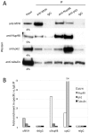
Mouse Nup98 interacts with the germ granule component MVH in testes. (A) Lysates of C57/Bl6 mouse testes were immunoprecipitated using the indicated antibodies (top) followed by western blot analysis. Input lanes contain 4 or 5% of the extract used for immunoprecipitation. (B) Quantitation of results shown in A. Fold enrichment was calculated by determining the ratio of band intensities in the specific immunoprecipitations over the IgG immunoprecipitation. Plotted values are derived from the experiment shown in A. A second independent immunoprecipitation experiment (not shown) yielded similar enrichment values: 11-fold enrichment of Nup98 in the MVH immunoprecipitate and a 46-fold enrichment of MVH in the Nup98 immunoprecipitate, as compared with the IgG immunoprecipitate.

Similar articles

Cited by

References

    1. Ageberg M., Drott K., Olofsson T., Gullberg U., Lindmark A. (2008). Identification of a novel and myeloid specific role of the leukemia-associated fusion protein DEK-NUP214 leading to increased protein synthesis. Genes Chromosomes Cancer 47, 276-287 - PubMed
    1. Anderson P., Kedersha N. (2009). RNA granules: post-transcriptional and epigenetic modulators of gene expression. Nat. Rev. Mol. Cell Biol. 10, 430-436 - PubMed
    1. Brangwynne C. P., Eckmann C. R., Courson D. S., Rybarska A., Hoege C., Gharakhani J., Julicher F., Hyman A. A. (2009). Germline P granules are liquid droplets that localize by controlled dissolution/condensation. Science 324, 1729-1732 - PubMed
    1. Capelson M., Hetzer M. W. (2009). The role of nuclear pores in gene regulation, development and disease. EMBO Rep. 10, 697-705 - PMC - PubMed
    1. Carmody S. R., Wente S. R. (2009). mRNA nuclear export at a glance. J. Cell Sci. 122, 1933-1937 - PMC - PubMed

Publication types

MeSH terms

Substances