Ectodermal Wnt6 is an early negative regulator of limb chondrogenesis in the chicken embryo
- PMID: 20334703
- PMCID: PMC2859743
- DOI: 10.1186/1471-213X-10-32
Ectodermal Wnt6 is an early negative regulator of limb chondrogenesis in the chicken embryo
Abstract
Background: Pattern formation of the limb skeleton is regulated by a complex interplay of signaling centers located in the ectodermal sheath and mesenchymal core of the limb anlagen, which results, in the forelimb, in the coordinate array of humerus, radius, ulna, carpals, metacarpals and digits. Much less understood is why skeletal elements form only in the central mesenchyme of the limb, whereas muscle anlagen develop in the peripheral mesenchyme ensheathing the chondrogenic center. Classical studies have suggested a role of the limb ectoderm as a negative regulator of limb chondrogenesis.
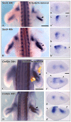
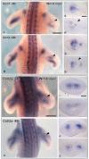
Results: In this paper, we investigated the molecular nature of the inhibitory influence of the ectoderm on limb chondrogenesis in the avian embryo in vivo. We show that ectoderm ablation in the early limb bud leads to increased and ectopic expression of early chondrogenic marker genes like Sox9 and Collagen II, indicating that the limb ectoderm inhibits limb chondrogenesis at an early stage of the chondrogenic cascade. To investigate the molecular nature of the inhibitory influence of the ectoderm, we ectopically expressed Wnt6, which is presently the only known Wnt expressed throughout the avian limb ectoderm, and found that Wnt6 overexpression leads to reduced expression of the early chondrogenic marker genes Sox9 and Collagen II.
Conclusion: Our results suggest that the inhibitory influence of the ectoderm on limb chondrogenesis acts on an early stage of chondrogenesis upsteam of Sox9 and Collagen II. We identify Wnt6 as a candidate mediator of ectodermal chondrogenic inhibition in vivo. We propose a model of Wnt-mediated centripetal patterning of the limb by the surface ectoderm.
Figures


Similar articles
-
Involvement of Wnt-5a in chondrogenic pattern formation in the chick limb bud.Dev Growth Differ. 1999 Feb;41(1):29-40. doi: 10.1046/j.1440-169x.1999.00402.x. Dev Growth Differ. 1999. PMID: 10445500
-
The pattern of expression of the chicken homolog of HOX1I in the developing limb suggests a possible role in the ectodermal inhibition of chondrogenesis.Dev Dyn. 1992 Jan;193(1):92-101. doi: 10.1002/aja.1001930112. Dev Dyn. 1992. PMID: 1347239
-
Frizzled-7 and limb mesenchymal chondrogenesis: effect of misexpression and involvement of N-cadherin.Dev Dyn. 2002 Mar;223(2):241-53. doi: 10.1002/dvdy.10046. Dev Dyn. 2002. PMID: 11836788
-
Ectoderm as a determinant of early tissue pattern in the limb bud.Cell Differ. 1984 Nov;15(1):17-24. doi: 10.1016/0045-6039(84)90025-3. Cell Differ. 1984. PMID: 6394145 Review.
-
The recombinant limb as a model for the study of limb patterning, and its application to muscle development.Cell Tissue Res. 1999 Apr;296(1):121-9. doi: 10.1007/s004410051273. Cell Tissue Res. 1999. PMID: 10199972 Review.
Cited by
-
Genome-Wide Specific Selection in Three Domestic Sheep Breeds.PLoS One. 2015 Jun 17;10(6):e0128688. doi: 10.1371/journal.pone.0128688. eCollection 2015. PLoS One. 2015. PMID: 26083354 Free PMC article.
-
Substrate specificity of human carboxypeptidase A6.J Biol Chem. 2010 Dec 3;285(49):38234-42. doi: 10.1074/jbc.M110.158626. Epub 2010 Sep 20. J Biol Chem. 2010. PMID: 20855895 Free PMC article.
-
Systems for intricate patterning of the vertebrate anatomy.Philos Trans A Math Phys Eng Sci. 2021 Dec 27;379(2213):20200270. doi: 10.1098/rsta.2020.0270. Epub 2021 Nov 8. Philos Trans A Math Phys Eng Sci. 2021. PMID: 34743605 Free PMC article.
-
miR-892b Inhibits Hypertrophy by Targeting KLF10 in the Chondrogenesis of Mesenchymal Stem Cells.Mol Ther Nucleic Acids. 2019 Sep 6;17:310-322. doi: 10.1016/j.omtn.2019.05.029. Epub 2019 Jun 13. Mol Ther Nucleic Acids. 2019. PMID: 31284128 Free PMC article.
-
Sost and its paralog Sostdc1 coordinate digit number in a Gli3-dependent manner.Dev Biol. 2013 Nov 1;383(1):90-105. doi: 10.1016/j.ydbio.2013.08.015. Epub 2013 Aug 29. Dev Biol. 2013. PMID: 23994639 Free PMC article.
References
Publication types
MeSH terms
Substances
LinkOut - more resources
Full Text Sources
Research Materials

