. 2010 May;11(5):427-34.
doi: 10.1038/ni.1856. Epub 2010 Mar 21.

Capture of influenza by medullary dendritic cells via SIGN-R1 is essential for humoral immunity in draining lymph nodes

Affiliations

Capture of influenza by medullary dendritic cells via SIGN-R1 is essential for humoral immunity in draining lymph nodes

Santiago F Gonzalez et al. Nat Immunol. 2010 May.

Abstract

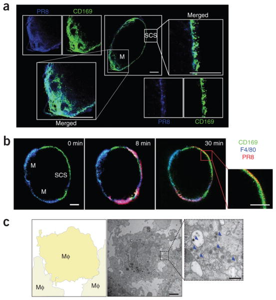
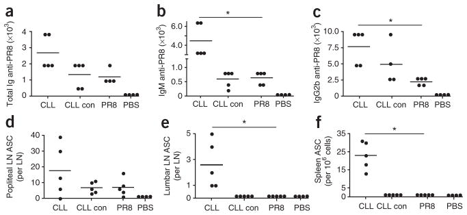
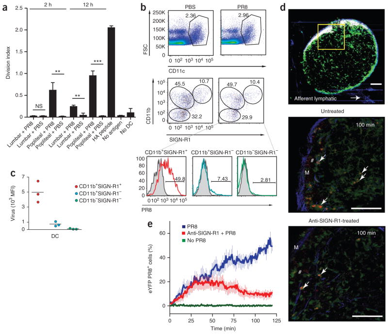
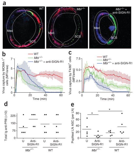
A major pathway for B cell acquisition of lymph-borne particulate antigens relies on antigen capture by subcapsular sinus macrophages of the lymph node. Here we tested whether this mechanism is also important for humoral immunity to inactivated influenza virus. By multiple approaches, including multiphoton intravital imaging, we found that antigen capture by sinus-lining macrophages was important for limiting the systemic spread of virus but not for the generation of influenza-specific humoral immunity. Instead, we found that dendritic cells residing in the lymph node medulla use the lectin receptor SIGN-R1 to capture lymph-borne influenza virus and promote humoral immunity. Thus, our results have important implications for the generation of durable humoral immunity to viral pathogens through vaccination.


Conflict of interest statement

COMPETING FINANCIAL INTERESTS

The authors declare no competing financial interests.

Figures

Figure 1
Figure 1
Lymph-borne influenza virus is captured by macrophages in the subcapsular sinus and medulla of the draining lymph node. (a) Confocal microscopy of the localization of PR8 (blue) in the popliteal lymph node 30 min after footpad injection, in a lymph node cryosection stained with anti-CD169 (green) to identify medullary (M) and subcapsular sinus (SCS) macrophages. Scale bars, 200 μm. Results are representative of four independent experiments with four mice. (b) MP-IVM of the entry of PR8 (red) into the popliteal lymph node after pretreatment with anti-CD169 (green; subcapsular sinus and medulla) and anti-F4/80 (blue; medulla). Numbers in top right corners indicate time after virus injection. Scale bars, 200 μm. Additional images, Supplementary Movie 1. Results are representative of six independent experiments with six mice. (c) Transmission electron microscopy (middle, right) of PR8 in the subcapsular sinus of a popliteal lymph node 30 min after injection. Left, diagram of middle image (boxed area is enlarged at right). Arrowheads (right) indicate phagocytosed virus particles. Scale bars, 2 μm (middle) or 500 nm (right). Mφ, macrophage. Results are representative of three independent experiments with three mice.
Figure 2
Figure 2
SSMs are not required for humoral immunity to influenza virus. (ac) MP-IVM of PR8 (red) in a popliteal lymph node after treatment with dendrimer and anti-CD169 (green) and anti-CD35 (blue). Numbers in top right corners indicate time after virus injection. Scale bar, 200 μm. Results are representative of four independent experiments with four mice. (df) Serum titers of PR8-specific IgG2a (d), IgG2b (e) or IgM (f) at 10 d in mice pretreated with PR8 alone (PR8) or dendrimer and PR8 (Den + PR8) or injected with PBS alone (PBS). Results are representative of three experiments with five mice per group. (gi) Antibody-secreting cells (ASC) in the popliteal lymph nodes (LN; g), lumbar lymph nodes (h) and spleen (per 1 × 10⁶ spleen cells; i) at 10 d after injection as described in df. *P = 0.005 (unpaired t-test). Results are representative of three experiments with five mice per group. In di, each symbol represents an individual mouse; small horizontal lines indicate the mean. (j) Frequency of B1–8 B cells (B220+IgMa+ immunoglobulin-λ1-positive (Igλ1+) cells) that bound NP-conjugated PR8 in dendrimer-treated mice at 2 h and 8 h after injection of virus. Each symbol represents an individual mouse. Results are from one experiment (mean of two mice per group at each time point).
Figure 3
Figure 3
Medullary macrophages are not required for humoral immunity to influenza virus. (ac) Serum titers of PR8-specific total immunoglobulin (Ig; a), IgM (b) or IgG2b (c) at 10 d after injection of PR8 into mice treated with CLLs (CLL), empty liposomes (CLL con) or PR8 alone (PR8) or injected with PBS only (PBS). (df) Antibody-secreting cells in the popliteal lymph nodes (d), lumbar lymph nodes (e) and spleen (per 1 × 10⁶ spleen cells; f) at 10 d after injection of PR8 into mice treated as described in ac. Each symbol represents an individual mouse; small horizontal lines indicate the mean. *P = 0.005 (unpaired t-test). Results are representative of three experiments with five mice per group.
Figure 4
Figure 4
CD11b+SIGN-R1+ lymph node–resident DCs bind lymph-borne PR8 in the medulla. (a) Proliferation of naive hemagglutinin-specific CD4+ T cells labeled with the cytosolic dye CFSE and cultured alone (No DC), with DCs purified from the popliteal or lumbar lymph nodes at 2 h and 12 h after PR8 or PBS injection or with DCs left untreated (No antigen) or pulsed with hemagglutinin (HA) peptide, assessed by CFSE dilution after 72 h (division index). *P < 0.05, **P < 0.005 and ***P < 0.0005 (unpaired t-test). Results are representative of four independent experiments (error bars, s.d. of three mice). (b) Cytofluorometry of the surface expression of CD11b and SIGN-R1 (middle) by CD11c+ cells (top) in popliteal lymph nodes 2 h after injection of PBS or PR8, identifying three distinct DC populations (bottom). Numbers adjacent to outlined areas indicate percent CD11c+ cells (top row), or percent CD11bSIGN-R1 cells (bottom left, middle row), CD11b+SIGN-R1 cells (top left, middle row) or CD11b+SIGN-R1+ cells (top right, middle row); numbers above bracketed lines (bottom) indicate DC populations that captured PR8 (solid lines) relative to those of PBS-injected mice (gray filled histograms). Results are representative of five independent experiments with five mice. (c) Virus uptake by DC subsets, presented as mean fluorescence intensity (MFI). Each symbol represents an individual mouse; small horizontal lines indicate the mean. Data are representative of three experiments with three mice per condition. (d) MP-IVM of the capture of PR8 by DCs in the medulla of popliteal lymph nodes from untreated and anti-SIGN-R1-treated mice at 100 min after virus injection (additional images, Supplementary Movie 3). Arrows indicate virus-bearing eYFP+ DCs; #, virus not bound by eYFP+ DCs. Scale bars, 200 μm (top) or 100 μm (middle and bottom). Results are representative of four independent experiments with eight mice. (e) Quantification of PR8 capture by DCs in the medulla of popliteal lymph nodes from mice left untreated (No PR8) or injected with PR8 alone (PR8) or after anti-SIGN-R1 treatment (Anti-SIGN-R1 + PR8). Results are representative of two independent experiments with two mice (untreated) or four independent experiments with four mice (PR8 and PR8 plus anti-SIGN-R1; error bars, s.e.m.).
Figure 5
Figure 5
CD11c+ cells bind lymph-borne influenza virus in the medulla of the lymph node and increase their velocity toward the FDC area. (a) Velocity of eYFP+ DCs in the medulla of popliteal lymph nodes 60 min before (Pre-virus) and after injection of PR8, for eYFP+ DCs that bound virus (Virus+ DCs) or did not bind virus (Virus DCs). Each symbol represents an individual cell; small horizontal lines indicate the mean. *P < 0.001 (unpaired t-test). Results are representative of four independent experiments with 200 cells per group. (bf) Vector analysis of the net and angular displacement of DCs in the medulla of popliteal lymph nodes 60 min before and after injection of PR8. (bd) Displacement of 50 randomly selected eYFP+ DCs per group from their point of origin (x,y = 0,0); blue lines indicate mean vector. Scale bar, 200 μm. (e,f) Summary of net displacement (e) and angular displacement (f) for the DCs described in a. Each symbol represents an individual cell; small horizontal lines indicate the mean. *P < 0.001 (unpaired t-test). Results are representative of four independent experiments with four mice (bd) or four mice and 200 cells per group (e,f).
Figure 6
Figure 6
MBL is required for PR8 capture in the lymph node subcapsular sinus, whereas SIGN-R1 mediates PR8 capture in the lymph node medulla. (a) MP-IVM of the capture of PR8 (red) at 45 min in wild-type mice (WT), Mbl−/− mice and anti-SIGN-R1-treated Mbl−/− mice after pretreatment with antibody to the metallophilic macrophage–specific marker MOMA-1 (green), anti-F4/80 (blue) and anti-CD35 (8C12; blue). Results are representative of three independent experiments per treatment. (b,c) Quantification of PR8 capture by MOMA-1+ cells (b) or F4/80+ cells (c) in the subcapsular sinus or medullary area, respectively, over the first 60 min after PR8 injection. Results are representative of three independent experiments with four mice. (d) Serum titers of PR8-specific total immunoglobulin at 10 d after injection of PR8 into untreated (U) and anti-SIGN-R1-treated Mbl−/− and wild-type mice. Results are representative of QQ experiments with five mice per group. (e) Antibody-secreting cells in the popliteal lymph node 10 d after footpad injection of PR8 into untreated and anti-SIGN-R1-treated Mbl−/− and wild-type mice. *P < 0.05 (unpaired t-test). Results are representative of one experiment with five mice per group.

Similar articles

Cited by

References

    1. Pulendran B, Ahmed R. Translating innate immunity into immunological memory: implications for vaccine development. Cell. 2006;124:849–863. - PubMed
    1. Gerhard W, Mozdzanowska K, Furchner M, Washko G, Maiese K. Role of the B-cell response in recovery of mice from primary influenza virus infection. Immunol Rev. 1997;159:95–103. - PubMed
    1. Gretz JE, Anderson AO, Shaw S. Cords, channels, corridors and conduits: critical architectural elements facilitating cell interactions in the lymph node cortex. Immunol Rev. 1997;156:11–24. - PubMed
    1. Kosco-Vilbois MH, Gray D, Scheidegger D, Julius M. Follicular dendritic cells help resting B cells to become effective antigen-presenting cells: Induction of B7/BB1 and upregulation of major histocompatibility complex class II molecules. J Exp Med. 1993;178:2055–2066. - PMC - PubMed
    1. Allen CD, Cyster JG. Follicular dendritic cell networks of primary follicles and germinal centers: phenotype and function. Semin Immunol. 2008;20:14–25. - PMC - PubMed

Publication types

MeSH terms

LinkOut - more resources