Review
. 2010 Apr;11(4):237-51.
doi: 10.1038/nrm2867. Epub 2010 Mar 18.

A nucleator arms race: cellular control of actin assembly

Affiliations
Review

A nucleator arms race: cellular control of actin assembly

Kenneth G Campellone et al. Nat Rev Mol Cell Biol. 2010 Apr.

Abstract

For over a decade, the actin-related protein 2/3 (ARP2/3) complex, a handful of nucleation-promoting factors and formins were the only molecules known to directly nucleate actin filament formation de novo. However, the past several years have seen a surge in the discovery of mammalian proteins with roles in actin nucleation and dynamics. Newly recognized nucleation-promoting factors, such as WASP and SCAR homologue (WASH), WASP homologue associated with actin, membranes and microtubules (WHAMM), and junction-mediating regulatory protein (JMY), stimulate ARP2/3 activity at distinct cellular locations. Formin nucleators with additional biochemical and cellular activities have also been uncovered. Finally, the Spire, cordon-bleu and leiomodin nucleators have revealed new ways of overcoming the kinetic barriers to actin polymerization.


Figures

Figure 1
Figure 1. Localization and function of actin nucleation factors in mammalian cells
Actin filaments (red lines) are nucleated and organized into branched networks by the Arp2/3 complex and its nucleation-promoting factors, or are generated in unbranched forms by formins and tandem WH2-domain nucleators. Functional roles for different nucleation factors are depicted during phagocytosis, cell junction assembly, endocytosis, membrane ruffling and lamellipodia dynamics, filopodia formation, Golgi and tubulo-vesicular membrane dynamics, and stress fiber formation in a generic mammalian cell. Question marks (?) indicate that the precise role for the depicted nucleation factor is unclear. Arp2/3, actin-related protein 2/3; Cobl, cordon-bleu; Daam, Dishevelled-associated activator of morphogenesis; FHOD, formin homology domain; FMN, formin; FRL, formin-related in leukocytes; INF, inverted formin; JMY, junction mediating regulatory; mDia, murine Diaphanous; N-WASP, neuronal-WASP; WASP, Wiskott-aldrich syndrome protein; WASH, WASP/Scar homolog; WAVE, WASP-verprolin homologous; WHAMM, WASP homologue associated with actin, membranes, and microtubules.
Figure 2
Figure 2. Structure of the Arp2/3 complex in Y-branches and model for nucleation and branching
A) The morphology of a y-branched actin filament and the Arp2/3 complex is shown, both in an electron micrograph and in structural models based on electron tomography (modified with permission from Ref.13). The complex consists of the actin-related proteins Arp2 and Arp3 plus the additional Arp complex subunits ARPC1-5. In this model, all seven subunits participate in binding to the existing filament, while Arp2 and Arp3 act as the first two subunits of the nascent filament. B) The Arp2/3 complex is recruited by the WCA domains of Class I NPFs in proximity to cellular membranes (1). The collective activities of WH2 and CA segments serve the basic purpose of bringing the Arp2/3 complex together with the first actin subunit in the new filament and generate a branch (2). Arp2/3 branchpoints can be stabilized by F-actin-binding Class II NPFs, like cortactin (3). Coronin-family proteins interact with the Arp2/3 complex and F-actin to prevent cofilin-mediated disassembly of newly-formed filaments (4, top). Coronin can also replace the Arp2/3 complex and synergize with cofilin to trigger debranching and disassembly of older ADP-actin filaments (4, middle). Disassembly of older branches can also occur spontaneously, following phosphate release from Arp2 and actin (4, bottom). A, acidic; β-P, beta-propellor; C, connector; CC, coiled-coil; R, repeat; W, WH2. Structural models in part a modified, with permission, from Ref.13 © (2008) Rockefeller University Press.
Figure 3
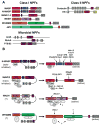
Figure 3. Different groups of NPFs possess distinct modes of regulation
A) Mammalian Class I NPFs contain C-terminal WCA domains that bind G-actin and the Arp2/3 complex, plus diverse N-terminal regulatory regions. Microbial pathogens also express Class I NPFs. These include ActA from Listeria, RickA from Rickettsia, and P78/83 from baculoviruses. Class II NPFs contain N-terminal acidic domains that bind the Arp2/3 complex, central F-actin-binding repeats, and regulatory C-terminal domains. A, acidic; AI, autoinhibitory; B, basic; C, connector; CC, coiled-coil; CRIB, Cdc42-Rac-interactive-binding; L, linker; N, amino-terminal; P, polyproline; PRD, proline-rich-domain; R, repeat; SH3, Src-homology-3; SHD, Scar-homology-domain; TBR, tubulin-binding region; W, WASP-homology-2 (WH2) domain; WH1, WASP-homology-1; WAHD1, WASH-homology-domain-1; WMD, WHAMM-membrane interaction-domain. B) N-WASP NPF activity is regulated both by autoinhibition and by interactions with proteins from the WIP family. It can be stimulated by direct binding of phosphoinositides, small GTPases, tyrosine phosphorylation, and SH2/SH3 domains. WAVE2 activity is controlled by a protein complex comprised of Brk1, Abi1, Nap1, and Sra1. It is stimulated by Nck-Nap1 and Rac-Sra1 interactions, or by binding of phosphoinositides or SH3 domains to WAVE2 itself. WASH activity is likely controlled by a multi-subunit complex that contains capping protein (CP) and FAM21. WHAMM NPF activity does not appear to be autoinhibited, and is likely to be controlled by factors that interact with its WMD and/or CC regions. Question marks (?) indicate that the depicted mechanism of activation is speculative.
Figure 4
Figure 4. FH2 domain structure and elongation model of formin-mediated actin polymerization
A) A ribbon diagram of the dimeric FH2 domain from S.cerevisiae Bni1 is shown (Ref.89). A “lasso” extends from the knob of one monomer and wraps around the “post” of the other monomer to stabilize this dimeric configuration. FH, formin homology. B) The Bni1p FH2 domain wrapped around a space-filling model of an actin dimer is shown. C) An FH2 dimer associates with the barbed end of an actin filament, while the FH1 domains recruit profilin-actin (1). The FH1 domain delivers profilin-actin to the barbed end, and this is either preceded by (Ref.92) or follows (Ref.93) the FH2 domain stepping towards the barbed end (2). The second FH2 repeats this process (3). The formin closed conformation prevents capping by other factors (4). Image in part a is modified, with permission, from Ref. 89 © (2004) Elsevier. Image in part b is modified, with permission, from Nature Ref. 92 © (2005) Macmillan publishers Ltd. All rights reserved.
Figure 5
Figure 5. Different formins possess distinct domain organizations
A) Mammalian formins share a conserved FH1-FH2 actin polymerizing module along with diverse regulatory motifs. The GBD-DID region of FHOD1-2 is structurally distinct from the analogous portions of other formins (non-bold typeface). CC, coiled-coil; DAD, diaphanous-autoinhibitory-domain; DID, diaphanous-inhibitory-domain; DD, dimerization-domain; FH, formin-homology; FSI, formin-spire-interaction; GBD, GTPase-binding-domain; PDZ, PSD95-DlgA-ZO1; W, WH2 domain. B) Diaphanous-related formins like mDia2 are dimeric and regulated by autoinhibition. Their actin nucleation functions are stimulated by binding to Rho family GTPases, such as RhoA-C and Cdc42.
Figure 6
Figure 6. WH2 domain-containing actin nucleators and models for polymerization
A) Members of the Spire, Cobl, and Lmod families use WH2 domains and additional actin monomer-binding sequences to nucleate and assemble unbranched actin filaments. They also contain other domains that likely regulate their nucleation activities. Bacterial pathogens also express WH2-based nucleators to induce plasma membrane remodeling. These include Tarp from Chlamydia and VopF/VopL from Vibrio. FYVE, Fab1-YOTB-Vac1-EEA1; h-b-h, helix-basic-helix; KIND, kinase noncatalytic domain; LRR, leucine-rich-repeats; P, polyproline; PRD, proline-rich-domain; sec, secretion signal; T, tropomyosin and actin-binding helices; W, WH2; Y, tyrosine-rich motifs. B) A ribbon diagram of three tandem WH2 domains bound to actin monomers is shown (modified with permission from Ref.151). How bound actin monomers are reorganized into a conformation that favors polymerization is not yet known. C) Spire is a versatile regulator of actin dynamics that can nucleate actin filaments (which might be further elongated by formins, as denoted by the question mark (?)), cap and sever existing filaments, and sequester monomers. D) Cobl- and Lmod-family nucleators use multiple monomer-binding sequences to assemble trimeric actin nuclei and may remain associated with their pointed ends. Image in part b is modified, with permission, from Ref.151© (2008) National Academy of Sciences.

Similar articles

Cited by

References

    1. Goley ED, Welch MD. The ARP2/3 complex: an actin nucleator comes of age. Nature Rev Mol Cell Biol. 2006;7:713–726. - PubMed
    1. Chesarone MA, DuPage AG, Goode BL. Unleashing formins to remodel the actin and microtubule cytoskeletons. Nature Rev Mol Cell Biol. 2010;11:62–74. - PubMed
    1. Stradal TE, Scita G. Protein complexes regulating Arp2/3-mediated actin assembly. Curr Opin Cell Biol. 2006;18:4–10. - PubMed
    1. Goley ED, Rodenbusch SE, Martin AC, Welch MD. Critical conformational changes in the Arp2/3 complex are induced by nucleotide and nucleation promoting factor. Mol Cell. 2004;16:269–279. - PubMed
    1. Zencheck WD, et al. Nucleotide- and activator-dependent structural and dynamic changes of arp2/3 complex monitored by hydrogen/deuterium exchange and mass spectrometry. J Mol Biol. 2009;390:414–427. - PMC - PubMed

Publication types

MeSH terms