

Potassium channels and neurovascular coupling
- PMID: 20234102
- PMCID: PMC4405141
- DOI: 10.1253/circj.cj-10-0174
Potassium channels and neurovascular coupling
Abstract
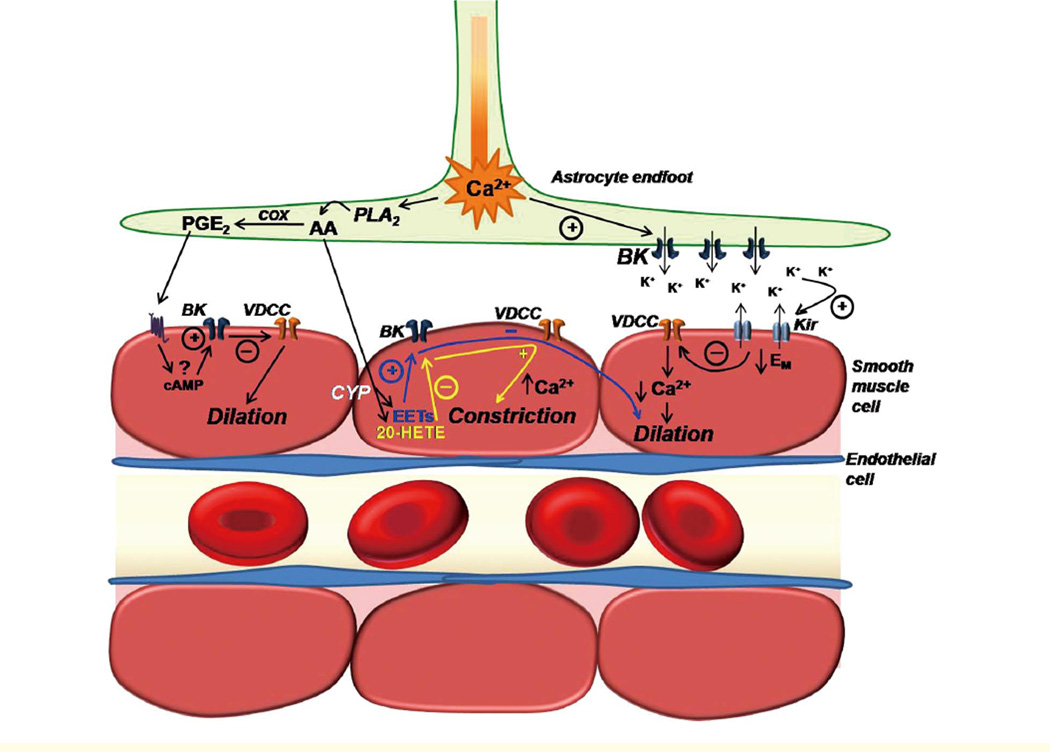
Neuronal activity is communicated to the cerebral vasculature so that adequate perfusion of brain tissue is maintained at all levels of neuronal metabolism. An increase in neuronal activity is accompanied by vasodilation and an increase in local cerebral blood flow. This process, known as neurovascular coupling (NVC) or functional hyperemia, is essential for cerebral homeostasis and survival. Neuronal activity is encoded in astrocytic Ca(2+) signals that travel to astrocytic processes (;endfeet') encasing parenchymal arterioles within the brain. Astrocytic Ca(2+) signals cause the release of vasoactive substances to cause relaxation, and in some circumstances contraction, of the smooth muscle cells (SMCs) of parenchymal arterioles to modulate local cerebral blood flow. Activation of potassium channels in the SMCs has been proposed to mediate NVC. Here, the current state of knowledge of NVC and potassium channels in parenchymal arterioles is reviewed.
Figures

Similar articles
-
Dynamic inositol trisphosphate-mediated calcium signals within astrocytic endfeet underlie vasodilation of cerebral arterioles.J Gen Physiol. 2006 Dec;128(6):659-69. doi: 10.1085/jgp.200609650. J Gen Physiol. 2006. PMID: 17130519 Free PMC article.
-
Astrocyte Ca2+ Signaling Drives Inversion of Neurovascular Coupling after Subarachnoid Hemorrhage.J Neurosci. 2015 Sep 30;35(39):13375-84. doi: 10.1523/JNEUROSCI.1551-15.2015. J Neurosci. 2015. PMID: 26424885 Free PMC article.
-
Inversion of neurovascular coupling by subarachnoid blood depends on large-conductance Ca2+-activated K+ (BK) channels.Proc Natl Acad Sci U S A. 2012 May 22;109(21):E1387-95. doi: 10.1073/pnas.1121359109. Epub 2012 Apr 30. Proc Natl Acad Sci U S A. 2012. PMID: 22547803 Free PMC article.
-
Ion channel networks in the control of cerebral blood flow.J Cereb Blood Flow Metab. 2016 Mar;36(3):492-512. doi: 10.1177/0271678X15616138. Epub 2015 Nov 9. J Cereb Blood Flow Metab. 2016. PMID: 26661232 Free PMC article. Review.
-
Vascular inward rectifier K+ channels as external K+ sensors in the control of cerebral blood flow.Microcirculation. 2015 Apr;22(3):183-96. doi: 10.1111/micc.12190. Microcirculation. 2015. PMID: 25641345 Free PMC article. Review.
Cited by
-
Learning on Jupiter, learning on the Moon: the dark side of the G-force. Effects of gravity changes on neurovascular unit and modulation of learning and memory.Front Behav Neurosci. 2012 Sep 24;6:64. doi: 10.3389/fnbeh.2012.00064. eCollection 2012. Front Behav Neurosci. 2012. PMID: 23015785 Free PMC article.
-
Fluid and ion transfer across the blood-brain and blood-cerebrospinal fluid barriers; a comparative account of mechanisms and roles.Fluids Barriers CNS. 2016 Oct 31;13(1):19. doi: 10.1186/s12987-016-0040-3. Fluids Barriers CNS. 2016. PMID: 27799072 Free PMC article. Review.
-
Role of peroxisome proliferator-activated receptor-γ in vascular muscle in the cerebral circulation.Hypertension. 2014 Nov;64(5):1088-93. doi: 10.1161/HYPERTENSIONAHA.114.03935. Epub 2014 Sep 2. Hypertension. 2014. PMID: 25185134 Free PMC article.
-
Changes in cortical microvasculature during misery perfusion measured by two-photon laser scanning microscopy.J Cereb Blood Flow Metab. 2014 Aug;34(8):1363-72. doi: 10.1038/jcbfm.2014.91. Epub 2014 May 21. J Cereb Blood Flow Metab. 2014. PMID: 24849667 Free PMC article.
-
Early effects of high-fat diet on neurovascular function and focal ischemic brain injury.Am J Physiol Regul Integr Comp Physiol. 2013 Jun 1;304(11):R1001-8. doi: 10.1152/ajpregu.00523.2012. Epub 2013 Apr 10. Am J Physiol Regul Integr Comp Physiol. 2013. PMID: 23576615 Free PMC article.
References
-
- Anderson CM, Nedergaard M. Astrocyte-mediated control of cerebral microcirculation. Trends Neurosci. 2003;26:340–344. author reply 344 – 345. - PubMed
-
- Iadecola C. Regulation of the cerebral microcirculation during neural activity: Is nitric oxide the missing link? Trends Neurosci. 1993;16:206–214. - PubMed
Publication types
MeSH terms
Substances
Grants and funding
- HL077378/HL/NHLBI NIH HHS/United States
- DK065947/DK/NIDDK NIH HHS/United States
- DK053832/DK/NIDDK NIH HHS/United States
- P01 HL077378/HL/NHLBI NIH HHS/United States
- R37 DK053832/DK/NIDDK NIH HHS/United States
- HL098243/HL/NHLBI NIH HHS/United States
- R01 DK053832/DK/NIDDK NIH HHS/United States
- HL44455/HL/NHLBI NIH HHS/United States
- R01 HL098243/HL/NHLBI NIH HHS/United States
- R01 DK065947/DK/NIDDK NIH HHS/United States
- T32 HL007944/HL/NHLBI NIH HHS/United States
- T32HL007944/6-10/HL/NHLBI NIH HHS/United States
- R01 HL044455/HL/NHLBI NIH HHS/United States
LinkOut - more resources
Full Text Sources
Miscellaneous