. 2010 Apr;11(4):303-12.
doi: 10.1038/ni.1853. Epub 2010 Mar 14.

CD169(+) macrophages present lipid antigens to mediate early activation of iNKT cells in lymph nodes

Affiliations

CD169(+) macrophages present lipid antigens to mediate early activation of iNKT cells in lymph nodes

Patricia Barral et al. Nat Immunol. 2010 Apr.

Abstract

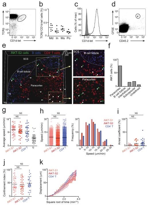
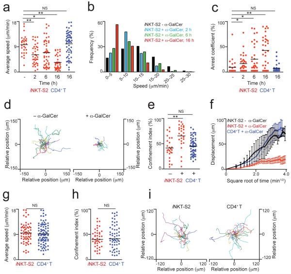
Invariant natural killer T cells (iNKT cells) are involved in the host defense against microbial infection. Although it is known that iNKT cells recognize glycolipids presented by CD1d, how and where they encounter antigen in vivo remains unclear. Here we used multiphoton microscopy to visualize the dynamics and activation of iNKT cells in lymph nodes. After antigen administration, iNKT cells became confined in a CD1d-dependent manner in close proximity to subcapsular sinus CD169(+) macrophages. These macrophages retained, internalized and presented lipid antigen and were required for iNKT cell activation, cytokine production and population expansion. Thus, CD169(+) macrophages can act as true antigen-presenting cells controlling early iNKT cell activation and favoring the fast initiation of immune responses.


Figures

Figure 1
Figure 1
Distribution and dynamics of iNKT cells in LNs. (a) iNKT cells in mediastinal LNs from WT animals were identified by flow cytometry as TCRβ+CD1d-tet+ cells. (b) Percentage of iNKT from TCRβ+B220 cells in various LNs. Data is from two independent experiments with 3-4 animals per group. Md, mediastinal; In, inguinal; Ms, mesenteric; Po, popliteal. (c) CD1d-tet staining of iNKT-S2 cells. (d) iNKT-S2 cells were labelled with CFSE, transferred into CD45.1 congenic mice and detected in LNs of recipient animals as CFSE+CD45.2+B220. (e,f) iNKT-S2 (green), CD4+ T (red) and B cells (blue) were adoptively transferred into WT recipients. LNs were fixed and imaged by multi-photon microscopy. (e) Three-dimensional reconstruction from a 200 μm section of a popliteal LN showing the distribution of iNKT cells (white arrowheads). (f) Percentage of iNKT cells in various locations in the LN. (g-k) iNKT-S1 (pale red) and iNKT-S2 (red) cells were adoptively transferred alongside CD4+ T (blue) and/or B cells (grey). Intact LNs were imaged by multi-photon microscopy. Average speed (g), instantaneous speed distribution (h), arrest coefficient (i) and confinement index (j) for individual cells are shown with mean values as black lines. Data were obtained in at least two independent experiments and were compared with two-tailed unpaired Mann-Whitney test. (NS, not significant) (k) Displacement (mean ± s.e.m) of iNKT and CD4+ T cells plotted against the square root of time. Mean motility coefficient (M) can be calculated as M=x2/6t, where x is the slope of the linear part of the graph. (g-i) Each symbol represents an individual cell.
Figure 2
Figure 2
Early activation of LN iNKT cells. (a-f) Animals were injected with α-GalCer or control particles and 3 days later LNs were analyzed. (a) Images of popliteal LNs from WT animals injected with control (top) or α-GalCer (bottom) particles. (b) Fold increase in total cell number (b) or in the depicted populations (c) in LNs of WT (b,c) and Jα18 (NKT KO, b) mice after injection with α-GalCer particles relative to animals injected with control particles; *p<0.05 (d) iNKT cells in mediastinal LNs after i.p. injection of control (left) or α-GalCer (right) particles (e,f) Percentage of iNKT cells in various LNs after i.p. (e) or s.c. foot-pad (f) injection of α-GalCer particles. +,draining; −,non-draining LNs. Each dot represents one LN, black lines represent mean. Data were compared with two-tailed t-test. (p**<0.005; ***p=0.0007). (g) CFSE-labelled adoptively transferred iNKT cells were detected as CFSE+CD1d-tet+TCRβ+B220DAPI two days after injection of α-GalCer (black) or control (grey) particles. (h-k) iNKT cell activation was measured as expression of CD25 (h-j) and CD69 (k) after i.p. injection of α-GalCer (black) or control particles (grey). MFI were normalized to mice injected with control particles. (l) NK1.1 expression in LN iNKT cells (left). Expression of CD25 (middle) and CD69 (right) in NK1.1+ (black) and NK1.1 (red) iNKT cells in mediastinal LNs 12 h after injection with α-GalCer (empty profiles) or control particles (filled profiles). (m) Intracellular IFN-γ staining for mediastinal LN-iNKT cells 12 h after injection of α-GalCer (black) or control (grey) particles.
Figure 3
Figure 3
iNKT cells arrest in the LNs in response to specific antigen. (a-i) iNKT-S2 cells (red), alongside CD4+ T cells (blue), were labelled and injected i.v. into recipient mice and allowed to home for 14-16 h. (a-f) WT recipient mice were injected i.p. with particles coated with α-GalCer (+ α-GalCer) or control lipids (− α-GalCer) and at various time points draining mediastinal LNs were removed and imaged by multi-photon microscopy. The average speed (a), instantaneous speed distribution (b) and arrest coefficient (c) of iNKT cells are shown at different time-points. (d,e) Representative migratory tracks (d) and confinement index (e) for iNKT cells at 16 h after injection of particulate lipids are shown. (f) Displacement (mean ± s.e.m.) of iNKT cells from mice injected with particles coated with α-GalCer (red) or control lipids (black) is plotted against the square root of time. Dynamic parameters for CD4+ T cells (blue, a,c,e,f) at 16 h after injection of particulate α-GalCer are shown. (g-i) CD1d-KO recipient mice were i.p. injected with particles coated with α-GalCer and mediastinal LNs were imaged at 16 h after antigen administration. The average speed (g), confinement index (h) and representative migratory tracks (i) of iNKT and CD4+ T cells are shown. Mean values are shown as black lines. Data were obtained from at least two independent experiments per condition and were compared with a two-tailed unpaired Mann-Whitney test. *p<0.01; **p<0.0005 NS, not significant. In a,c,e,g,h, each symbol represents one cell
Figure 4
Figure 4
iNKT cells arrest on SCS CD169+ macrophages (a,b) iNKT-S2 (red) and B cells (blue), were transferred into WT recipients prior to i.p. injection of α-GalCer particles (green). Mediastinal LNs were imaged by multi-photon microscopy. (a) Time-lapse images from a movie acquired 15-65 μm below the LN surface showing an iNKT cell arrested in the SCS (white circle) 2 h after injection of particulate α-GalCer. Scale bar, 20 μm (b) Three-dimensional reconstruction of the SCS 16 h after injection of particulate α-GalCer, including tracks of B and iNKT cells (20 min movie). (c) Confocal microscopy images of mediastinal LNs 2 h after i.p. injection of α-GalCer particles (green), stained with CD169 (blue, left and middle) or CD11c (blue, right). Scale bar, 300 μm (left), 5 μm (middle), 15 μm (right) (d) Flow cytometry analyses of lipid uptake by CD169+ macrophages (left) or CD11chi DCs (right) in mediastinal LNs 4 h after injection of α-GalCer particles (black). Grey, no injected (e) Three-dimensional reconstruction of a LN (100 μm section) after injection of α-GalCer (top) or control (bottom) particles, showing iNKT cells (blue), particulate lipids (red) and CD169 cells (green). Long ticks, 50 μm. (f) Percentage of iNKT cells in direct contact with CD169+ macrophages after injection of α-GalCer or control particles. (g) Time-lapse images from two different movies (top and bottom) showing iNKT cells (arrowheads) in contact with CD169+ macrophages (green) 6 h after α-GalCer (red) injection. Time stamp is in mm:ss. Scale bar, 20 μm (top), 15 μm (bottom). Data are representative of three (a-d) or six (e-g) experiments.
Figure 5
Figure 5
Macrophages are required for early iNKT cell activation in the LNs (a-f) WT mice were injected with CLL (+CLL) or not (−CLL) 6 days prior to removal and analysis of draining LNs. (a) Confocal microscopy images of LNs from CCL treated (rigth) and untreated (left) mice stained for B220 (blue) and CD169 (red). (b) Percentage of CD169+ macrophages (left) and iNKT cells (right) from total mononuclear cells in LNs of CLL treated or untreated mice. *p<0.0001 (c-f) iNKT cell activation was assessed by flow cytometry after injection with α-GalCer (linear profiles) or control (grey filled profile) particles. MFI were normalized to mice injected with control particles. (c) CD25 expression in LN iNKT cells was assessed 12 h after injection of α-GalCer particles into CLL treated (red) or untreated (black) mice. *p=0.0023 (d-f) Frequency of iNKT cells in draining LNs was assessed 3 days after injection of particles into CLL treated or untreated mice. Representative FACS profiles (d) and quantification of iNKT cell frequency (e, *p=0.0025, **p=0.0002) and numbers (f, **p=0.0015; *p=0.0408) are shown. Each data point represents one LN from individual mice. Black lines represent mean values. Data were pooled from two independent experiments and compared with a two-tailed t-test. NS, not significant. Error bars (f), s.e.m.
Figure 6
Figure 6
LN CD169+ macrophages present lipids to iNKT cells. (a) CD1d expression in CD169+-CD11b+ macrophages (black). Grey, isotype control. (b) CD11b+ cells before (left, middle) or after (right) sort stained with anti-CD169 antibody (middle, right) or isotype control (left). (c-d) Purified CD169+ cells from b were incubated with α-GalCer (+,c), Gal(α1→2)α-GalCer (+,d) or control (−) particles before co-culture with DN32.D3 cells. IL-2 was the read-out for lipid presentation by CD169+ cells. (e) LN CD169+ macrophages (Mφ filled squares), CD11chi DCs (triangles) and B cells (empty squares) were incubated with α-GalCer particles before co-culture (1-5 × 10⁴) with 5 × 10⁴ DN32.D3 cells. (f) CD169+CD11b+CD11cint cells were stained with anti-CD11b and F4/80 antibodies before (left) and after (right) purification of SCS macrophages. (g) SCS macrophages were incubated with α-GalCer (+) or control (−) particles before co-culture with DN32.D3 cells (h,i) Mice were injected with α-GalCer (+,h), Gal(α1→2)α-GalCer (+,i) or control (−) particles and 2 h later CD169+ cells from draining LNs were purified and co-cultured with DN32.D3 cells. (j) Mice were injected with α-GalCer particles and CD169+ macrophages (Mφ), CD11chi DCs and B cells were purified and co-cultured with DN32.D3 cells. (k-l) CD169+ cells were pulsed with α-GalCer (+) or control (−) particles before co-culture (2 or 3 days) with CFSE labelled-primary iNKT cells purified from LNs. iNKT cells cultured without macrophages (No Mφ) were included as control. (k) IFN-γ was measured as a read-out for lipid presentation. (l) iNKT cell proliferation was detected as CFSE dilution. Data represent one experiment from at least two performed independently. Statistical analyses, two-tailed t-test. *p<0.0001. Error bars (c,d,g-k), s.e.m.
Figure 7
Figure 7
Bacterial glycolipids stimulate macrophage-dependent iNKT cell activation (a-b) Fold increase in total cell number (a) or in the depicted populations (b) in LNs of WT (a,b) and Jα18 (NKT KO, a) mice after injection with GSL-1′ particles relative to animals injected with control particles, *p<0.05; NS, no significant (c-f) iNKT cell activation was assessed by flow cytometry after i.p. injection of GSL-1′ (black) or control particles (grey). (c,d) CD25 expression in mediastinal LN iNKT cells at various times after injection. MFI were normalized to mice injected with control particles. (e,f) Expression of CD69 (e) and intracellular IFN-γ (f) in iNKT cells from mediastinal LNs 12 h after injection. Data are representative of 3 independent experiments. (g) CD25 expression in LN iNKT cells was assessed 12 h after injection of GSL-1′ particles into CLL treated (red) or untreated (black) mice. MFI were normalized to mice injected with particles containing control lipids. Black lines indicate mean values. Data points represent one mediastinal LN from individual mice and representative from two independent experiments with at least 2 animals per experiment. Data were compared with two-tailed t-test, ***p=0.0006. (h-k) iNKT-S1 (red) and CD4+T cells (blue) were transferred into WT recipients injected i.p. with GSL-1′ particles. Average speed (h), instantaneous speed distribution (i), arrest coefficient (j) and mean displacement (± s.e.m., k) for iNKT and CD4+T cells are shown at 16 h after antigen administration. Data were obtained from at least two independent experiments and compared with a two-tailed unpaired Mann-Whitney test. **p<0.005 Error bars (a,b), s.e.m.

Similar articles

Cited by

References

    1. Cerundolo V, Silk J, Masri SH, Salio M. Harnessing invariant NKT cells in vaccination strategies. Nat Rev Immunol. 2009;9:28–38. - PubMed
    1. Bendelac A, Savage PB, Teyton L. The biology of NKT cells. Annu Rev Immunol. 2007;25:297–336. - PubMed
    1. Brigl M, Brenner MB. CD1: antigen presentation and T cell function. Annu Rev Immunol. 2004;22:817–890. - PubMed
    1. Kinjo Y, et al. Natural killer T cells recognize diacylglycerol antigens from pathogenic bacteria. Nat Immunol. 2006;7:978–986. - PubMed
    1. Kinjo Y, et al. Recognition of bacterial glycosphingolipids by natural killer T cells. Nature. 2005;434:520–525. - PubMed

Publication types

MeSH terms