Nano-Graphene Oxide for Cellular Imaging and Drug Delivery
- PMID: 20216934
- PMCID: PMC2834318
- DOI: 10.1007/s12274-008-8021-8
Nano-Graphene Oxide for Cellular Imaging and Drug Delivery
Abstract
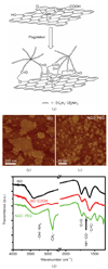
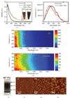
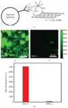
Two-dimensional graphene offers interesting electronic, thermal, and mechanical properties that are currently being explored for advanced electronics, membranes, and composites. Here we synthesize and explore the biological applications of nano-graphene oxide (NGO), i.e., single-layer graphene oxide sheets down to a few nanometers in lateral width. We develop functionalization chemistry in order to impart solubility and compatibility of NGO in biological environments. We obtain size separated pegylated NGO sheets that are soluble in buffers and serum without agglomeration. The NGO sheets are found to be photoluminescent in the visible and infrared regions. The intrinsic photoluminescence (PL) of NGO is used for live cell imaging in the near-infrared (NIR) with little background. We found that simple physisorption via pi-stacking can be used for loading doxorubicin, a widely used cancer drug onto NGO functionalized with antibody for selective killing of cancer cells in vitro. Owing to its small size, intrinsic optical properties, large specific surface area, low cost, and useful non-covalent interactions with aromatic drug molecules, NGO is a promising new material for biological and medical applications.
Figures


Similar articles
-
Supramolecular nanomedicine derived from cucurbit[7]uril-conjugated nano-graphene oxide for multi-modality cancer therapy.Biomater Sci. 2021 May 18;9(10):3804-3813. doi: 10.1039/d1bm00426c. Biomater Sci. 2021. PMID: 33881050
-
PEGylated doxorubicin cloaked nano-graphene oxide for dual-responsive photochemical therapy.Int J Pharm. 2019 Feb 25;557:66-73. doi: 10.1016/j.ijpharm.2018.12.037. Epub 2018 Dec 21. Int J Pharm. 2019. PMID: 30580088
-
A novel intracellular pH-responsive formulation for FTY720 based on PEGylated graphene oxide nano-sheets.Drug Dev Ind Pharm. 2018 Jan;44(1):99-108. doi: 10.1080/03639045.2017.1386194. Epub 2017 Oct 17. Drug Dev Ind Pharm. 2018. PMID: 28956455
-
Nano-graphene in biomedicine: theranostic applications.Chem Soc Rev. 2013 Jan 21;42(2):530-47. doi: 10.1039/c2cs35342c. Chem Soc Rev. 2013. PMID: 23059655 Review.
-
Graphene Oxide-Based Nanocarriers for Cancer Imaging and Drug Delivery.Curr Pharm Des. 2015;21(22):3215-22. doi: 10.2174/1381612821666150531170832. Curr Pharm Des. 2015. PMID: 26027564 Review.
Cited by
-
PEGylated single-walled carbon nanotubes as nanocarriers for cyclosporin A delivery.AAPS PharmSciTech. 2013 Jun;14(2):593-600. doi: 10.1208/s12249-013-9944-2. Epub 2013 Mar 12. AAPS PharmSciTech. 2013. PMID: 23479049 Free PMC article.
-
Molecular simulations of conformation change and aggregation of HIV-1 Vpr13-33 on graphene oxide.Sci Rep. 2016 Apr 21;6:24906. doi: 10.1038/srep24906. Sci Rep. 2016. PMID: 27097898 Free PMC article.
-
Graphene oxide scaffold accelerates cellular proliferative response and alveolar bone healing of tooth extraction socket.Int J Nanomedicine. 2016 May 24;11:2265-77. doi: 10.2147/IJN.S104778. eCollection 2016. Int J Nanomedicine. 2016. PMID: 27307729 Free PMC article.
-
Nano-carbons as theranostics.Theranostics. 2012;2(3):235-7. doi: 10.7150/thno.4156. Epub 2012 Mar 1. Theranostics. 2012. PMID: 22448193 Free PMC article.
-
Nanocomposites of Nitrogen-Doped Graphene Oxide and Manganese Oxide for Photodynamic Therapy and Magnetic Resonance Imaging.Int J Mol Sci. 2022 Dec 1;23(23):15087. doi: 10.3390/ijms232315087. Int J Mol Sci. 2022. PMID: 36499412 Free PMC article.
References
-
- Geim AK, Novoselov KS. The rise of graphene. Nat. Mater. 2007;6:183–191. - PubMed
-
- Kopelevich Y, Esquinazi P. Graphene physics in graphite. Adv. Mater. 2007;19:4559–4563.
-
- Li X, Wang X, Zhang L, Lee S, Dai H. Chemically derived, ultrasmooth graphene nanoribbon semiconductors. Science. 2008;319:1229–1232. - PubMed
-
- Stankovich S, Dikin DA, Dommett GHB, Kohlhaas KM, Zimney EJ, Stach EA, Piner RD, Nguyen ST, Ruoff RS. Graphene-based composite materials. Nature. 2006;442:282–286. - PubMed
-
- Dikin DA, Stankovich S, Zimney EJ, Piner RD, Dommett GHB, Evmenenko G, Nguyen ST, Ruoff RS. Preparation and characterization of graphene oxide paper. Nature. 2007;448:457–460. - PubMed
Grants and funding
LinkOut - more resources
Full Text Sources
Other Literature Sources
Research Materials
Miscellaneous
