. 2010 May 14;285(20):15220-15233.
doi: 10.1074/jbc.M109.063750. Epub 2010 Mar 9.

Differential effects of sumoylation on transcription and alternative splicing by transcription elongation regulator 1 (TCERG1)

Affiliations

Differential effects of sumoylation on transcription and alternative splicing by transcription elongation regulator 1 (TCERG1)

Miguel Sánchez-Álvarez et al. J Biol Chem. .

Abstract

Modification of proteins by small ubiquitin-like modifier (SUMO) is emerging as an important control of transcription and RNA processing. The human factor TCERG1 (also known as CA150) participates in transcriptional elongation and alternative splicing of pre-mRNAs. Here, we report that SUMO family proteins modify TCERG1. Furthermore, TCERG1 binds to the E2 SUMO-conjugating enzyme Ubc9. Two lysines (Lys-503 and Lys-608) of TCERG1 are the major sumoylation sites. Sumoylation does not affect localization of TCERG1 to the splicing factor-rich nuclear speckles or the alternative splicing function of TCERG1. However, mutation of the SUMO acceptor lysine residues enhanced TCERG1 transcriptional activity, indicating that SUMO modification negatively regulates TCERG1 transcriptional activity. These results reveal a regulatory role for sumoylation in controlling the activity of a transcription factor that modulates RNA polymerase II elongation and mRNA alternative processing, which are discriminated differently by this post-translational modification.


Figures

FIGURE 1.
FIGURE 1.
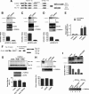
TCERG1 is a sumoylation substrate in vivo and interacts directly with human Ubc9. A, Western blotting (WB) analyses of endogenous TCERG1 from HeLa cells with or without 25 nm NEM and/or 1% SDS. Specific antibodies against TCERG1 and cyclin T1 (as a loading control) were used to localize the proteins. The brace indicates the positions of the two predominant slow migrating bands in the samples treated with 25 nm NEM and 1% SDS buffer. Molecular masses in kDa are indicated to the left. B, HeLa cells were cotransfected with a plasmid encoding T7-tagged TCERG1 along with empty vector (lane 1) or vectors encoding HA-SUMO-1 (lane 2), HA-SUMO-2 (lane 3), or HA-SUMO-3 (lane 4). WCE were directly analyzed by Western blotting or subjected to immunoprecipitation (IP) with T7-specific antibodies followed by SDS-PAGE and Western blotting with anti-T7, anti-HA, anti-SUMO-1, anti-SUMO-2, or anti-cyclin T1 antibodies. Specific higher molecular weight forms of TCERG1 are visible above the position of the 175-kDa marker and are indicated by arrows and bracket (see description in text). The unmodified form of TCERG1 is indicated by a closed arrowhead. C and D, same experiment described for B was repeated but using plasmids encoding T7-tagged amino- (C) and carboxyl-terminal (D) regions of TCERG1. *, nonspecific band present in WCE and reactive to anti-T7 antibodies. **, immunoglobulin fraction eluted from the matrix. E, purification of HA-tagged Ubc9. Lanes 1 and 2, crude E. coli lysates with (+) or without (−) isopropyl 1-thio-β-d-galactopyranoside (IPTG); lane 3, elution fraction after thrombin cleavage (HA-hUbc9). F, amino-terminal region of TCERG1 interacts directly with human Ubc9. HA-tagged Ubc9 was incubated with equal amounts of GST or GST-TCERG1(234–662) and GST-TCERG1(631–1098) bound to glutathione-Sepharose beads. Bound proteins (lanes 2–5) were eluted with SDS-PAGE loading buffer and resolved in a 15% SDS-polyacrylamide gel along a sample of the input (lane 1). The upper part of the gel was stained with Coomassie Blue R-250 to visualize the eluted GST fusion proteins. and the lower part was transferred to a membrane and incubated with an anti-HA-specific antibody to detect Ubc9. Molecular masses (M) in kDa are indicated to the left.
FIGURE 2.
FIGURE 2.
Lys-503 and Lys-608 are the major sites of sumoylation of TCERG1 and are conserved among species. A shows a ClustalW amino acid alignment of TCERG1 homologs that include the putative SUMO acceptor motifs. Conserved acceptor lysines within the core consensus motif (in yellow) and NDSM consensus motif (in blue) features are highlighted. Hs, Homo sapiens; Mm, Mus musculus; Gg, Gallus gallus; Xl, Xenopus laevis; Dr, Danio rerio; Sm, Schistosoma mansoni; Ae, Aedes aegypti; NLS, nuclear localization signal. B–D, HEK293T cells were cotransfected with expression constructs of HA-SUMO-1 (A), HA-SUMO-2 (B), or HA-SUMO-3 (D) and T7-tagged wild-type (WT) TCERG1 or the indicated mutants, and lysates subjected to immunoblotting with anti-T7 and anti-cyclin T1 antibodies. The sumoylated forms of TCERG1 are indicated by arrows (see description in text). The unmodified form of TCERG1 is indicated by a closed arrowhead. WB, Western blot. E–G, HEK293T cells were cotransfected with expression constructs of HA-SUMO-1 (E), HA-SUMO-2 (F), or HA-SUMO-3 (G) and T7-tagged WT TCERG1 or the indicated mutants, and lysates were subjected to immunoprecipitation (IP) with anti-T7 antibodies and Western blotting analysis with anti-HA antibodies. The sumoylated forms of TCERG1 are indicated by arrows. H, in vitro expressed GST-TCERG1(134–662) fusion protein, either wild type or the indicated mutants, was incubated with SUMO-1 reaction components, and the reaction products were analyzed by SDS-PAGE and immunoblotting with specific antibodies against GST. The bands corresponding to the SUMO-conjugated forms of TCERG1(134–662) are indicated by arrows. The unmodified form of recombinant TCERG1 is indicated by a close arrowhead. I, the same experiment described in H was carried out with SUMO-2. Quantification of the experimental data obtained with the double mutant variant from two independent experiments were quantified and are shown in graphic form at the right of the panel. K503,608R is K503R,K608R.
FIGURE 3.
FIGURE 3.
Nuclear localization of TCERG1 is independent from modification at Lys-503 and Lys-608 by sumoylation. A, immunofluorescence analysis of HEK293T cells transfected with ECFP-TCERG1-WT or the indicated mutant constructs (ECFP, blue). TOPRO-3 labeling was used to stain chromatin (red). Individual and merge images are shown. B, colocalization of the indicated TCERG1 constructs with the essential splicing factor SC35 that commonly serves to define nuclear speckles. Dual labeling of cells with either ECFP-TCERG-WT or SUMO mutants (ECFP, blue) and with the anti-SC35 antibody (SC-35, red) was performed. Line scans showing local intensity distributions of TCERG1 and of SC35 are shown to the right of the panels. Bars indicate the position of the line scans. K503,608R is K503R,K608R.
FIGURE 4.
FIGURE 4.
Effect of wild-type TCERG1 and sumoylation mutants on EDI alternative splicing. A, schemes of the minigenes carrying the different promoters. Exons are represented by boxes and introns by lines. Alternative EDI isoforms generated by inclusion (EDI+, 500 bp) or skipping (EDI−, 230 bp) of EDI exon are indicated. Also indicated are the position of the primers used to amplify the mRNA splicing variants. α-globin, human α-globin; mFN, human fibronectin; HSV-TK, herpes simplex virus-thymidine kinase; ss, single strand. B–D, effect of TCERG1 overexpression on alternative splicing elicited by minigenes carrying the α-globin (A), fibronectin (B), and HSV-TK (C) promoters. RNA splicing variants were detected by radioactive RT-PCR, and the products of amplification were separated by polyacrylamide gels. β-Actin was amplified as a control. A sample of cell lysate was immunoblotted with anti-TCERG1 antibody to demonstrate the expression levels. Ratios between radioactivity in EDI+ bands and EDI− bands from two independent experiments are shown below the panels. Ctl, control. E, quantitative analysis of the amount of nascent transcript and total mRNA species derived from the α-globin EDI minigene upon cotransfecting with wild-type TCERG1 by real time PCR. Quantification of the experimental data from four independent experiments is shown in graphic form. F, schematic representation of the tau exon 10 minigene system. Alternative spliced mRNAs containing tau exon 10 (Tau 10+, 245 bp) and mRNAs with tau exon 10 skipped (Tau 10−, 153 bp) are shown in the figure. G, effect of TCERG1 overexpression in the regulation of tau exon 10. RT-PCR analysis of the cotransfections of tau 10 minigene with TCERG1. β-Actin was amplified as a control. A sample of cell lysate was immunoblotted with anti-TCERG1 antibody to demonstrate the expression levels. Ratios between radioactivity in tau 10+ bands and tau− bands from two independent experiments are shown below the panel. H, cotransfection assays were carried out with wild type and sumoylation mutants of TCERG1 and the α-globin EDI minigene. Patterns of EDI minigene splicing were analyzed as in previous panels. I, curve dose-response for wild-type (WT) TCERG1 and double mutant variant (K503R,K608R (K503,608R)) on EDI alternative splicing. The α-globin EDI minigene and the indicated TCERG1 constructs were cotransfected at 100 (lanes 2 and 4) and 500 (lanes 3 and 5) ng. RNA splicing variants were detected by radioactive RT-PCR, and the products of amplification were separated by polyacrylamide gels (top panel). Experimental data from two independent experiments were quantified and are shown in graphic form (middle panel). A sample of cell lysates were immunoblotted with anti-TCERG1 antibody to demonstrate the expression levels (bottom panel).
FIGURE 5.
FIGURE 5.
SUMO modification sites negatively regulate the TCERG1-mediated transcriptional activation. A, HEK293T cells were cotransfected with a Gal4-luciferase reporter plasmid (Gal4E1B-LUC), a control plasmid expressing the Gal4 DNA binding domain (Gal4-DBD, 1st lane), the wild-type amino-terminal domain of TCERG1(134–662) (lane 2), or the SUMO mutant variants (K503R, 3rd lane; K608R, 4th lane; K503R,K608R (K503,608R), lane 5) fused to the Gal4 DNA-binding domain. The luciferase activity is shown relative to the Gal4-DBD that was set at 1. The data shown are from four independent experiments performed in triplicate (p (wild type (WT) versus Gal4-DBD) = 0.02; p (wild type versus K503R) = 0.0058; p (wild type versus K608R) = 0.11; p (wild type versus K503R,K608R) = 0.01; paired Student's t test, set at p < 0.05). A sample of cell lysate was immunoblotted with anti-Gal4 antibody to demonstrate the expression levels. B, HEK293T cells were cotransfected with a Gal4-luciferase reporter plasmid (Gal4E1B-LUC) and a control plasmid (lane 1) or increasing amounts of TCERG1(134–662) without the Gal4 DNA-binding domain (lanes 2 and 3). The luciferase activity is shown relative to the control that was set at 1. The data shown are from four independent experiments performed in triplicate. C, HEK293T cells were cotransfected with a Gal4-luciferase reporter plasmid lacking a functional TATA-box (Gal4ΔE1B-LUC), a control plasmid expressing the Gal4 DNA binding domain (Gal4-DBD, 1st lane), the wild-type amino-terminal domain of TCERG1(134–662) (2nd lane) or the SUMO mutant variants (K503R, 3rd lane; K608R, 4th lane; K503,608R, 5th lane) fused to the Gal4 DNA-binding domain. The luciferase activity is shown relative to the Gal4-DBD that was set at 1. The data shown are from four independent experiments performed in triplicate. D, HEK293T cells were cotransfected with the p3X-kb-L reporter plasmid together with increasing concentrations of wild-type TCERG1 expression plasmid. The luciferase activity was calculated relative to the control that was cotransfected with the reporter and empty plasmids. The total DNA amount for transfection was kept the same in each sample by normalizing with empty vector. E, HEK293T cells were cotransfected with the p3X-kb-L reporter plasmid together with empty vector (1st lane), wild-type TCERG1 (2nd lane), and SUMO variants (K503R, 3rd lane; K608R, 4th lane; K503,608R, 5th lane) DNA constructs. The luciferase activity was calculated relative to the control. The data shown are from four independent experiments performed in triplicate (*, p < 0.01; **, p < 0.02; ***, p < 0.04). Cell lysates were analyzed by immunoblotting with the indicated antibodies to detect the TCERG1 and CDK9 proteins.

Similar articles

Cited by

References

    1. Kornblihtt A. R., de la Mata M., Fededa J. P., Munoz M. J., Nogues G. (2004) RNA 10, 1489–1498 - PMC - PubMed
    1. Proudfoot N. J., Furger A., Dye M. J. (2002) Cell 108, 501–512 - PubMed
    1. Auboeuf D., Hönig A., Berget S. M., O'Malley B. W. (2002) Science 298, 416–419 - PubMed
    1. Nogues G., Kadener S., Cramer P., Bentley D., Kornblihtt A. R. (2002) J. Biol. Chem. 277, 43110–43114 - PubMed
    1. Kornblihtt A. R. (2005) Curr. Opin. Cell Biol. 17, 262–268 - PubMed

Publication types

Substances

LinkOut - more resources