. 2010 Mar 15;207(3):521-34.
doi: 10.1084/jem.20091711. Epub 2010 Mar 8.

IL-15 trans-presentation by pulmonary dendritic cells promotes effector CD8 T cell survival during influenza virus infection

Affiliations

IL-15 trans-presentation by pulmonary dendritic cells promotes effector CD8 T cell survival during influenza virus infection

Jodi McGill et al. J Exp Med. .

Abstract

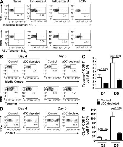
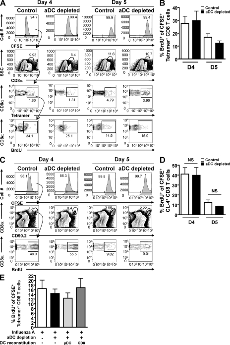
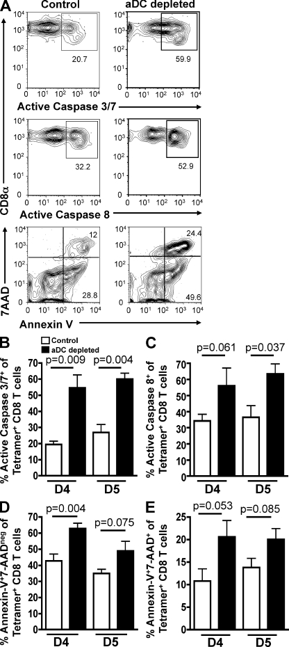
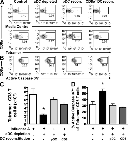
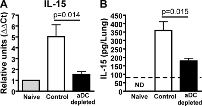
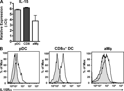
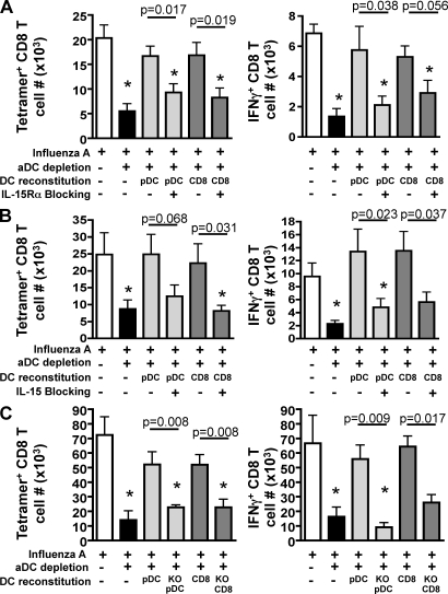
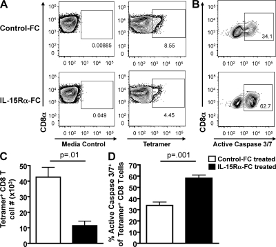
We have recently demonstrated that peripheral CD8 T cells require two separate activation hits to accumulate to high numbers in the lungs after influenza virus infection: a primary interaction with mature, antigen-bearing dendritic cells (DCs) in the lymph node, and a second, previously unrecognized interaction with MHC I-viral antigen-bearing pulmonary DCs in the lungs. We demonstrate that in the absence of lung-resident DC subsets, virus-specific CD8 T cells undergo significantly increased levels of apoptosis in the lungs; however, reconstitution with pulmonary plasmacytoid DCs and CD8alpha(+) DCs promotes increased T cell survival and accumulation in the lungs. Further, our results show that the absence of DCs after influenza virus infection results in significantly reduced levels of IL-15 in the lungs and that pulmonary DC-mediated rescue of virus-specific CD8 T cell responses in the lungs requires trans-presentation of IL-15 via DC-expressed IL-15Ralpha. This study demonstrates a key, novel requirement for DC trans-presented IL-15 in promoting effector CD8 T cell survival in the respiratory tract after virus infection, and suggests that this trans-presentation could be an important target for the development of unique antiviral therapies and more effective vaccine strategies.


Figures

Figure 1.
Figure 1.
aDC depletion at 48 h p.i. results in reduced numbers of IAV-specific CD8 T cells in the lungs on days 4 and 5 p.i. (A) Groups of mice were infected with either a sublethal dose of IAV, influenza type B, or the A2 strain of RSV. One group of mice remained uninfected. On day 4 p.i., the mice were sacrificed and their lungs were analyzed by flow cytometry for the frequencies of influenza NP147-specific and RSV M282-specific tetramer+ CD8 T cells. Representative FACS plots are gated on CD3+CD8+ T cells. Data are representative of one experiment (n = 2–3 mice/group). (B and C) Groups of BALB/c mice were infected with a sublethal dose of IAV. Half of the mice were aDC depleted at 48 h p.i. (aDC depleted), whereas the other half remained nondepleted (control). On days 4 and 5 p.i., the frequency (B) and number (C) of antigen-specific tetramer+ CD8 T cells in the lungs were enumerated. Representative FACS plots are gated on CD3+CD8+ T cells. Numbers of tetramer+ CD8 T cells in the lungs were determined by subtracting background staining using the media control (B, top). Data are pooled from two separate experiments and represent means ± SEM (n = 5–9 mice/group). (D and E) Influenza-specific, CD90.2+ CL-4 T cells were adoptively transferred to groups of CD90.1+ BALB/c mice as described in Materials and methods. 24 h later, mice were infected with a sublethal dose of IAV. Half of the mice were aDC depleted at 48 h p.i. (aDC depleted), whereas the other half remained nondepleted (control). On days 4 and 5 p.i., the frequency (D) and number (E) of CD90.2+, adoptively transferred CL-4 T cells were determined by flow cytometry. Representative FACS plots are gated on CD3+CD8+ T cells. Data are representative of three separate experiments and represent means ± SEM (n = 3–4 mice/group).
Figure 2.
Figure 2.
aDC depletion at 48 h p.i. does not alter IAV-specific CD8 T cell proliferation in the lungs. (A and B) Groups of BALB/c mice were infected with a sublethal dose of IAV. Half of the mice were subsequently aDC depleted at 48 h p.i. (aDC depleted), whereas the other half remained nondepleted (control). On days 4 and 5 p.i., mice were administered CFSE i.n, followed 2 h later by BrdU i.n. 4 h after BrdU administration, the proliferation, as measured by BrdU incorporation, of lung-resident CFSE+tetramer+ CD8 T cells was assessed by flow cytometry using the gating strategy outlined in A. Data in A and B are representative of two to three separate experiments (n = 5–8 mice/group). (C and D) Influenza-specific, CD90.2+ CL-4 T cells were adoptively transferred to groups of CD90.1+ BALB/c mice as in Fig. 1 (D and E). 24 h later, mice were infected with a sublethal dose of IAV with or without aDC depletion. On days 4 and 5 p.i., mice were treated with i.n. CFSE and BrdU. 4 h after BrdU administration, mice were sacrificed and their lungs were analyzed by flow cytometry for the frequency of BrdU+ cells among CFSE+CD8+CD90.2+ T cells using the representative gating strategy shown in C. Data in C and D are representative of one experiment (n = 3–5 mice/group). (E) Mice were infected and aDC depleted as in A and B; however, groups of aDC-depleted mice were subsequently reconstituted with purified pulmonary pDCs (light gray bars) or CD8α+ DCs (dark gray bars) on day 3 p.i. (i.e., 24 h after depletion). On day 5 p.i., virus-specific CD8 T cell proliferation was determined as in A and B. Data in E are representative of two to three separate experiments (n = 5–8 mice/group). Means ± SEM are shown. No statistical difference was observed between the analyzed groups. SSC, side scatter.
Figure 3.
Figure 3.
aDC depletion at 48 h p.i. results in increased apoptosis of IAV-specific CD8 T cells in the lungs. Groups of BALB/c mice were infected with a sublethal dose of IAV with or without aDC depletion as in Fig. 1. On days 4 and 5 p.i., mice were sacrificed and their lungs were analyzed by flow cytometry for apoptosis of tetramer+ CD8 T cells as measured by the frequency of active caspase 3/7+ (A, top; and B), active caspase 8+ (A, middle; and C), and annexin V+7-AADneg and annexin V+7-AAD+tetramer+ CD8 T cells (A, bottom; and D and E). Representative FACS plots from day 4 p.i. are gated on CD3+CD8+tetramer+ CD8 T cells. Data are representative of three to four separate experiments (n = 3–6 mice/group). Means ± SEM are shown.
Figure 4.
Figure 4.
Pulmonary DC reconstitution of aDC-depleted mice results in decreased apoptosis of IAV-specific CD8 T cells in the lungs. Groups of BALB/c mice were infected with a sublethal dose of IAV with or without aDC depletion as in Fig. 1. On day 3 p.i., groups of aDC-depleted mice were reconstituted i.n. with 2.5 × 10⁴ pulmonary pDCs (light gray bars) or CD8α+ DCs (dark gray bars) purified from the lungs of IAV-infected donors. On day 5 p.i., the frequency (A) and number (C) of tetramer+ CD8 T cells, and the frequency of apoptotic, tetramer+ CD8 T cells (B and D) per lung were measured. Data in A are representative FACS plots gated on CD3+CD8+ T cells. Numbers of tetramer+ CD8 T cells in the lungs were determined by subtracting background staining using the media control (A, top). Data in B are representative FACS plots gated on CD3+CD8+tetramer+ T cells. Data in C and D are pooled from two separate experiments (n = 6 mice/group). Means ± SEM are shown. *, P ≤ 0.05 relative to undepleted control group.
Figure 5.
Figure 5.
aDC depletion at 48 h p.i. results in decreased pulmonary IL-15 mRNA and protein expression. Groups of BALB/c mice were infected with a sublethal dose of IAV with or without aDC depletion as in Fig. 1. (A) On day 6 p.i., lungs were removed and analyzed using qRT-PCR analysis for IL-15 expression. Results were normalized to GAPDH expression, and then to expression of IL-15 mRNA by naive lungs to calculate the ΔΔCt. Data are pooled from two separate experiments and represent means ± SEM (n = 3–6 mice/group). (B) On day 6 p.i., lung homogenates from groups of naive, control, and aDC-depleted mice were analyzed for IL-15 protein expression by ELISA as described in Materials and methods. The dashed line represents the limit of detection of the assay. Data are representative of four separate experiments and represent means ± SEM (n = 5 mice/group). ND, not detected.
Figure 6.
Figure 6.
Pulmonary pDCs and CD8α+ DCs express IL-15 and IL-15Rα. (A) Groups of BALB/c mice were infected with a sublethal dose of IAV. On day 6 p.i., lungs were pooled and pDCs, CD8α+ DCs, and aMϕs were FACS purified and analyzed by qRT-PCR analysis for IL-15 expression. Results were normalized to GAPDH expression. Data are pooled from three separate experiments and represent means ± SEM. (B) Groups of BALB/c mice were infected with a sublethal dose of IAV. On day 6 p.i., pDCs, CD8α+ DCs, and aMϕs in the lungs were analyzed for surface expression of IL-15Rα (continuous line) versus isotype control (shaded) by flow cytometry. Data are representative of three separate experiments.
Figure 7.
Figure 7.
Pulmonary pDC– and CD8α+ DC–mediated rescue of IAV-specific CD8 T cell responses from aDC-depleted mice requires IL-15 trans-presentation. Groups of BALB/c mice were infected with a sublethal dose of IAV with or without aDC depletion as in Fig. 1. On day 3 p.i., groups of aDC-depleted mice were reconstituted i.n. with 2.5 × 10⁴ purified pulmonary pDCs (light gray bars) or CD8α+ DCs (dark gray bars) that were left untreated or blocked in vitro with (A) anti–IL-15Rα antibody or (B) anti–IL-15 antibody before adoptive transfer. On day 6 p.i., the number of pulmonary antigen-specific CD8 T cells was enumerated by tetramer staining (left) or ICS for IFN-γ (right). Data are pooled from three separate experiments and represent means ± SEM (n = 9–12 mice/group). (C) Groups of C57BL/6 mice were infected with a sublethal dose of IAV and aDCs depleted at 48 h p.i. (black bars), whereas controls remained undepleted (white bars). On day 3 p.i., groups of aDC-depleted mice were then reconstituted i.n. with 2.5 × 10⁴ purified pulmonary pDCs (light gray bars) or CD8α+ DCs (dark gray bars) that were purified from IAV-infected C57BL/6 IL-15−/− (KO) or wild-type IL-15+/+ donors. On day 7 p.i., the number of pulmonary IAV-specific CD8 T cells was enumerated by tetramer staining (left) or ICS for IFN-γ (right). Data are pooled from two separate experiments and represent means ± SEM (n = 6–7 mice/group). *, P ≤ 0.05 relative to undepleted control group.
Figure 8.
Figure 8.
IL-15Rα–Fc administration results in reduced IAV-specific CD8 T cell responses and increased CD8 T cell apoptosis in the lungs after IAV infection. Groups of BALB/c mice were infected with a sublethal dose of IAV. On days 3, 4, and 5 p.i., mice were i.n. administered IL-15Rα–Fc (black bars) or control human Fc fragments (white bars). On day 6 p.i., the lungs were analyzed for (A) the frequency and (C) number of tetramer+ CD8 T cells or (B and D) for apoptosis of tetramer+ CD8 T cells. Representative FACS plots are gated on (A) CD3+CD8+ T cells or (B) CD3+CD8+tetramer+ T cells. Numbers of tetramer+ CD8 T cells in the lungs were determined by subtracting background staining using the media control (A, left). Data are representative of two separate experiments and represent means ± SEM (n = 3–5 mice/group).

Similar articles

Cited by

References

    1. Abebe F., Mustafa T., Nerland A.H., Bjune G.A. 2006. Cytokine profile during latent and slowly progressive primary tuberculosis: a possible role for interleukin-15 in mediating clinical disease. Clin. Exp. Immunol. 143:180–192 10.1111/j.1365-2249.2005.02976.x - DOI - PMC - PubMed
    1. Akbar A.N., Borthwick N.J., Wickremasinghe R.G., Panayoitidis P., Pilling D., Bofill M., Krajewski S., Reed J.C., Salmon M. 1996. Interleukin-2 receptor common gamma-chain signaling cytokines regulate activated T cell apoptosis in response to growth factor withdrawal: selective induction of anti-apoptotic (bcl-2, bcl-xL) but not pro-apoptotic (bax, bcl-xS) gene expression. Eur. J. Immunol. 26:294–299 10.1002/eji.1830260204 - DOI - PubMed
    1. Alam A., Cohen L.Y., Aouad S., Sékaly R.P. 1999. Early activation of caspases during T lymphocyte stimulation results in selective substrate cleavage in nonapoptotic cells. J. Exp. Med. 190:1879–1890 10.1084/jem.190.12.1879 - DOI - PMC - PubMed
    1. Aldridge J.R., Jr., Moseley C.E., Boltz D.A., Negovetich N.J., Reynolds C., Franks J., Brown S.A., Doherty P.C., Webster R.G., Thomas P.G. 2009. TNF/iNOS-producing dendritic cells are the necessary evil of lethal influenza virus infection. Proc. Natl. Acad. Sci. USA. 106:5306–5311 10.1073/pnas.0900655106 - DOI - PMC - PubMed
    1. Bansal-Pakala P., Halteman B.S., Cheng M.H., Croft M. 2004. Costimulation of CD8 T cell responses by OX40. J. Immunol. 172:4821–4825 - PubMed

Publication types