. 2010 Apr 30;285(18):13839-49.
doi: 10.1074/jbc.M109.097345. Epub 2010 Mar 5.

Structural requirements for VAP-B oligomerization and their implication in amyotrophic lateral sclerosis-associated VAP-B(P56S) neurotoxicity

Affiliations

Structural requirements for VAP-B oligomerization and their implication in amyotrophic lateral sclerosis-associated VAP-B(P56S) neurotoxicity

SoHui Kim et al. J Biol Chem. .

Abstract

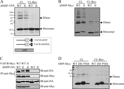
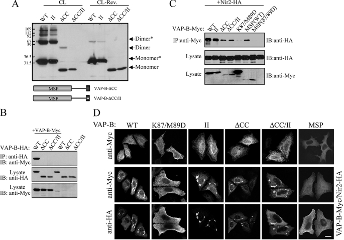
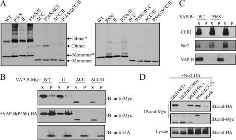
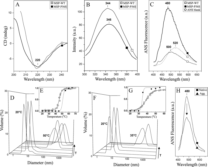
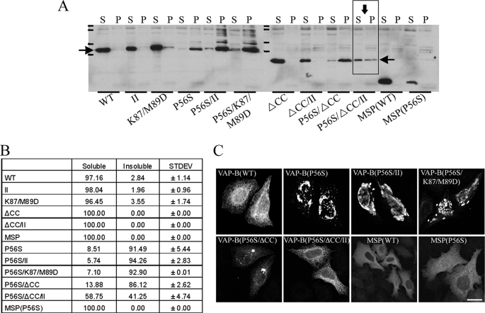
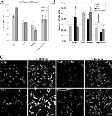
The integral endoplasmic reticulum (ER)-membrane protein VAP-B interacts with various lipid-transfer/binding proteins containing an FFAT motif through its N-terminal MSP domain. A genetic mutation within its MSP domain, P56S, was identified in familial forms of motor neuron diseases. This mutation induces the formation of insoluble VAP-B(P56S) protein aggregates by an unknown mechanism. In this study, we defined the structural requirements for VAP-B oligomerization and demonstrated their contribution for VAP-B(P56S) aggregation and neurotoxicity. We show that the oligomerization of VAP-B is mainly mediated by its coiled-coil domain and that the GXXXG dimerization motif within the transmembrane domain mediates transmembrane domains self-association but is insufficient to drive VAP-B oligomerization. We further show that the oligomerization of the wild-type VAP-B is independent of its MSP domain. However, we found that the P56S mutation induces conformational changes within the MSP domain and facilitates its propensity to aggregate by exposing hydrophobic patches to the solvent. These conformational changes have no direct effect on FFAT binding. Rather, they enhance VAP-B(P56S) oligomerization driven by the combined contributions of the coiled-coil and the transmembrane domains, thereby preventing accessibility to FFAT-binding site, facilitating the production of VAP-B(P56S)-insoluble aggregates and consequently its neurotoxicity. These results shed light on the mechanism by which VAP-B(P56S) aggregates are formed and induce familial motor neuron diseases.


Figures

FIGURE 1.
FIGURE 1.
The GXXXG motif mediates self-assembly of the TMD but is insufficient to drive VAP-B oligomerization. A, the oligomerization of Myc-tagged wild-type and VAP-B mutants was assessed following cross-linking (CL) of HEK293 cells expressing the indicated VAP-B proteins, using Western blotting with anti-Myc antibody. Arrows mark the positions of the monomer, dimer, and tetramer. The following VAP-B proteins were examined: wild-type (WT), the G235I (I) mutant, the G235/239I (II) double mutant, the FFAT-binding mutant K87/M89D, the K87D/M89D/G235I; K87/M89D/(I) triple mutant, and the K87D/M89D/G235I/G239I; K87/M89D/(II) mutant. B, the indicated HA-tagged VAP-B proteins were expressed in HEK293 cells either alone or together with the indicated Myc-tagged VAP-B. The interaction between the VAP-B proteins was determined by immunoprecipitation with anti-Myc antibody followed by immunoblotting with anti-HA antibody. C, HEK293 cells expressing the TMD of VAP-B (WT) or the II mutant fused to monomeric YFP (A206K) were cross-linked by formaldehyde, and their oligomerization was assessed by Western blotting using anti-GFP antibody before and after cross-linker cleavage, CL and CL-Rev., respectively. Arrows mark the positions of the monomer and dimer. The weak band that appears above the dimer might represent a complex between YFP-TMD and endogenous VAP-B (or -A). Positions of prestained molecular mass markers, in kDa, are indicated on the left.
FIGURE 2.
FIGURE 2.
The MSP domain does not contribute to VAP-B oligomerization. The indicated VAP-B mutants lacking the MSP domain (ΔMSP) of either the WT or the VAP-B(II) mutant were expressed HEK293 cells. Their self-assembly was assessed following cross-linking by Western blotting using anti-GFP (A) or anti-Myc (B) antibodies. The position of the monomer and the dimer is marked by an arrow. C, the indicated HA-tagged ΔMSP mutants were expressed either alone or together with the indicated Myc-tagged VAP-B proteins, and their interaction was assessed by co-immunoprecipitation studies using anti-Myc antibody for immunoprecipitation and anti-HA antibody for immunoblotting. D, the Myc-tagged MSP domains of the wild-type VAP-B, the K87/M89D (DD) double mutant, or the P56S mutant were expressed in HEK293 cells. Their self-assembly was assessed following cross-linking (CL) by Western blotting using anti-Myc antibody. Cross-linker cleavage: CL-Rev. Note that the expression level of the MSP-P56S mutant is relatively low, implying that it is not as stable as the other mutants. The position of the monomer and the dimer is marked by an arrow. Positions of prestained molecular mass markers, in kDa, are indicated on the left.
FIGURE 3.
FIGURE 3.
The coiled-coil domain is critical for VAP-B oligomerization. Deletion of the coiled-coil (ΔCC) domain markedly reduces VAP-B oligomerization and together with the II mutant apparently abolishes VAP-B homo- (A) and hetero-dimerization (B) as determined by cross-linking experiments (A) and coimmunoprecipitation studies (B), respectively. C, deletion of the coiled-coil domain has no effect on Nir2 binding. The indicated Myc-tagged VAP-B proteins were co-expressed with HA-tagged Nir2, and their association was assessed by immunoprecipitation using anti-Myc antibody following by immunoblotting using anti-HA antibody. D, the indicated Myc-tagged VAP-B proteins were expressed in HeLa cells either alone or together with HA-tagged Nir2. The localization of the indicated proteins was determined by indirect immunofluorescence analysis. Representative confocal images demonstrating the formation of stacked ER-membrane arrays upon co-expression with Nir2 are shown in the lower panels. Scale bar, 10 μm.
FIGURE 4.
FIGURE 4.
The P56S mutation enhances VAP-B(P56S) dimerization thereby preventing FFAT binding. A, cross-linking of HeLa cells expressing the indicated Myc-tagged VAP-B proteins before (left panel) and after (right panel) cross-linker cleavage. The ability of each mutant to undergo dimerization was assessed by Western blotting using anti-Myc antibody. As shown, the dimerization of the P56S is stronger than the wild-type protein. Densitometric analysis (software: ImageJ) of four different experiments indicated that the ratio between dimer to monomer plus dimer in the WT protein is 47.8 ± 5.7%, whereas in the P56S mutant the ratio is 66.0 ± 4.7%. Likewise, the dimerization of the P56S/ΔCC mutant is stronger than the ΔCC mutant, yet the P56S/ΔCC/II is mainly monomeric. B, recruitment of VAP-B proteins into VAP-B(P56S) aggregates. The indicated Myc-tagged VAP-B proteins were transiently expressed in HeLa cells either alone or together with the HA-tagged VAP-B(P56S) mutant. The distribution of the different VAP-B proteins between the Triton X-100-soluble (S) and insoluble (P) fractions was assessed by Western blotting using the indicated antibodies. C, recruitment of FFAT motif-containing proteins into VAP-B(P56S) aggregates. HA-tagged Nir2 or ceramide transfer protein was transiently expressed in HeLa cells, either alone or together with Myc-tagged wild-type VAP-B or the P56S mutant. Distribution of Nir2 and ceramide transfer protein between the detergent-soluble (S) and insoluble (P) fractions was assessed by Western blotting using anti-HA antibody. D, the P56S mutation does not affect FFAT binding. The indicated Myc-tagged VAP-B mutants were transiently co-expressed with HA-Nir2, and their association was determined by immunoprecipitation with anti-Myc antibody followed by immunoblotting with anti-HA antibody. As shown the MSP domain of the P56S mutant interacts with Nir2 in a similar manner to that of the wild-type domain.
FIGURE 5.
FIGURE 5.
Effects of the P56S mutation on the MSP domain. Purified MSP-WT (open square) and MSP-P56S (closed square) were analyzed using different biophysical methodologies: A, far-UV CD (0.1 mg/ml of protein). B, Trp-emission fluorescence (0.05 mg/ml protein). C, 1,8-ANS fluorescence emission (0.05 mg/ml protein). D, DLS distribution of MSP-WT at different temperatures (20, 25, 35, 50, 55, 60, and 65 °C, [protein] = 0.4 mg/ml). E, MSP-WT thermal unfolding curve determined by CD (open circles, Tm = 47 °C) and aggregation profile determined from DLS (closed circles, Tagg > 50 °C). F, DLS distribution of MSP-P56S at different temperatures (25, 25, 30, 35, and 45 °C, [protein] = 0.4 mg/ml). G, MSP-P56S thermal unfolding curve determined by CD (open circles, Tm = 39 °C) and aggregation profile determined from DLS (closed circles, Tagg > 35 °C). H, 1,8-ANS fluorescence emission spectra of native (solid square) and thermally aggregated (solid triangle) MSP-P56S. The spectra were recorded at 20 °C, and the arrow denotes the decrease in the emission of the 1,8-ANS fluorescence observed in the aggregated sample. (0.05 mg/ml of protein). In E and G, the y axis is the fractional change in respect to protein unfolding and aggregation; the solid line is a fit of the unfolding curve to a two-state model, whereas the dashed line on the thermal aggregation profiles is merely represented to guide the eye. See “Experimental Procedures” for detailed conditions.
FIGURE 6.
FIGURE 6.
Oligomerization of VAP-B enhances the production of VAP-B(P56S)-insoluble aggregates. A, the indicated Myc-tagged VAP-B proteins were expressed in HeLa cells and their distribution between the Triton X-100-soluble (S) and insoluble (P) fractions was assessed by Western blotting using anti-Myc antibody. Densitometric analysis (software: ImageJ) of the Western blot signals was used to estimate the relative distribution of each mutant between the soluble and insoluble fractions (B). C, the P56S/ΔCC/II mutant did not form visible aggregates. The indicated mutants were expressed in HeLa cells, fixed in 4% paraformaldehyde, immunostained with anti-Myc antibody, and analyzed by confocal microscope. Bar, 10 μm.
FIGURE 7.
FIGURE 7.
Disruption of VAP-B(P56S) dimerization attenuates its neurotoxic effects. A, the VAP-B(P56S) mutant affects cell viability. Neuro2a cells were infected with lentivirus encoding the indicated proteins. After 72 h, the cells were incubated in serum-free medium (0.2% bovine serum albumin) to induce neuronal differentiation. MTT assays were performed at time 0, and at 24 and 48 h following serum withdrawal. The relative MTT values obtained at 24 or 48 h compared with time 0 are shown. Data are mean ± S.D. values of triplicates from two independent experiments. Statistical significance was assessed by Student's t test. **, p < 0.0005 and *, p < 0.005, respectively, obtained using Student's t test of comparing P56S mutant versus either the WT protein or the P56S/ΔCC/II mutant for each time point. B, effect of VAP-B(P56S) mutant on neurite outgrowth of Neuro2a cells. Neuro2A cells were infected as above and seeded on polylysine-coated glass coverslips. 72 h after infection, the cells were incubated in serum-free media for 24 h, fixed in 4% paraformaldehyde, and immunostained with anti-Myc antibody (for VAP-B proteins) and with α-tubulin to assess neurite length. The cells were then analyzed by confocal microscopy. Z-stack images of 25 random areas were acquired in blind, and cell morphology was classified into three different categories: round, undifferentiated cells possessing short neurites, and differentiated cells exhibiting long neurites (3-fold of cell body). Approximately 500 cells of each infection were analyzed. The relative distribution of the three phenotypes obtained from two different experiments is shown ± S.D. Representative confocal images of these infections are shown in C. Scale bar, 20 μm. Statistical analysis was performed as above. ***, p values < 0.0005 and *, p < 0.05, respectively, comparing each mutant in each group with GFP.

Similar articles

Cited by

References

    1. Ross C. A., Poirier M. A. (2004) Nat. Med. 10, (suppl.) S10–S17 - PubMed
    1. Amarilio R., Ramachandran S., Sabanay H., Lev S. (2005) J. Biol. Chem. 280, 5934–5944 - PubMed
    1. Brown R. H., Jr., Robberecht W. (2001) Semin. Neurol. 21, 131–139 - PubMed
    1. Orrell R. W. (2000) Neuromuscul. Disord. 10, 63–68 - PubMed
    1. Gros-Louis F., Gaspar C., Rouleau G. A. (2006) Biochim. Biophys. Acta 1762, 956–972 - PubMed

Publication types

MeSH terms

Substances