

Correlation effect of EGFR and CXCR4 and CCR7 chemokine receptors in predicting breast cancer metastasis and prognosis
- PMID: 20181250
- PMCID: PMC2845107
- DOI: 10.1186/1756-9966-29-16
Correlation effect of EGFR and CXCR4 and CCR7 chemokine receptors in predicting breast cancer metastasis and prognosis
Abstract
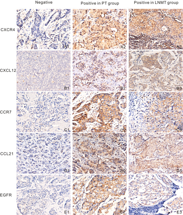
Background: The chemokine receptors CXCR4 and CCR7 play an important role in cancer invasion and metastasis. This study investigated the expression of CXCR4, CCR7, CXCL12, CCL21, and EGFR to illustrate the role of these biomarkers in breast cancer metastasis and prognosis.
Methods: The CXCR4, CCR7, CXCL12, CCL21, and EGFR biomarkers were analyzed along with ER, PR, and HER-2/neu in breast cancer tissue microarray (TMA) specimens, including 200 primary breast cancer specimens by immunohistochemistry. Corresponding lymph nodes from the same patients were also examined using the same method.
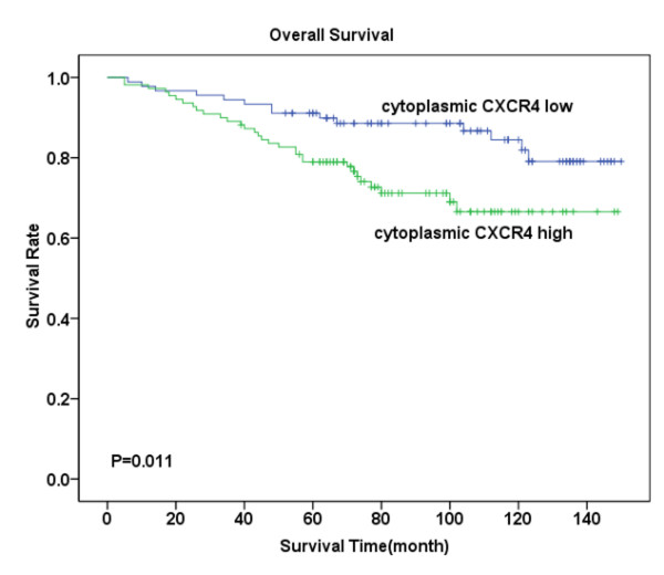
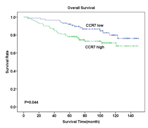
Results: Together with their CXCL12 and CCL21 ligands, CXCR4 and CCR7 were significantly highly expressed in tumor cells with lymph node (LN) metastasis. Similarly, EGFR was expressed highly in tumors with LN metastasis. The ligands were especially expressed in metastatic tumors than in primary tumors from the same patients. Moreover, the expression of both CXCR4 accompanied by CCR7 and CXCL12 accompanied by CCL21 were up-regulated. Kaplan-Meier survival analysis revealed that patients exhibiting high CXCR4, CCR7, and EGFR expression experienced a shorter survival period compared with those with low expression.
Conclusions: The expression of CXCR4, CCR7, and EGFR may be associated with LN metastasis. Moreover, the expression of these receptors can serve as an indicator of undesirable prognosis in patients with breast cancer.
Figures


Similar articles
-
CCR7 and CXCR4 as novel biomarkers predicting axillary lymph node metastasis in T1 breast cancer.Clin Cancer Res. 2005 Aug 15;11(16):5686-93. doi: 10.1158/1078-0432.CCR-05-0014. Clin Cancer Res. 2005. PMID: 16115904
-
Association of CXCR4 and CCR7 chemokine receptor expression and lymph node metastasis in human cervical cancer.Ann Oncol. 2007 Jan;18(1):70-76. doi: 10.1093/annonc/mdl342. Epub 2006 Oct 10. Ann Oncol. 2007. PMID: 17032700
-
CXCL12 promotes CCR7 ligand-mediated breast cancer cell invasion and migration toward lymphatic vessels.Cancer Sci. 2022 Apr;113(4):1338-1351. doi: 10.1111/cas.15293. Epub 2022 Feb 14. Cancer Sci. 2022. PMID: 35133060 Free PMC article.
-
Organ selectivity in metastasis: regulation by chemokines and their receptors.Clin Exp Metastasis. 2008;25(4):345-56. doi: 10.1007/s10585-007-9097-3. Epub 2007 Sep 21. Clin Exp Metastasis. 2008. PMID: 17891505 Review.
-
Expression of CXCR4 and breast cancer prognosis: a systematic review and meta-analysis.BMC Cancer. 2014 Jan 29;14:49. doi: 10.1186/1471-2407-14-49. BMC Cancer. 2014. PMID: 24475985 Free PMC article. Review.
Cited by
-
CXCR4 expression in feline mammary carcinoma cells: evidence of a proliferative role for the SDF-1/CXCR4 axis.BMC Vet Res. 2012 Mar 14;8:27. doi: 10.1186/1746-6148-8-27. BMC Vet Res. 2012. PMID: 22417013 Free PMC article.
-
Investigation of CCR7 Marker Expression Using Immunohistochemical Method and Its Association with Clinicopathologic Properties in Patients with Breast Cancer.Int J Hematol Oncol Stem Cell Res. 2018 Apr 1;12(2):103-110. Int J Hematol Oncol Stem Cell Res. 2018. PMID: 30233771 Free PMC article.
-
Evolution of the Tumor Microenvironment toward Immune-Suppressive Seclusion during Brain Metastasis of Breast Cancer: Implications for Targeted Therapy.Cancers (Basel). 2021 Sep 29;13(19):4895. doi: 10.3390/cancers13194895. Cancers (Basel). 2021. PMID: 34638378 Free PMC article.
-
Meta-analysis of the prognostic value of C-C chemokine receptor type 7 in patients with solid tumors.Cancer Manag Res. 2019 Feb 26;11:1881-1892. doi: 10.2147/CMAR.S190510. eCollection 2019. Cancer Manag Res. 2019. PMID: 30881115 Free PMC article. Review.
-
β-Arrestin1 and distinct CXCR4 structures are required for stromal derived factor-1 to downregulate CXCR4 cell-surface levels in neuroblastoma.Mol Pharmacol. 2014 Apr;85(4):542-52. doi: 10.1124/mol.113.089714. Epub 2014 Jan 22. Mol Pharmacol. 2014. PMID: 24452472 Free PMC article.
References
-
- Paget S. The distribution of secondary growths in cancer of the breast. Cancer Metastasis Rev. 1989;8:98–101. - PubMed
-
- Hassan S, Ferrario C, Saragovi U, Quenneville L, Gaboury L, Baccarelli A, Salvucci O, Basik M. The influence of tumor-host interactions in the stromal cell-derived factor-1/CXCR4 ligand/receptor axis in determining metastatic risk in breast cancer. Am J Pathol. 2009;175:66–73. doi: 10.2353/ajpath.2009.080948. - DOI - PMC - PubMed
-
- Cabioglu N, Gong Y, Islam R, Broglio KR, Sneige N, Sahin A, Gonzalez-Angulo AM, Morandi P, Bucana C, Hortobagyi GN, Cristofanilli M. Expression of growth factor and chemokine receptors: new insights in the biology of inflammatory breast cancer. Ann Oncol. 2007;18:1021–1029. doi: 10.1093/annonc/mdm060. - DOI - PubMed
Publication types
MeSH terms
Substances
LinkOut - more resources
Full Text Sources
Other Literature Sources
Medical
Research Materials
Miscellaneous
