. 2010 Mar;120(3):894-906.
doi: 10.1172/JCI40104. Epub 2010 Feb 22.

Chikungunya disease in nonhuman primates involves long-term viral persistence in macrophages

Affiliations

Chikungunya disease in nonhuman primates involves long-term viral persistence in macrophages

Karine Labadie et al. J Clin Invest. 2010 Mar.

Abstract

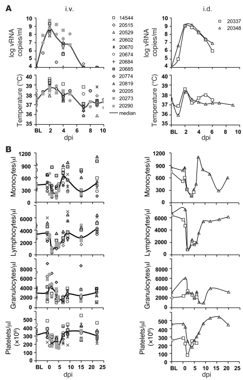
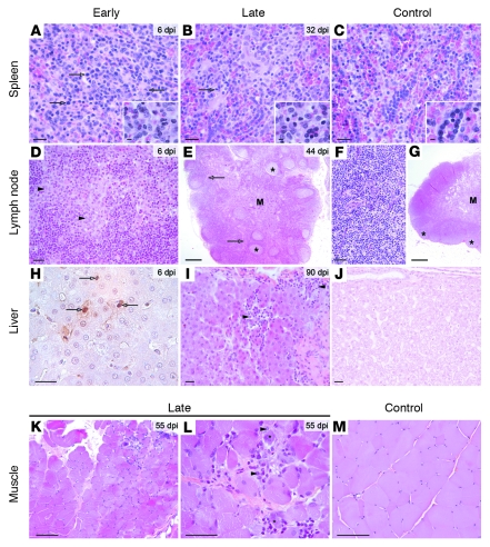
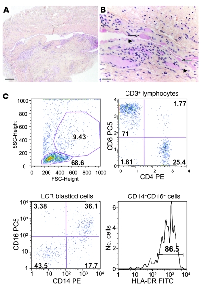
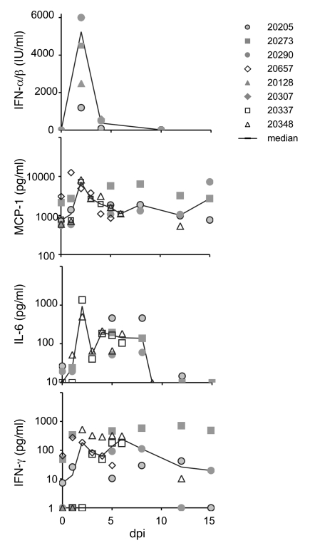
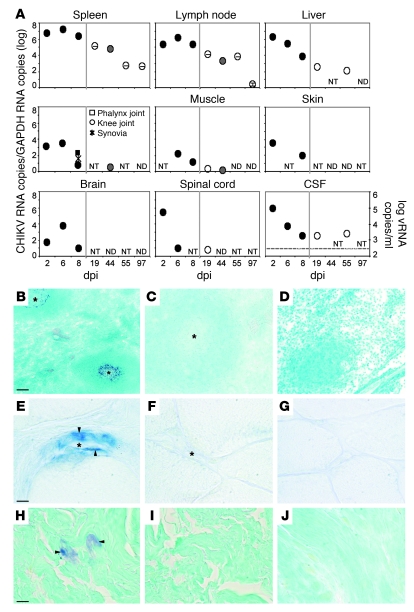
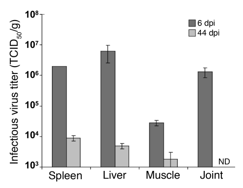
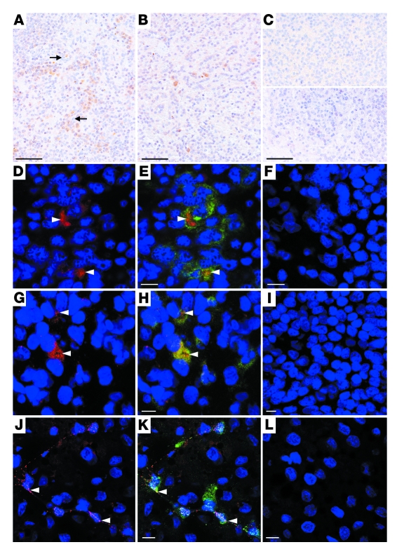
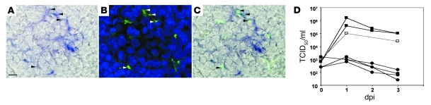
Chikungunya virus (CHIKV) is a mosquito-borne alphavirus that induces in humans a disease characterized by fever, rash, and pain in muscles and joints. The recent emergence or reemergence of CHIKV in the Indian Ocean Islands and India has stressed the need to better understand the pathogenesis of this disease. Previous CHIKV disease models have used young or immunodeficient mice, but these do not recapitulate human disease patterns and are unsuitable for testing immune-based therapies. Herein, we describe what we believe to be a new model for CHIKV infection in adult, immunocompetent cynomolgus macaques. CHIKV infection in these animals recapitulated the viral, clinical, and pathological features observed in human disease. In the macaques, long-term CHIKV infection was observed in joints, muscles, lymphoid organs, and liver, which could explain the long-lasting CHIKV disease symptoms observed in humans. In addition, the study identified macrophages as the main cellular reservoirs during the late stages of CHIKV infection in vivo. This model of CHIKV physiopathology should allow the development of new therapeutic and/or prophylactic strategies.


Figures

Figure 1
Figure 1. CHIKV infection in adult cynomolgus macaques.
(A) Kinetics of viral replication and rectal temperatures of 15 animals following i.v. (n = 13) or i.d. (n = 2) inoculation of 103 PFU CHIKV strain LR2006-OPY1. Viral load was evaluated by quantitative RT-PCR. BL, baseline. (B) Kinetics of blood cell counts in animals following i.v. or i.d. inoculation of 103 PFU CHIKV.
Figure 2
Figure 2. Lesions observed in tissues collected from 12 macaques inoculated with intermediate doses of CHIKV.
Histology of tissues from CHIKV-infected macaques. (A) Spleen, 6 dpi. Density of mononuclear cells was diffusely increased in the red pulp. These mononuclear cells corresponded mostly to macrophages with abundant cytoplasm and large nucleolated nucleus (inset). Some mononuclear cells were undergoing mitosis (arrows). (B) Spleen, 32 dpi. Macrophages were still numerous in the red pulp; a few mitotic cells were visible (arrow). (C) Normal spleen. Red pulp contained numerous red blood cells. (D) Lymph node, 6 dpi. The cortex was distended by numerous macrophages (some denoted by arrowheads). (E) Lymph node, 44 dpi. Severe follicular enlargement (asterisks) was associated with macrophage infiltration. Postcapillary venules of the T-dependent area (arrows) and medulla (M) are indicated. (F and G) Normal lymph node. Lymphoid follicles (asterisks) and medulla are indicated. (H) Liver, 6 dpi. The number of apoptotic hepatocytes with nuclei (arrows), detected by TUNEL assay, increased. (I) Liver, 90 dpi. Multifocal interstitial mononuclear cell infiltration (arrowheads) was observed in the liver parenchyma. (J) Normal liver. (K and L) Skeletal muscle, 55 dpi. Mild multifocal necrosis of muscle fibers (asterisk) was associated with infiltration by mononuclear cells, including macrophages (arrowheads). (M) Normal muscle. (AG and IM) Hematoxylin eosin safran stain. (H) In situ detection of cell death using TUNEL staining. Scale bars: 100 μm (AD, F, and HM); 10 μm (insets); 1 mm (E and G).
Figure 3
Figure 3. Mononuclear cell infiltration in tissues of macaques inoculated with high-dose CHIKV.
(A and B) Tissues lesions in ankle joint collected from a 7-dpi macaque inoculated with 108 PFU CHIKV. Mild fibrinous exudate (arrows) was associated with mononuclear cell infiltration of the synovial tissue (arrowheads). Hematoxylin eosin safran staining. Scale bars: 300 μm (A); 100 μm (B). (C) Staining of cerebrospinal fluid cells collected from a macaque inoculated with 108 PFU showing clinical signs of meningoencephalitis. Infiltrating cells were mainly CD8+ T cells and activated monocyte/macrophages (CD14+CD16+HLA-DR+). Numbers denote percent of population in the respective gate or quadrant.
Figure 4
Figure 4. Kinetics of plasma inflammatory mediators.
Macaques were inoculated with 103 PFU CHIKV. Plasma cytokines and chemokines induced by the CHIKV infection were assayed by a bioassay for IFN-α/β or using Luminex assays for CCL2 (MCP-1), IL-6, and IFN-γ.
Figure 5
Figure 5. CHIKV distribution in organs of CHIKV-infected macaques, 2–97 dpi.
(A) Relative quantitative RT-PCR in spleen, lymph nodes, liver, joint, and muscle of macaques inoculated with 103 PFU (white symbols), 106 PFU (gray symbols), or more than 107 PFU (black symbols). The CHIKV RNA copy number was normalized to GAPDH copy number. CHIKV RNA was also quantified in CSF. For each sample, the relative copy number represents the mean of at least 3 independent RT-PCR amplifications carried out on 2 independent RNA extractions. Vertical error bar denotes SEM. NT, not tested; ND, not detected. (BJ) In situ hybridization assays in spleen (BD), muscle (EG), and joint tissue (HJ) of macaques infected with 103 PFU, 6 dpi. (B, E, and H) CHIKV 26S RNA was detected in the cytoplasm of many mononuclear cells surrounding follicular centers (asterisks) of the splenic white pulp (B), and in some endothelial cells (arrowheads) surrounding vascular lumen (asterisk) of the muscular endomysial tissue (E). (H) Some vRNA was also detected in the cytoplasm of a few cells in the synovial tissue (arrowheads). (C, F, and I) Control assay using probe specific for RRV 26S RNA. (D, G, and J) Control assay using CHIKV 26S RNA on tissues of an uninfected macaque. Scale bars: 200 μm (BD); 20 μm (EJ).
Figure 6
Figure 6. Infectious virus titers in spleen, liver, muscle, and joint collected from CHIKV-infected macaques, 6 and 44 dpi.
Tissues were collected at 6 dpi from macaques inoculated i.v. with 107 PFU CHIKV, or at 44 dpi from macaques inoculated i.v. with 106 PFU CHIKV, and the amount of infectious virus present in tissues was quantified by TCID50. Data are mean ± SEM of 2 independent virus titrations. The detection threshold was 700 TCID50/g.
Figure 7
Figure 7. Immunohistochemical findings in spleen (AI) and liver (JL) collected from CHIKV-infected macaques at various times.
(A) Viral antigens were detected by immunohistochemistry at 6 dpi in the cytoplasm of numerous mononuclear cells (brown staining) of the splenic red pulp. Staining was associated with pronounced macrophage infiltration (see Figure 4). Macrophage sheathed capillaries of the red pulp are denoted by arrows. (B) Viral antigen detected in mononuclear cells of the enlarged follicles of the splenic white pulp at 44 dpi. (C) Isotypic control assay (top) and control assay using splenic tissue of an uninfected macaque (bottom). (DL) CHIKV antigen-positive cells are indicated (arrowhead). (D and E) CHIKV antigen in CD68+ macrophages of the spleen, 44 dpi. (G and H) CHIKV antigen in S100+ splenic dendritic cells, 6 dpi. (J and K) CHIKV antigen in FVIII+ endothelial cells lining the sinusoids of the liver, 44 dpi. (D, G, and J) CHIKV antigen labeling is shown in red; nuclei are stained in blue. (E, H, K) Merged images, with cell-specific markers in green. (F, I, and L) Isotype control assays. Scale bars: 200 μm (AC); 20 μm (DL).
Figure 8
Figure 8. CHIKV infects macrophages.
(A) Coupled in situ hybridization for CHIKV and immunohistochemistry for CD68 in spleen of macaque infected with 103 PFU, 6 dpi. Most mononuclear cells in which CHIKV 26S RNA was detected (A, arrowheads) were identified as macrophages, as they stained positive for CD68 (B). (C) Merged image. Scale bar: 20 μm. (D) Macrophages (squares) or dendritic cells (circles) were infected with the CHIKV LR2006-OPY1 at MOI 10 (black symbols) or MOI 5 (white symbols). Viral production was expressed as virus titers in TCID50/ml on BHK-21 cells.

Comment in

Similar articles

Cited by

References

    1. Mason PJ, Haddow AJ. An epidemic of virus disease in Southern Province, Tanganyika Territory, in 1952–53; an additional note on Chikungunya virus isolations and serum antibodies. Trans R Soc Trop Med Hyg. 1957;51(3):238–240. doi: 10.1016/0035-9203(57)90022-6. - DOI - PubMed
    1. Brighton SW, Prozesky OW, de la Harpe AL. Chikungunya virus infection. A retrospective study of 107 cases. S Afr Med J. 1983;63(9):313–315. - PubMed
    1. McGill PE. Viral infections: alpha-viral arthropathy. Baillieres Clin Rheumatol. 1995;9(1):145–150. doi: 10.1016/S0950-3579(05)80151-7. - DOI - PubMed
    1. Powers AM, Logue CH. Changing patterns of chikungunya virus: re-emergence of a zoonotic arbovirus. J Gen Virol. 2007;88(Pt 9):2363–2377. doi: 10.1099/vir.0.82858-0. - DOI - PubMed
    1. Mavalankar D, Shastri P, Raman P. Chikungunya epidemic in India: a major public-health disaster. Lancet Infect Dis. 2007;7(5):306–307. doi: 10.1016/S1473-3099(07)70091-9. - DOI - PubMed

Publication types