. 2010 Feb 22;188(4):595-609.
doi: 10.1083/jcb.200906044.

Anchorage of VEGF to the extracellular matrix conveys differential signaling responses to endothelial cells

Affiliations

Anchorage of VEGF to the extracellular matrix conveys differential signaling responses to endothelial cells

Tom T Chen et al. J Cell Biol. .

Abstract

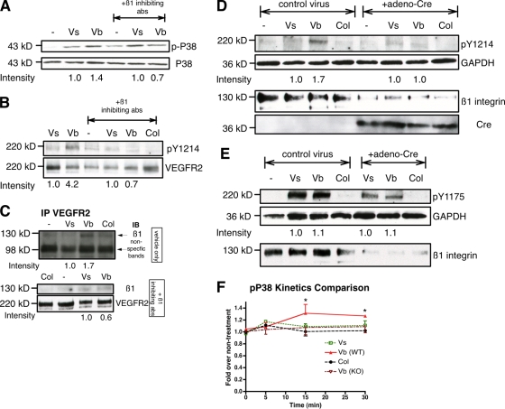
VEGF can be secreted in multiple isoforms with variable affinity for extracellular proteins and different abilities to induce vascular morphogenesis, but the molecular mechanisms behind these effects remain unclear. Here, we show molecular distinctions between signaling initiated from soluble versus matrix-bound VEGF, which mediates a sustained level of VEGFR2 internalization and clustering. Exposure of endothelial cells to matrix-bound VEGF elicits prolonged activation of VEGFR2 with differential phosphorylation of Y1214, and extended activation kinetics of p38. These events require association of VEGFR2 with beta1 integrins. Matrix-bound VEGF also promotes reciprocal responses on beta1 integrin by inducing its association with focal adhesions; a response that is absent upon exposure to soluble VEGF. Inactivation of beta1 integrin blocks the prolonged phosphorylation of Y1214 and consequent activation of p38. Combined, these results indicate that when in the context of extracellular matrix, activation of VEGFR2 is distinct from that of soluble VEGF in terms of recruitment of receptor partners, phosphorylation kinetics, and activation of downstream effectors.


Figures

Figure 1.
Figure 1.
Matrix-bound VEGF165 is able to phosphorylate VEGFR2. (A) Confluent PAE-KDR cultures were exposed for 5 min to matrix-bound VEGF165 (Vb), soluble VEGF165 (Vs; 100 ng/ml and 200 ng/ml), vehicle only (−), or polymerized collagen gels. Level of VEGFR2 (210 kD) phosphorylation was determined by Western blot analysis using a pan-phosphotyrosine antibody (4G10). The bottom panels are blots reprobed with antibodies against VEGFR2, as control. (B) Confluent HUVECs, HSVECs, and HAECs were exposed to Vb, Vs, or matrix-collagen gel controls. The blots were stripped and reprobed with antibodies against VEGFR2 as controls. (C) Similar experiment settings as A and B using 200 ng/ml VEGF, but in the presence of a nylon membrane when indicated. (D) VEGFR2 phosphorylation was induced by different concentrations of Vs or Vb, as indicated. (E) VEGF165 (200ng/ml) from two different sources was compared under bound and soluble conditions. BB94 (MMP inhibitor, 2 µM) was incorporated within the collagen gel together with VEGF165. The arrows indicate different phosphorylation patterns of VEGFR2 caused by Vb treatment. The densitometry of D and E were calculated by p-VEGFR2 over VEGFR2 and using vehicle (−) as 1.0. (F) HUVECs exposed to either soluble (200 ng/ml) or matrix-bound VEGF165 and at the indicated times. Phosphorylation levels of VEGFR2 were then determined by Western blot using 4G10 antibody. Note that prolonged phosphorylation of VEGFR2 in bound when compared with soluble VEGF165. (G) Densitometric analysis of same experiments as F (n = 4–5), shown as ratio of Vb over Vs in each time point (*, P < 0.05 for 10- and 20-min time point between Vb and Vs).
Figure 2.
Figure 2.
VEGFR2 clustering and internalization induced by soluble and matrix-bound VEGF165. (A) HUVECs were treated with vehicle (−), Vs (200 ng/ml), Vb (200 ng/ml), or collagen (col) for 15 min. Cells were fixed and stained using either rabbit Ig control (top) or antibody against extracellular domain of VEGFR2 in the absence of permeabilization. Computer-generated images (black and white on the right) were used for quantitation of positive staining of VEGFR2 (VEGFR2 distribution) and to identify particle size over 10-fold larger than controls (VEGFR2 clustering). (B) Statistical analysis for VEGFR2 distribution and VEGFR2 clustering are shown. For VEGFR2 clustering, baseline (=1) size was assessed when cells were exposed to vehicle only (*, P < 0.05, between Vb to either −, Vs, or Col). (C) Internalization of VEGFR2 was analyzed in confluent HUVECs treated with Vs (200 ng/ml), Vb (200 ng/ml), and vehicle controls for 15 min. To detect the internalized receptor, cells were treated first with a recombinant single-chain antibody against human VEGFR2 (scFvA7), subsequently exposed to Vs or Vb, followed by acid washes before fixation. Internalized VEGFR2 appears in a vesicular pattern. (D) Same experiments as C but analyzed by Western blots using VEGFR2 or 4G10 antibodies. Total VEGFR2 was measured from the same lysate used for immunoprecipitation used for internalized VEGFR2. (E) Statistics analysis of C. The results (referred to as counts per cell) were reported in the bar graph of three independent experiments. Six random fields were analyzed in each experiment. (F) Densitometric analysis of D. The results were from six independent experiments. Internalized VEGFR2 by Vs and Vb were normalized with total VEGFR2 in the lysate, and then compare normalized Vb over Vs in each experiment. (G) Confluent HUVECs were pretreat with Dynosore or DMSO at 37°C for 15 min. Cells were then treated with Vs, Vb, or vehicles (−) for 5 min. Phosphorylation assays were performed and the cell lysates were then immunoprecipitated with 4G10 and then blotted with total VEGFR2 antibody. Total VEGFR2 was determined by using the same lysate used for immunoprecipitation. In A and C, nuclei stained with TOPRO3 appears in blue. *, P < 0.05; **, P < 0.005.
Figure 3.
Figure 3.
Prolonged phosphorylation induced by matrix-bound VEGF165 is specific to tyrosine 1214. (A) HUVECs were incubated with Vs or Vb (200 ng/ml) for either 5 or 15 min, as indicated. Phosphorylation assays were performed and 4G10 was used as pan-phosphotyrosine antibody. Densitometric analysis of phospho-VEGFR2 by Vs and Vb were normalized with total VEGFR2 and then compare Vb over Vs as indicated. (B) Phosphorylation assays were performed using phosphotyrosine antibodies specific for the indicated residues on VEGFR2. (C) HEK 293 cell lines expressing Y1214F VEGFR2 mutant were incubated with soluble (Vs; 200 ng/ml) or bound VEGF165 (Vb) for 5 min and 15 min, vehicle (−), or collagen only (col) for 5 min. Equal protein levels were separated by gel electrophoresis and probed with 4G10. The blot was then reprobed with VEGFR2 antibody for normalization. Densitometric analysis of phosphorylation status induced by Vb to Vs is indicated. (D) Similar experiments were performed using HEK 293 stably transfected with native VEGFR2 (293NR).
Figure 4.
Figure 4.
p38 and Akt pathways are differentially induced by soluble and matrix-bound VEGF165. (A) Confluent HUVEC cultures were incubated in the presence or absence of soluble (Vs) or matrix-bound VEGF165 (Vb; 200 ng/ml) for 5 min. Cell lysates were obtained, resolved by SDS-PAGE, and probed with antibody against phospho-p38 (43 kD). The blot was then stripped and reprobed with total p38 antibodies for normalization. (B) HUVECs were exposed to Vs or Vb (200 ng/ml) at the indicated times. The blot was then stripped and reprobed with total p38 antibody for normalization. (C) Same settings as B, but membrane was immunoblotted with phospho-Akt (S473) and total Akt (60 kD). Densitometry was determined by phospho-p38 over total p38 (n = 6) (D) or phospho-Akt over total Akt (n = 6) (E).
Figure 5.
Figure 5.
Prolonged activation of VEGFR2 requires matrix-binding motifs in VEGF. (A) Various VEGF isoforms (165, 121, 113; 200 ng/ml) were polymerized with collagen as indicated. The gel was prepared and washed as described in Materials and methods and placed on confluent monolayers for 5 min. Phosphorylation assays were performed and total VEGFR2 levels were determined by reprobing the same membrane. (B) Same as A, but treated for 15 min and probed with antibodies against pY1214 (top) or pP38 (bottom). Numbers on the bottom of the Western blot indicate densitometric quantification. (C) Different collagen types were used to anchor VEGF (Vb, 200 ng/ml) as indicated. Exposure to collagen matrices for 15 min resulted in prolonged phosphorylation of Y1214. (D) Dose curve of VEGFR2 activation by VEGF. Detection of phosphorylation was performed using 4G10 antibodies. Numbers below the Western blotndicate densitometric quantification at 15 min using 100 ng/ml VEGF165 treatment as 1.0. (E) 500 ng/ml of VEGF165 was used on cells for 5, 15, and 30 min in the presence or absence of a carrier to stabilize the protein. On the right, 500 ng/ml of VEGF was used at time 0, and an additional fresh aliquot at 500 ng/ml was added at indicated time points for another 5 min. Phosphorylation assay was performed using 4G10 antibody.
Figure 6.
Figure 6.
VEGFR2 associates with β1 integrin when exposed to matrix-bound but not soluble VEGF165. (A) Confluent HUVEC monolayers were incubated with soluble (200 ng/ml) or bound VEGF165 (200 ng/ml) and collagen for 5, 15, and 30 min. Cultures were then lysed and equal cell lysates were used for immunoprecipitation with VEGFR2 antibodies. The precipitated protein was then resolved on 4–12% SDS-PAGE and probed with a β1 integrin antibody (130 kD). The blot was then reprobed with anti-VEGFR2. (B) Representative images of proximity ligation assays with quantification (C) after treatment with the indicated growth factors. Red dots denote regions of signal amplification consistent with VEGFR2/β1 integrin interactions. Nuclear stain is TOPRO3 (blue) (n = 4; *, P < 0.05 between Vb to either −, Vs, or Col).
Figure 7.
Figure 7.
β1 integrin redistributes to focal adhesion upon stimulation by matrix-bound VEGF165. HUVECs treated at the conditions indicated for 15 min were fixed and stained for β1 integrin (red), paxillin (green), and TOPRO3 (blue). Arrows (A and B) indicate colocalization of β1 integrin and paxillin, arrowheads in B show focal adhesions without β1 integrin–positive staining. Asterisks in A indicate cells with higher expression of β1 integrin, to indicate that regardless of the diffused expression of β1 integrin only, Vb, but not Vs, resulted in redistribution of the integrin to focal adhesions. (A) Lower magnification; (B) higher magnification of independent experiments.
Figure 8.
Figure 8.
Blocking β1 integrin dampens activation of pY1214 and P38 of matrix-bound VEGF165. (A) Confluent HUVEC monolayers were starved for 6 h and pretreated with β1-inhibiting antibodies for 15 min, then treated with the indicated reagents for an additional 15 min. Cell lysates were collected, resolved on 4–12% SDS-PAGE, probed with phopho-p38 antibody, and reprobed with total p38 antibody. (B) Same as A, treated with indicated reagents for 15 min and probed with antibody against pY1214. Membranes were then stripped off and reprobed with total VEGFR2 antibody. (C) HUVECs were treated as in A. Cell lysates were collected and immunoprecipiated with VEGFR2 antibodies. Immunoprecipitated proteins were probed with anti-β1 integrin or anti-VEGFR2. The top panel shows cells without preincubation with β1-inhibiting antibodies. The bottom panel shows cells preincubated with β1-inhibiting antibodies. (D) Mouse liver ECs from β1fl/fl mice were isolated and treated with adeno-Cre virus to induce deletion of the gene. After 3 d, cultures were treated with indicated growth factors for 15 min and cell lysates were probed with pY1214, GAPDH (36 kD), and β1 antibodies. Blots were then stripped and reprobed with anti-Cre (35 kD). (E) Similar experimental setting as D but probed with pY1175. (F) Confluent β1fl/fl or β1ko MLEC monolayers in 96-well plates were starved overnight and exposed to the indicated treatments for 5, 15, or 30 min. p38 activation was determined by cell-based ELISA and the fold change at each time point was calculated by phospho-p38 over total p38 compared with no treatment. The difference between pP38 activation in WT and KO cells is statistically significant at 15 and 30 min. *, P < 0.05 (n = 4).
Figure 9.
Figure 9.
β1 integrin is required for clustering and continued internalization of VEGFR2. (A) Internalization of VEGFR2 was analyzed in confluent HUVECs pretreated with β1 inhibitory antibodies or Ig control for 15 min, and then treated with Vs (200 ng/ml), Vb (200 ng/ml), and vehicle controls for another 15 min. To detect the internalized receptor, cells were treated with a recombinant single-chain antibody against human VEGFR2 (scFvA7), subsequently exposed to the indicated treatment followed by acid washes before lysis. Internalized VEGFR2 was analyzed by Western blot using VEGFR2 or 4G10 antibodies. Total VEGFR2 was determined in the same lysates. The numbers indicate densitometric analysis comparing Vs and Vb. (B) HUVECs pretreated with β1 inhibitory antibodies or Ig control for 15 min were exposed to vehicle (−), Vs (200 ng/ml), Vb (200 ng/ml), or collagen (col) for 15 min. Cells were fixed and stained using recombinant single-chain antibody against human VEGFR2 (scFvA7) with E-tag in the absence of permeabilization. FITC-conjugated anti–E-tag was used for detection of positive staining. Nuclei stained with TOPRO3 appears in blue. (C) Schematic model depicting the interaction of soluble and matrix-bound VEGF with VEGFR2. Soluble VEGF (left) results in rapid binding, internalization, and activation of VEGFR2. Matrix-bound VEGF (right) interacts easily with receptors as the cells migrate into the matrix. The affinity for the matrix is two orders of magnitude lower than its receptor. Over time, the progressive recruitment of integrins facilitates clustering of VEGFR2 and results in binding between integrins and VEGFR2. This interaction maintains the phosphorylation status of Y1214.

Similar articles

Cited by

References

    1. Abraham S., Yeo M., Montero-Balaguer M., Paterson H., Dejana E., Marshall C.J., Mavria G. 2009. VE-Cadherin-mediated cell-cell interaction suppresses sprouting via signaling to MLC2 phosphorylation. Curr. Biol. 19:668–674 10.1016/j.cub.2009.02.057 - DOI - PubMed
    1. Abu-Ali S., Fotovati A., Shirasuna K. 2008. Tyrosine-kinase inhibition results in EGFR clustering at focal adhesions and consequent exocytosis in uPAR down-regulated cells of head and neck cancers. Mol. Cancer. 7:47 10.1186/1476-4598-7-47 - DOI - PMC - PubMed
    1. Anderson S.M., Chen T.T., Iruela-Arispe M.L., Segura T. 2009. The phosphorylation of vascular endothelial growth factor receptor-2 (VEGFR-2) by engineered surfaces with electrostatically or covalently immobilized VEGF. Biomaterials. 30:4618–4628 10.1016/j.biomaterials.2009.05.030 - DOI - PMC - PubMed
    1. Ashikari-Hada S., Habuchi H., Kariya Y., Kimata K. 2005. Heparin regulates vascular endothelial growth factor165-dependent mitogenic activity, tube formation, and its receptor phosphorylation of human endothelial cells. Comparison of the effects of heparin and modified heparins. J. Biol. Chem. 280:31508–31515 10.1074/jbc.M414581200 - DOI - PubMed
    1. Bates D.O., Cui T.G., Doughty J.M., Winkler M., Sugiono M., Shields J.D., Peat D., Gillatt D., Harper S.J. 2002. VEGF165b, an inhibitory splice variant of vascular endothelial growth factor, is down-regulated in renal cell carcinoma. Cancer Res. 62:4123–4131 - PubMed

Publication types

MeSH terms