. 2010 May;30(9):2193-205.
doi: 10.1128/MCB.01510-09. Epub 2010 Feb 22.

The SUMO E3 ligase activity of Pc2 is coordinated through a SUMO interaction motif

Affiliations

The SUMO E3 ligase activity of Pc2 is coordinated through a SUMO interaction motif

Shen-hsi Yang et al. Mol Cell Biol. 2010 May.

Abstract

Protein modification by SUMO conjugation has emerged to be an important regulatory event. Recently, the mechanisms through which SUMO elicits its effects on target proteins have been elucidated. One of these is the noncovalent association between SUMO and coregulatory proteins via SUMO interaction motifs (SIMs). We therefore searched for additional binding proteins to elucidate how SUMO acts as a signal to potentiate novel noncovalent interactions with SUMO-binding proteins. We identified an E3 ligase, Pc2, as a SUMO-binding protein with two functionally distinct SIMs. Here, we focus on the role of SIM2 and demonstrate that it is crucial for many of the documented Pc2 functions, which converge on determining its E3 ligase activity. One role of SUMO binding in this context is the subnuclear partitioning of the active form of Ubc9 (SUMO approximately Ubc9) by Pc2. The significance of the SIM2-dependent functions of Pc2 is demonstrated in the control of the precise expression of lineage-specific genes during embryonic stem cell differentiation.


Figures

FIG. 1.
FIG. 1.
Identification of a SIM in Pc2 by yeast two-hybrid assays. (A) The domain structure of Pc2 is shown schematically: the chromodomain is indicated by a black box, the polyhistidine stretch is indicated by a striped box, and a white box indicates the region identified by a yeast two-hybrid screen. The binding regions of Ubc9 in Pc2 are indicated. (B) Schematic representation of a series of truncated Pc2 proteins fused to the Gal4(AD) in yeast expression vectors, which were used in yeast two-hybrid assays (left). The amino acids at each end of the deletion mutants are numbered. Amino acid sequences targeted for point mutation or deletion are highlighted in a black box, and their relative positions within the constructs are indicated by asterisks. The nomenclature of Pc2 expression constructs is indicated. A qualitative assessment of the relative binding of SUMO-1Δgg fused to the GAL4(DBD) is shown. “−” represents no growth, and “+” indicates growth and, hence, binding. (C) Amino acid sequence alignment of known SIMs with putative SIMs in Pc2. SIMs are shaded. The core consensus SIMs, ΨΨXΨ and ΨXΨΨ, are indicated.
FIG. 2.
FIG. 2.
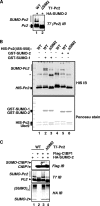
In vitro binding between Pc2 and SUMO. (A and B) Nickel affinity pulldown assays of the indicated His-tagged proteins. Schematic diagrams show series of bacterially expressed GST-Pc2 (A) and His6-Pc2 (B) fusion proteins used in the in vitro pulldown assays. The nomenclature of Pc2 constructs is indicated on the left. The relative positions within the constructs targeted for mutation or deletion are indicated by asterisks. A qualitative assessment of relative binding of GST-Pc2 or SUMO fusion proteins to the His6-SUMO-1 or His6-Pc2 fusion proteins is indicated on the right. “−” represents no binding, “+” illustrates weak binding, and “++” indicates strong binding. A representative immunoblot from the pulldown assays is shown underneath the schematics. (A) Interactions between Pc2 and SUMO-1 with the indicated GST-Pc2 fusion proteins and His6-tagged SUMO-1 (top) (anti-GST immunoblot [IB]). Inputs of His6-SUMO-1 (middle) (Ponceau stained) and GST-Pc2 fusion proteins (bottom) (Coomassie stained) are shown. (B) Interactions between Pc2 and SUMO-1 with the indicated GST-SUMO fusion proteins (top) (anti-GST IB). Inputs of His6-Pc2 fusion proteins (bottom) (Ponceau stained) and GST-SUMO fusion proteins (right) (Coomassie stained) are shown. (C) Concentration-dependent interactions between WT and ΔSIM2 His6-Pc2 fusion proteins (indicated on the right) and His6-SUMO-1 were analyzed by fluorescence spectrometry. The data are plotted as a change in fluorescence intensity due to SUMO binding, ΔI [I(Pc2 + SUMO-1) − (IPc2 + ISUMO-1)] normalized to ΔI at 1 μM Pc2, versus concentrations of Pc2 (x axis [Pc2]) and are presented as the averages of data from three independent experiments (standard errors of the means [SEMs] are shown; n = 3). (D) GST pulldown assays of the indicated Pc2 derivatives with GST-SUMO-1. The source of Pc2 derivatives was lysates from 293T cells transfected with the indicated full-length Pc2 proteins. Binding of Pc2 was detected by IB (top) (anti-Flag). Inputs of GST-SUMO-1 (middle) (Ponceau stained) and Pc2 (bottom) (anti-Flag immunoblot) proteins are shown. A schematic diagram showing the locations of SIM1 and SIM2 in Pc2 is shown at the top.
FIG. 3.
FIG. 3.
The SIM2 motif is important for Pc2 autosumoylation and substrate sumoylation. (A) The indicated WT or ΔSIM2 mutant versions of T7-Pc2(1-558) were coexpressed with HA-SUMO-2. Cell lysates were analyzed by immunoblotting (IB) with an anti-T7 antibody to detect SUMO-modified (bracket) and unmodified (arrows) Pc2. (B) In vitro sumoylation assays were performed with the indicated combinations of GST-SUMO isoforms and either WT or ΔSIM2 mutant versions of Pc2. Pc2 and its modified forms were detected by IB using anti-His antibodies (top), and input proteins were visualized by Ponceau staining of the membrane prior to IB (bottom). The asterisk likely represents anomalously migrating forms of Pc2. (C) Pc2-mediated sumoylation of CtBP1 in vivo. Wild-type or ΔSIM2 mutant versions of T7-Pc2(1-558) were coexpressed with HA-SUMO-2 and Flag-CtBP1 as indicated. Total lysates were analyzed by IB to detect total and sumoylated CtBP1 (Flag) and Pc2 (T7) or total sumoylated cellular proteins (HA).
FIG. 4.
FIG. 4.
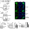
SIM2 in Pc2 is essential for the subcellular relocalization of Ubc9. (A) Schematic representation of T7-tagged Pc2 constructs used in subcellular fractionation and fluorescent microscopy studies. (B, D, and E) Wild-type, ΔSIM2, or K492R mutant versions of T7-Pc2(1-558) were expressed in combination with or without HA-SUMO-2 and Myc6-Ubc9 as indicated. Lysates were fractionated into NP-40-soluble (“S”) and insoluble (“I”) fractions, which were further analyzed by IB. The subcellular distribution of Ubc9 was analyzed by IB with an anti-Myc antibody (top panels). Total Pc2 and sumoylated Pc2 were detected by IB with an anti-T7 antibody (middle). Erk2 levels were used as a “loading control” (bottom). The relocalization of Ubc9 in D was reduced to 47.5% ± 13.4% (n = 3) with the ΔSIM2 mutant compared to WT Pc2. (C) Fluorescence images of YFP-Ubc9 fusion proteins. 293 cells were cotransfected with YFP-Ubc9 and vectors expressing the indicated versions of Pc2 and SUMO-2. Fluorescence images of cells expressing YFP-tagged Ubc9 fusion proteins (green channel) are indicated. DNA was stained with DAPI (blue channel). A merge of signals is shown on the right (over 80% of cells exhibited this colocalization). (F and G) Subcellular localization assays of endogenous Ubc9 in 293T cells (F) and mESCs (G). (F) Wild-type or ΔSIM2 mutant versions of T7-Pc2(1-558) were expressed in combination with HA-SUMO-2. (G) Pc2 levels were depleted by treating mESCs with siRNA duplexes against mouse Pc2 (mPc2). Graphs show the quantification of the amount of Ubc9 in the insoluble fraction and represent the averages of data from two independent experiments.
FIG. 5.
FIG. 5.
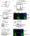
Thioester-bound and non-covalently-associated SUMO in Ubc9 are important for its redistribution by Pc2. (A) Schematic representation of constructs used in biochemical fractionation assays and cell imaging experiments. (B, C, E, F, and G) Fractionation experiments were performed as described in the legend of Fig. 4B with the exception that they were carried out in the presence and absence of T7-Pc2 and the presence of WT or C93S versions of Ubc9 (B), WT or Δgg versions of HcRed-SUMO-2 (C), WT or R13E/K14E mutant versions (E) or C93S and K14R mutant versions (F) of Ubc9, and combinations of WT or mutant SUMO-2 and Ubc9 (G), as indicated. (D and H) Localization of YFP-Ubc9 fusion proteins. The experiments were performed as described in the legend of Fig. 4C, with the exception that YFP-Ubc9, T7-Pc2(1-558), and WT or Δgg versions of HcRed-SUMO-2 (D) or WT or D62R versions of HA-SUMO-2 (H) were expressed in cells as indicated.
FIG. 6.
FIG. 6.
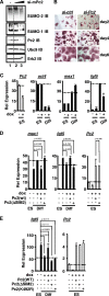
SUMO binding via SIM2 is important for Pc2 to regulate lineage-specific gene expression in mouse embryonic stem cells. (A) Effects of Pc2 depletion on total cellular protein sumoylation by either SUMO-1 or SUMO-2 in mESCs. Cells were transfected with increasing concentrations of siRNA (0, 10, and 50 nM) against mouse Pc2 (mPc2) and analyzed by IB with the indicated antibodies. IB with an anti-Erk antibody was used as a loading control (bottom). (B) mESCs were maintained in ES cell medium and stained for alkaline phosphatase activity (red staining) at the indicated times after the transfection of control (si-ctrl) or Pc2 (si-Pc2) RNAi duplexes. (C) RT-PCR analysis of gene expression in a stable mESC line that inducibly expresses RNAi against mPc2 in the presence and absence of doxycycline (dox). The data show the relative mRNA expression levels of the mESC pluripotent marker oct4 and early differentiation markers msx1 and fgf5. “ES” and “diff” indicate that mESCs were cultured in ESC medium for 1 day (ES) or grown in the absence of LIF for 5 days (diff). Data are the averages of data from three independent experiments (SEMs are shown; n = 3). (D and E) Experiments were performed as described above (C) except that siRNA-resistant WT, ΔSIM2, or K492R versions of human Pc2 were transfected. Data are averages of data from three (D) or two (E) independent experiments (SEMs are shown, and statistical significance is shown).
FIG. 7.
FIG. 7.
Model illustrating the role of SIM2 (dark gray rectangle) in Pc2 that coordinates the SUMO machinery-substrate association. Pc2 utilizes its SIM2 motif to coordinate substrate (Sub)-Ubc9 interactions in the context of PcG bodies. The SIM2 motif in Pc2 binds to SUMO linked to Ubc9 through a thioester bond and thereby recruits Ubc9 into PcG bodies.

Similar articles

Cited by

References

    1. Baba, D., N. Maita, J. G. Jee, Y. Uchimura, H. Saitoh, K. Sugasawa, F. Hanaoka, H. Tochio, H. Hiroaki, and M. Shirakawa. 2005. Crystal structure of thymine DNA glycosylase conjugated to SUMO-1. Nature 435:979-982. - PubMed
    1. Bossis, G., and F. Melchior. 2006. Regulation of SUMOylation by reversible oxidation of SUMO conjugating enzymes. Mol. Cell 21:349-357. - PubMed
    1. Cão, R., Y. Tsukada, and Y. Zhang. 2005. Role of Bmi-1 and Ring1A in H2A ubiquitylation and Hox gene silencing. Mol. Cell 20:845-854. - PubMed
    1. Capili, A. D., and C. D. Lima. 2007. Structure and analysis of a complex between SUMO and Ubc9 illustrates features of a conserved E2-Ubl interaction. J. Mol. Biol. 369:608-618. - PMC - PubMed
    1. Chalkiadaki, A., and I. Talianidis. 2005. SUMO-dependent compartmentalization in promyelocytic leukemia protein nuclear bodies prevents the access of LRH-1 to chromatin. Mol. Cell. Biol. 12:5095-5105. - PMC - PubMed

Publication types

MeSH terms