. 2010 Feb 22:9:42.
doi: 10.1186/1476-4598-9-42.

Antineoplastic effects of an Aurora B kinase inhibitor in breast cancer

Affiliations

Antineoplastic effects of an Aurora B kinase inhibitor in breast cancer

Christopher P Gully et al. Mol Cancer. .

Abstract

Background: Aurora B kinase is an important mitotic kinase involved in chromosome segregation and cytokinesis. It is overexpressed in many cancers and thus may be an important molecular target for chemotherapy. AZD1152 is the prodrug for AZD1152-HQPA, which is a selective inhibitor of Aurora B kinase activity. Preclinical antineoplastic activity of AZD1152 against acute myelogenous leukemia, multiple myeloma and colorectal cancer has been reported. However, this compound has not been evaluated in breast cancer, the second leading cause of cancer deaths among women.

Results: The antineoplastic activity of AZD1152-HQPA in six human breast cancer cell lines, three of which overexpress HER2, is demonstrated. AZD1152-HQPA specifically inhibited Aurora B kinase activity in breast cancer cells, thereby causing mitotic catastrophe, polyploidy and apoptosis, which in turn led to apoptotic death. AZD1152 administration efficiently suppressed the tumor growth in a breast cancer cell xenograft model. In addition, AZD1152 also inhibited pulmonary metastatic nodule formation in a metastatic breast cancer model. Notably, it was also found that the protein level of Aurora B kinase declined after inhibition of Aurora B kinase activity by AZD1152-HQPA in a time- and dose-dependent manner. Investigation of the underlying mechanism suggested that AZD1152-HQPA accelerated protein turnover of Aurora B via enhancing its ubiquitination.

Conclusions: It was shown that AZD1152 is an effective antineoplastic agent for breast cancer, and our results define a novel mechanism for posttranscriptional regulation of Aurora B after AZD1152 treatment and provide insight into dosing regimen design for this kinase inhibitor in metastatic breast cancer treatment.


Figures

Figure 1
Figure 1
AZD1152-HQPA inhibits breast cancer cell proliferation. Live cells were measured by MTT assay. Log(dose)-response curves for HER18, MDA-MB-468, MDA-MB-435, MDA-MB-231, MDA-MB-361 and BT474 are shown as labeled. Each data point represents the mean of at least 4 replicates; error bars represent 95% confidence intervals. Each IC50 was calculated based on sigmoidal curve fitting to the respective data set.
Figure 2
Figure 2
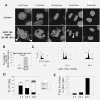
AZD1152-HQPA causes mitotic defects, aneuploidy and polyploidy in breast cancer cells. (A) HER18 cells were treated with control culture medium or 20 nM AZD1152-HQPA for 48 hours, stained with DAPI and examined using fluorescence microscopy. Interphase cells showed bi-nucleation (arrow) and micronuclei (arrow heads). Metaphase cells showed misaligned chromosomes (arrow). Anaphase cells showed misegregated chromosomes (arrow) and chromosomal bridges (arrow head). (B) Percentage of cells from A with mitotic catastrophe is plotted for vehicle and 20 nM AZD1152-HQPA treatments. (C) HER18 cells were treated with AZD1152-HQPA (100 nM) for indicated times and stained with propidium iodide prior to analysis of DNA content by flow cytometry. Gating indicates 2N (G1), 4N (G2/M) and 8N (polyploid) cells. Aneuploid cells were present between 4N and 8N after 48 h treatment. (D) Percentages of 2N, 4N and 8N cells are plotted at 0, 24 and 48 hour time points based on the data in C. (E) Number of 8N cells relative to 0 hours.
Figure 3
Figure 3
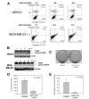
AZD1152-HQPA induces apoptosis and reduces clonogenic potential in breast cancer cells. (A) HER18 and MDA-MB-231 cells were exposed to AZD1152-HQPA (100 nM and 105 nM) for up to 72 hours and cells were stained with Annexin V-FITC plus propidium iodide. Samples were analyzed by flow cytometry for PI (y-axis) and Annexin V (x-axis) at 3 time points. Representative scattergrams are shown with percentages of cells displayed for each quadrant: Lower Left - live cells, Lower Right - early apoptosis, Upper Right - late apoptosis and Upper Left - necrotic cells. Overall percent apoptosis was calculated for each time point by adding Lower Right and Upper Right quadrants. (B) Apoptosis of HER18 and MDA-MB-231 cells was analyzed by Western blotting with anti-PARP antibody. PARP appears at 115 KD while the apoptotic indicator, cleaved PARP, appears at 85 KD. (C) HER18 cells were plated in triplicate at 5000 cells/plate in 6 cm tissue culture dishes and treated with either vehicle or 40 nM AZD1152-HQPA. Representative plates are shown after incubation for 12 days and staining with crystal violet. (D) Mean colony numbers from C are plotted for AZD1152-HQPA versus control. (E) Soft agar clonogenic assay was performed using HER18 cells. Mean soft agar colony numbers from triplicate plates are plotted for AZD1152-HQPA versus control. All Error bars represent 95% confidence intervals.
Figure 4
Figure 4
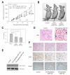
AZD1152 inhibits orthotopically xenografted HER18 cells in nude mice. (A) Tumor growth curves for orthotopically implanted HER18 cells are shown. Control mice received vehicle (0.3 M Tris pH 9.0), low dose mice received 62.5 mg/kg/dose AZD1152, and high dose mice received 125 mg/kg/dose AZD1152. Treatment began when tumors were measurable (approximately 50 mm3). Injections were given IP on days 1 and 2 of 7-day cycles, indicated by horizontal bars just above the x-axis. * P < 0.001 calculated as a mixed linear model. Error bars represent SE. (B) Representative mice bearing xenografts (arrows) from each treatment group. (C) Weights of dissected xenografts are plotted as means for each treatment group. Error bars represent 95% confidence intervals. P values were determined by one-way ANOVA. (D) H&E stained control and high dose (125 mg/kg/day) tumor samples are shown. Multi-nucleate cells (circles) were observed in tumor sections from AZD1152-treated mice but not in control mice. (E) Western blots using anti-phospho-Histone H3 and anti-actin are shown as labeled. (F) Histology and immunohistochemistry of xenografts are shown: top panels- H&E staining; middle panels- immunohistochemistry with antibodies for Ki-67; lower panels- cleaved Caspase 3.
Figure 5
Figure 5
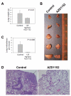
AZD1152 inhibits growth of lung tumor nodules in a lung metastasis nude mouse model. (A) Two million MDA-MB-231 human breast cancer cells were injected via tail veins of nude mice. AZD1152 (125 mg/kg/dose) or vehicle were administered IP on days 1 and 2 of 7-day repeating cycle for 4 cycles starting 2 days after cancer cell injection. Ten weeks after cancer cell injection, the mice were sacrificed and lungs were weighed and plotted as the means for each treatment group. (B) Matched lobes of the lungs from a representative mouse from each group are shown as labeled. (C) The number of macroscopic tumor nodules in the lungs was counted in each mouse. The mean number of nodules per mouse was plotted for each group. Error bars represent 95% confidence intervals. (D) H&E stained slides of lungs from control and drug treated mice are shown at the same magnification. Arrows indicate metastatic lung tumors.
Figure 6
Figure 6
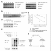
AZD1152 reduces Aurora B protein level by increasing poly-ubiquitination and degradation via the proteasome. (A) Western blots were performed for Aurora B, phospho-Histone H3 and actin levels. HER18 cells were treated with increasing duration of AZD1152 20 nM (left panel) and with increasing concentration of AZD1152 for 48 hours (right panel) as labeled. (B) HER18 cells were treated with or without 20 nM AZD1152 for 48 hours and then in the presence of cycloheximide for up to four hours. Immunoblots of Aurora B and actin are shown. (C) The integrated optical densities of the protein bands in B. The amount of Aurora B protein relative to 0 h of cycloheximide treatment was calculated for each group and corrected for gel loading differences based on actin. The relative rates of Aurora B turnover were estimated by the slope of the linear regression line for the control and AZD1152-HQPA-treated cells. (D) MDA-MB-231 cells show increased Aurora B protein levels when treated with the proteasome inhibitor MG132 and AZD1152-HQPA versus AZD1152-HQPA treatment alone. (E) AZD1152-HQPA causes increased poly-ubiquitinated Aurora B versus control. HER18 cells were transfected with the plasmids (Flag-Aurora B and HA-ubiquitin) and treated with the drugs (AZD1152-HQPA and the proteasome inhibitor MG132) as labeled above the top panel. Top panel: anti-HA immunoblot of anti-Flag-immunoprecipitates. Bottom panels: Anti-Flag immunoblot of whole cell lysates. (F) AZD1152-HQPA may disturb mitosis in at least two pathways: 1) through inhibition of Aurora B kinase activity, and 2) through increasing poly-ubiquitination and proteasomal degradation of Aurora B protein leading to decreased Aurora B function.

Similar articles

Cited by

References

    1. Yang J, Ikezoe T, Nishioka C, Tasaka T, Taniguchi A, Kuwayama Y, Komatsu N, Bandobashi K, Togitani K, Koeffler HP. AZD1152, a novel and selective aurora B kinase inhibitor, induces growth arrest, apoptosis, and sensitization for tubulin depolymerizing agent or topoisomerase II inhibitor in human acute leukemia cells in vitro and in vivo. Blood. 2007;110:2034–2040. doi: 10.1182/blood-2007-02-073700. - DOI - PubMed
    1. Tao Y, Zhang P, Girdler F, Frascogna V, Castedo M, Bourhis J, Kroemer G, Deutsch E. Enhancement of radiation response in p53-deficient cancer cells by the Aurora-B kinase inhibitor AZD1152. Oncogene. 2008;27:3244–3255. doi: 10.1038/sj.onc.1210990. - DOI - PubMed
    1. Katayama H, Brinkley WR, Sen S. The Aurora kinases: role in cell transformation and tumorigenesis. Cancer Metastasis Rev. 2003;22:451–464. doi: 10.1023/A:1023789416385. - DOI - PubMed
    1. Fu J, Bian M, Jiang Q, Zhang C. Roles of Aurora kinases in mitosis and tumorigenesis. Mol Cancer Res. 2007;5:1–10. doi: 10.1158/1541-7786.MCR-06-0208. - DOI - PubMed
    1. Ducat D, Zheng Y. Aurora kinases in spindle assembly and chromosome segregation. Exp Cell Res. 2004;301:60–67. doi: 10.1016/j.yexcr.2004.08.016. - DOI - PubMed

Publication types

MeSH terms

Substances