. 2010 Feb 11;5(2):e9175.
doi: 10.1371/journal.pone.0009175.

CXCR7 functions as a scavenger for CXCL12 and CXCL11

Affiliations

CXCR7 functions as a scavenger for CXCL12 and CXCL11

Ulrike Naumann et al. PLoS One. .

Abstract

Background: CXCR7 (RDC1), the recently discovered second receptor for CXCL12, is phylogenetically closely related to chemokine receptors, but fails to couple to G-proteins and to induce typical chemokine receptor mediated cellular responses. The function of CXCR7 is controversial. Some studies suggest a signaling activity in mammalian cells and zebrafish embryos, while others indicate a decoy activity in fish. Here we investigated the two propositions in human tissues.

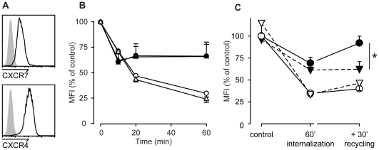
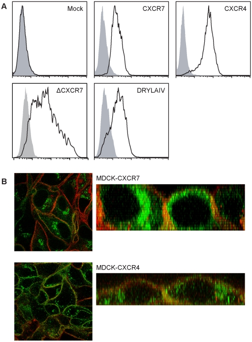
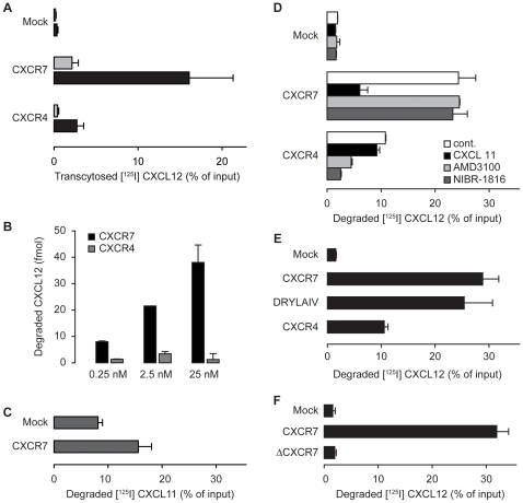
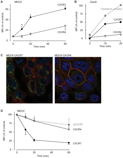
Methodology/principal findings: We provide evidence and mechanistic insight that CXCR7 acts as specific scavenger for CXCL12 and CXCL11 mediating effective ligand internalization and targeting of the chemokine cargo for degradation. Consistently, CXCR7 continuously cycles between the plasma membrane and intracellular compartments in the absence and presence of ligand, both in mammalian cells and in zebrafish. In accordance with the proposed activity as a scavenger receptor CXCR7-dependent chemokine degradation does not become saturated with increasing ligand concentrations. Active CXCL12 sequestration by CXCR7 is demonstrated in adult mouse heart valves and human umbilical vein endothelium.

Conclusions/significance: The finding that CXCR7 specifically scavenges CXCL12 suggests a critical function of the receptor in modulating the activity of the ubiquitously expressed CXCR4 in development and tumor formation. Scavenger activity of CXCR7 might also be important for the fine tuning of the mobility of hematopoietic cells in the bone marrow and lymphoid organs.


Conflict of interest statement

Competing Interests: The authors have declared that no competing interests exist.

Figures

Figure 1
Figure 1. CXCL12 induced internalization of CXCR7 and CXCR4.
(A) FACS analysis of resting Daudi B cells expressing endogenous CXCR4 and CXCR7. (B) Summary from three independent observations showing the time course of receptor expression of Daudi B cells expressing endogenous CXCR4 (open symbols) and CXCR7 (closed symbols) incubated with 100 nM (circles) or 1 µM (triangles) CXCL12. Extracellular bound chemokine was removed by an acidic wash (see Methods) and receptor surface expression was measured at the indicated times by FACS analysis. P<0.5 for ligand-induced internalization of CXCR7 and CXCR4. (C) Daudi B cells were incubated for 1 h at 37°C with 250 nM CXCL12. Extracellular bound chemokine was removed by an acidic wash and samples were split. Surface expression of CXCR7 (closed symbols) and CXCR4 (open symbols) was measured by FACS analysis directly (60′ internalization), or following an additional incubation for 30 min at 37°C to allow receptor reexpression (+30′ recycling). DMSO vehicle (circles) and 2 µM Bafilomycin A1 (triangles) were present during the entire procedure and a 60 min pre-treatment of the cells. MFIs are shown as percent of DMSO control without chemokine. Data from three independent observations; * P<0.05.
Figure 2
Figure 2. Ectopic expression of CXCR7 and CXCR4 on MDCK cells.
(A) MDCK were stably transfected with empty vector (Mock), with CXCR7, CXCR4, a vector coding for a CXCR7 lacking the cytoplasmic C-terminus (ΔCXCR7), and a vector coding for a chimeric CXCR7 containing the DRYLAIV motive of CXCR4. Receptor expression was determined by FACS analysis using saturating antibody concentrations (see Methods). (B) Confocal immunofluorescence analysis of unfixed MDCK cells expressing CXCR7 (upper panels) or CXCR4 (lower panels). Cells also expressed N-ter-Lck mCherry as membrane marker (red fluorescence). Left panels: confocal images of planes cut through intracellular regions of MDCK monolayers. Right panels: x-z planes reconstructed from confocal x-y stacks. For receptor (green) detection anti-CXCR7 (11G8 R&D) or anti CXCR4 (MAB173 R&D) were used. Receptor-bound primary antibodies were revealed with goat anti mouse IgG conjugated with Alexa488 (green fluorescence).
Figure 3
Figure 3. Degradation of CXCL12 mediated by CXCR7 and CXCR4.
(A) MDCK cells stably transfected with empty vector (Mock), with CXCR7, and CXCR4 were incubated with 0.25 nM [125I]-CXCL12 for 3 h at 37°C added to the apical side. Black bars, TCA resistant radioactivity recovered from the lower part of the transwell; grey bars, radioactivity which was precipitated with TCA. Data from 5 experiments performed in triplicates. (B) Degradation of CXCL12 mediated by CXCR7 is not saturated by increasing concentrations of ligand. MDCK cells stably expressing CXCR7 or CXCR4 were incubated with increasing concentrations of [125I]-CXCL12 as in (A). Total TCA resistant counts were used to calculate the amount of degraded chemokine. Representative data shown from one experiment performed in triplicates. (C) MDCK stably transfected with empty vector (Mock) or CXCR7 were exposed to 0.25 nM [125I]-CXCL11 as in B. In three independent experiments more pronounced spontaneous degradation of CXCL11 in Mock-transfected cells compared to CXCL12 degradation was observed. (D) MDCK stably transfected with empty vector (Mock), with CXCR7, and CXCR4 were exposed to 0.25 nM [125I]-CXCL12 as in A in the presence of medium (cont.), 1 µM CXCL11 or the CXCR4 inhibitors AMD3100 (10 µM) and NIBR-1816 (10 µM). (E) The DYLAIV motif is not critical for CXCR7 activity. MDCK stably transfected with empty vector (Mock), with CXCR7, CXCR4 and a vector coding for a chimeric CXCR7 containing the DRYLAIV motive of CXCR4 were incubated as in B. Data from experiments that were performed in triplicates (n = 5, except DRYLAIV n = 2). (F) MDCK stably transfected with empty vector (Mock), with CXCR7, or a vector coding for a CXCR7 lacking the cytoplasmic C terminus (ΔCXCR7) were exposed to [125I]-CXCL12 as in B.
Figure 4
Figure 4. Ligand-independent receptor cycling.
(A) MDCK cells expressing CXCR7 or CXCR4 were treated on ice with proteinase K to eliminate receptor epitopes from the surface. Cells were washed and incubated at 37°C in medium. At the indicated times aliquots were removed from the incubation and receptor surface expression measured by FACS analysis. (B) Daudi B cells expressing endogenous CXCR7, CXCR4, and transferrin receptor were treated with proteinase K as in (A), and subjected to FACS analysis. (C) MDCK cells expressing membrane-anchored mCherry (red color) and CXCR7 or CXCR4 were incubated with receptor specific monoclonal antibodies at 37°C for 30 min. Cells were fixed, permeabilized and stained with goat anti-mouse F(ab)2 Alexa 488. Red: Actin stained with Alexa Fluor 594 phalloidin; blue Nuclei stained with DAPI. Ligand-independent receptor internalization was visualized by confocal microscopy. Representative data shown from one experiment out of 5. (D) MDCK cells expression CXCR7, CXCR4, or a vector coding for a CXCR7 lacking the cytoplasmic C terminus (ΔCXCR7) were incubated on ice with receptor-specific antibodies. After removal of unbound antibodies cells were shifted to 37°C. At the indicated times aliquots were withdrawn and incubated with secondary goat anti-mouse F(ab)2 Alexa 488 at 0°C. Receptor surface expression was measured by FACS analysis.
Figure 5
Figure 5. Ligand-independent cycling of CXCR7 in zebrafish embryos.
In embryos expressing CXCR7-EGFP the receptor localizes at the plasma membrane (grey mark in lower panels) and intracellular vesicles (white dot). Time laps (time stamps) images shows that the vesicles are in contact with the membrane internalize and recycle back to the membrane. Internalization and cycling of CXCR7-EGF is similar in the presence (control–MO) or absence of CXCL12 (CXCL12–MO), underling the ligand independent mechanism. For the complete time laps see supplementary movies S1 cont and S2 CXCL12.
Figure 6
Figure 6. Uptake of CXCL12 by mouse heart valve.
The aortic heart valves of wild type mice internalize CXCL12 tagged with a fluorescent protein (CXCL12-mCherry, upper right, red dots marked with arrowheads). Uptake of CXCL12-mCherry is partially competed with unlabeled 1.5 µM CXCL12 (lower left), and is partially resistant to administration of 10 µM AMD3100 (lower right). Pictures were taken in the indicated area (white frame upper left).
Figure 7
Figure 7. Scavenging activity of CXCR7 in primary human endothelium.
(A) Human umbilical vein endothelial cells (HUVECs) were exposed for 3 h at 37°C to [125I]-CXCL12 in the presence of 1 µM CXCL11 or 10 µM of the CXCR4 inhibitor AMD3100. TCA resistant counts represent degraded chemokine and were plotted as percent of total added counts (cont). (B) Vein endothelial cells in fresh human umbilical cord slices express CXCR7 (purple upper left, blue nuclei DAPI), vein endothelial cells (CD31 marker red) internalize CXCL12 tagged with a fluorescent protein (CXCL12-venus, upper right, green dots marked with arrowheads), uptake of CXCL12-venus is not affected by 10 µM AMD3100 (lower left), but is competed with 1.5 µM CXCL11 (lower right). (C) Several images (512×512 pixels) from 3 independent experiments as shown in (B) were analyzed using Metamorph software. Total pixel areas showing fluorescence of the CXCL12-venus per image are shown. P<0.05 between cont. and CXCL11, not significant between cont. and AMD3100.

Similar articles

Cited by

References

    1. Thelen M, Stein JV. How chemokines invite leukocytes to dance. Nat Immunol. 2008;9:953–959. - PubMed
    1. Nagasawa T, Hirota S, Tachibana K, Takakura N, Nishikawa S, et al. Defects of B-cell lymphopoiesis and bone-marrow myelopoiesis in mice lacking the CXC chemokine PBSF/SDF-1. Nature. 1996;382:635–638. - PubMed
    1. Zou YR, Kottmann AH, Kuroda M, Taniuchi I, Littman DR. Function of the chemokine receptor CXCR4 in haematopoiesis and in cerebellar development. Nature. 1998;393:595–599. - PubMed
    1. Ma Q, Jones D, Borghesani PR, Segal RA, Nagasawa T, et al. Impaired B-lymphopoiesis, myelopoiesis, and derailed cerebellar neuron migration in CXCR4- and SDF-1-deficient mice. Proc Natl Acad Sci USA. 1998;95:9448–9453. - PMC - PubMed
    1. Balabanian K, Lagane B, Infantino S, Chow KY, Harriague J, et al. The chemokine SDF-1/CXCL12 binds to and signals through the orphan receptor RDC1 in T lymphocytes. J Biol Chem. 2005;280:35760–35766. - PubMed

Publication types

MeSH terms