. 2010 Feb 18;7(2):128-39.
doi: 10.1016/j.chom.2010.01.004.

Enhanced infection of liver sinusoidal endothelial cells in a mouse model of antibody-induced severe dengue disease

Affiliations

Enhanced infection of liver sinusoidal endothelial cells in a mouse model of antibody-induced severe dengue disease

Raphaël M Zellweger et al. Cell Host Microbe. .

Abstract

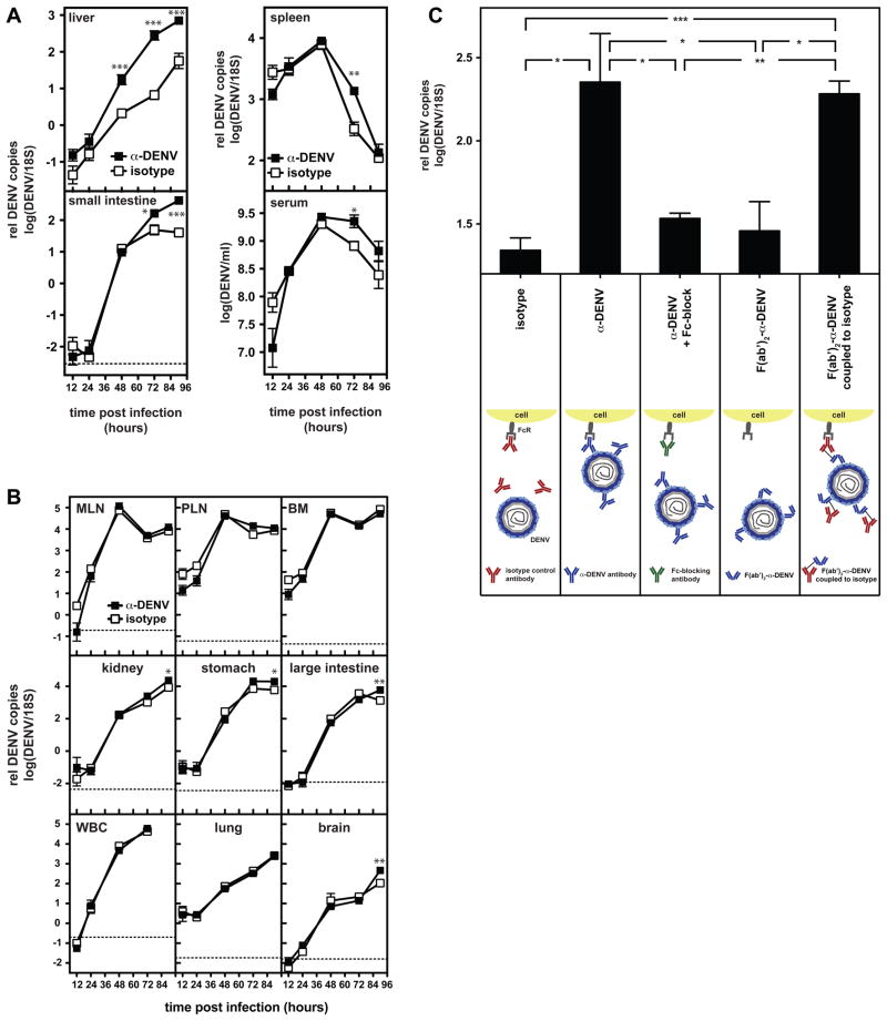
Dengue virus (DENV) causes disease ranging from dengue fever (DF), a self-limited febrile illness, to the potentially lethal dengue hemorrhagic fever and dengue shock syndrome (DHF/DSS). DHF/DSS usually occurs in patients who have acquired DENV-reactive antibodies prior to infection, either from a previous infection with a heterologous DENV serotype or from an immune mother. Hence, it has been hypothesized that subneutralizing levels of antibodies exacerbate disease, a phenomenon termed antibody-dependent enhancement (ADE). However, given the lack of suitable animal models for DENV infection, the mechanism of ADE and its contribution to pathology remain elusive. Here we demonstrate in mice that DENV-specific antibodies can sufficiently increase severity of disease so that a mostly nonlethal illness becomes a fatal disease resembling human DHF/DSS. Antibodies promote massive infection of liver sinusoidal endothelial cells (LSECs), resulting in increased systemic levels of virus. Thus, a subprotective humoral response may, under some circumstances, have pathological consequences.


Conflict of interest statement

The authors declare that they have no competing financial interests.

Figures

Figure 1
Figure 1. Heterologous immune serum decreases mean survival time of DENV-infected mice, homologous serum protects 50% of the mice from disease
A) Survival of DENV-infected AG129 mice (5×10⁸ GE S221 i.v.) in the presence of heterologous DENV1-, 3- or 4-immune serum, homologous DENV2-immune serum (black triangles) or naïve serum (white triangles); 200 μl of serum was administered i.p. on day -1 and 1. P-values from Gehan-Breslow-Wilcoxon test, n=4–6 mice per group. B) DENV2-reactive IgG in the sera used in A was measured by ELISA on plates coated with sucrose gradient-purified S221. C) Biological activity of the sera used in A was assessed by measuring their ability to reduce infection of C6/36 cells by DENV2. For clarity, a dotted line has been added at 50% infection. For comparison purposes, the naïve serum curve is depicted in all panels.
Figure 2
Figure 2. Subprotective levels of anti-DENV monoclonal antibodies decrease mean survival time of DENV-infected mice
A) Survival of DENV-infected AG129 mice treated on day -1 and 1 with 15 μg of anti-DENV antibody (black squares, n=7–12) or an isotype control (white squares, n=12–20). Various anti-DENV antibodies were used, see figure and text for details. As the same isotype control was used for both IgG2a antibodies, the same isotype control curve is depicted in the left and middle panels. B) Mean survival time of DENV-infected AG129 mice treated with 0.5, 2, 15, 200 or 500 μg of IgG2a against DENV E (clone 4G2, black bars, n=4–7), 500 μg of an isotype control (white bar, n=7) or PBS (grey bar, n=11). Statistically significant differences are indicated only between the PBS-treated group and the anti-DENV-treated groups for simplicity. C) Survival of AG129 mice infected intradermally (i.d.) with 5×10⁸ GE S221 and treated with anti-DENV antibodies (black squares, n=7) or isotype control (white squares, n=7). 15 μg of IgG2a against DENV prM/M were administered i.p. on day -1 and 1. D) Survival of AG129 mice infected with 5×10⁷ or 5×10⁶ GE S221 i.v. and treated with anti-DENV antibodies (black squares, n=4) or isotype control (white squares, n=4). Antibody was administered as in C. E) Survival of A129 mice infected on day 0 with 1×10¹¹ GE S221 i.v. and treated on day -1, 1, 2 and 3 with 50 μg of IgG2a against DENV prM/M (black squares, n=8) or an isotype control (white squares, n=9). P-values from Gehan-Breslow-Wilcoxon test (A-C-D-E) or two-tailed unpaired t-test with Welch’s correction, c.i. 95% (B): * P≤0.05, ** P≤0.01, *** P≤0.001, error bars represent SEM, n is the number of mice per group.
Figure 3
Figure 3. Administration of anti-DENV antibody (15 μg IgG2a against DENV prM/M) induces a DHF/DSS-like disease in AG129 mice infected with 5×10⁸ GE S221
A) IL-6, IL-10 and TNF levels in the serum of DENV-infected mice 48, 72 and 90 hours after infection (n=3–9). B) Mean survival time of anti-DENV-treated mice in which IL-6, IL-10R or TNF were neutralized blocked (black bars, n=4–6); control groups were treated with the respective isotype controls of the IL-6, IL-10R or TNF blocking antibodies (white bars, n=4–6). C) Platelet counts 90 hours after infection (n=6–7). D) Hematocrit 90 hours after infection (n=6–7). E) Vascular permeability in the liver of naïve mice and DENV-infected mice in the presence of isotype control or anti-DENV antibody 90 hours after infection (n=3–4). Evans Blue in tissues was extracted with formamide and quantified spectrophotometrically. The experiment was repeated 72 hours post-infection and similar results were obtained. F) Gastrointestinal bleeding in the small intestine 84 hours after infection in the presence of anti-DENV antibodies. Mice were perfused with PBS before pictures were taken. Tissues from one representative animal per group are shown. P-values from two-tailed unpaired t-test with Welch’s correction, c.i. 95%: * P≤0.05, ** P≤0.01, *** P≤0.001, error bars represent SEM, n is the number of mice per group. See also supplementary figure S1.
Figure 4
Figure 4. Administration of anti-DENV antibody results in an FcγR-dependent increase in viral load in DENV-infected mice
A and B) Viral RNA levels were quantified by qRT-PCR 12, 24, 48, 72 and 90 hours after infection of AG129 mice (5×10⁸ GE S221 i.v. on day 0) in the presence or absence of DENV-specific antibody (15 μg clone 2H2, administered d-1 and 1). Symbols represent mean ± SEM, n=3–6. C) DENV-infected AG129 mice (5×10⁸ GE S221 i.v. on day 0) were treated on day 1 with equimolar amounts of intact anti-DENV antibody (15 μg, n=8), intact anti-DENV antibody (15 μg) in the presence of FcγR-blocking antibody (500 μg clone 24G2 i.v. on d0 and 1, n=7), anti-DENV F(ab′)2 fragment (10 μg, n=4), or anti-DENV F(ab′)2 fragment chemically coupled 1:1 to an isotype control (25 μg n=4). As a control, one group was treated with an isotype control (n=4). P-values from two-tailed unpaired t-test with Welch’s correction, c.i. 95%: * P≤0.05, ** P≤0.01, *** P≤0.001, n is the number of mice per group, MLN: mesenteric lymph node, PLN: peripheral lymph nodes (axillary, brachial and inguinal), BM: bone marrow. See also supplementary figures S2 and S3.
Figure 5
Figure 5
Anti-DENV antibodies increase infection of liver sinusoidal endothelial cells (LSECs) in DENV-infected mice. Percentage of DENV antigen-positive cells in different liver cell populations. Gating is indicated on the left, population statistics are indicated in the bar graph on the right. Representative dot plots of DENV antigen staining of LSECs (R3 gate) and immunohistochemical staining of liver sections, stained for DENV NS3 (green), CD31 (red) and CD68 (blue) (low and high magnification images are on the left and right, respectively, and the size is indicated by the scale bars). n=3 per group; representative FACS plots and immunohistochemistry images are shown; P-values from two-tailed unpaired t-test with Welch’s correction, c.i. 95%: * P≤0.05, ** P≤0.01, *** P≤0.001.
Figure 6
Figure 6
An increased percentage of lamina propria macrophages are infected following elevated infection of LSECs in mice treated with antibodies. Percentage of DENV antigen-positive cells in MHC-II+ lamina propria cell populations. Gating is indicated on the top (the grey histogram depicts cells stained with an isotype control antibody) and population statistics are indicated in the bar graph below. Representative dot plots of DENV antigen staining of lamina propria macrophages (R3 gate) and immunohistochemical staining of small intestine sections, stained for DENV NS3 (green), CD68 (red) and F4/80 (blue) (low and high magnification images are on the left and right, respectively, and the size is indicated by the scale bars). n=3 per group; representative FACS plots and immunohistochemistry images are shown; P-values from two-tailed unpaired t-test with Welch’s correction, c.i. 95%: * P≤0.05, ** P≤0.01, *** P≤0.001. See also supplementary figure S4.
Figure 7
Figure 7
Antibodies enhance infection of LSECs by DENV clinical isolate strains of each serotype. AG129 mice were infected with following clinical isolates: DENV1 Julia (5×10⁶ FIU), DENV2 PL046 (1.5×10⁷ FIU), DENV3 UNC3001 (2.5×10⁵ FIU) or DENV4 H241 (3.5×10⁶ FIU) on day 0, and 50 μg of anti-DENV antibody 2H2 were administered on day -1 and 1. On day 3, liver sections were stained for DENV NS3 (green), CD31 (red) and CD68 (blue). Low and high magnification images are on the left and right, respectively, and the size is indicated by the scale bars.

Comment in

Similar articles

Cited by

References

    1. Balsitis SJ, Coloma J, Castro G, Alava A, Flores D, McKerrow JH, Beatty PR, Harris E. Tropism of dengue virus in mice and humans defined by viral nonstructural protein 3-specific immunostaining. The American journal of tropical medicine and hygiene. 2009;80:416–424. - PubMed
    1. Bashirova AA, Geijtenbeek TB, van Duijnhoven GC, van Vliet SJ, Eilering JB, Martin MP, Wu L, Martin TD, Viebig N, Knolle PA, et al. A dendritic cell-specific intercellular adhesion molecule 3-grabbing nonintegrin (DC-SIGN)-related protein is highly expressed on human liver sinusoidal endothelial cells and promotes HIV-1 infection. The Journal of experimental medicine. 2001;193:671–678. - PMC - PubMed
    1. Blackley S, Kou Z, Chen H, Quinn M, Rose RC, Schlesinger JJ, Coppage M, Jin X. Primary human splenic macrophages, but not T or B cells, are the principal target cells for dengue virus infection in vitro. Journal of virology. 2007;81:13325–13334. - PMC - PubMed
    1. Boonnak K, Slike BM, Burgess TH, Mason RM, Wu SJ, Sun P, Porter K, Rudiman IF, Yuwono D, Puthavathana P, et al. Role of dendritic cells in antibody-dependent enhancement of dengue virus infection. Journal of virology. 2008;82:3939–3951. - PMC - PubMed
    1. Brooks DG, Trifilo MJ, Edelmann KH, Teyton L, McGavern DB, Oldstone MB. Interleukin-10 determines viral clearance or persistence in vivo. Nature medicine. 2006;12:1301–1309. - PMC - PubMed

Publication types

Substances