. 2010 Aug;12(8):1083-97.
doi: 10.1111/j.1462-5822.2010.01452.x. Epub 2010 Feb 9.

LnaB: a Legionella pneumophila activator of NF-kappaB

Affiliations

LnaB: a Legionella pneumophila activator of NF-kappaB

Vicki P Losick et al. Cell Microbiol. 2010 Aug.

Abstract

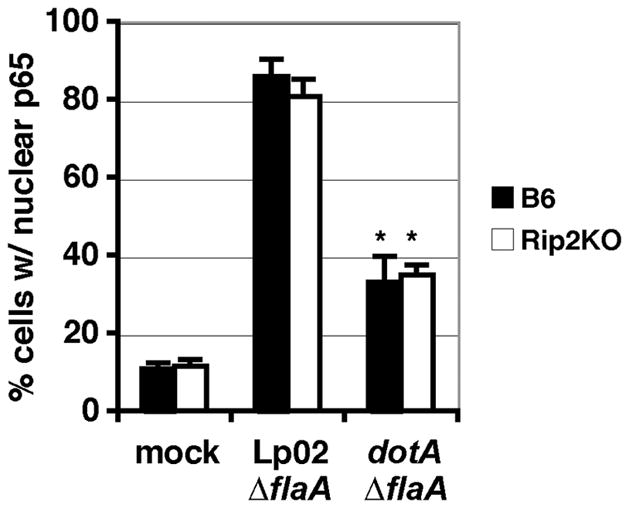
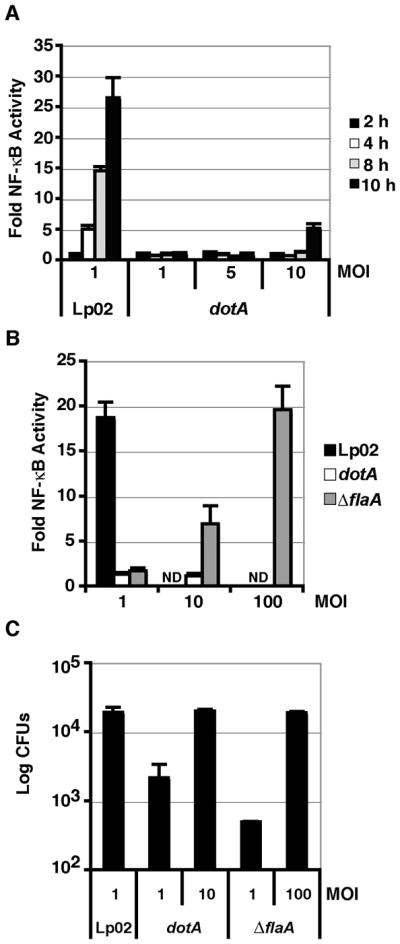
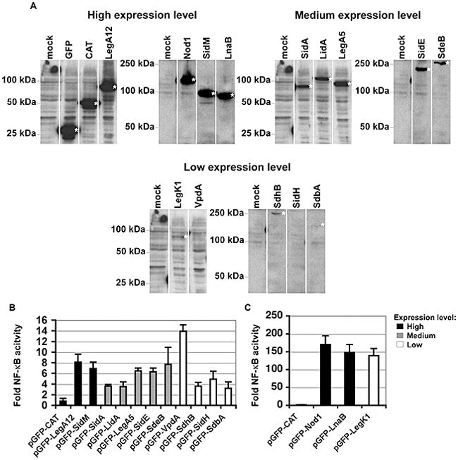
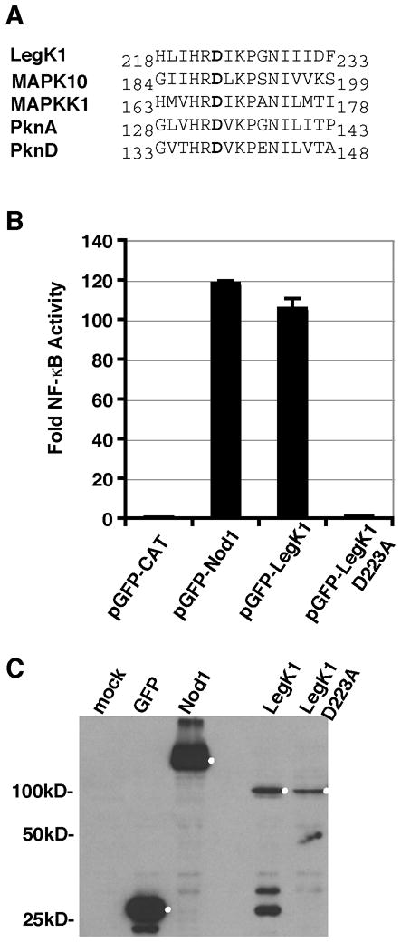
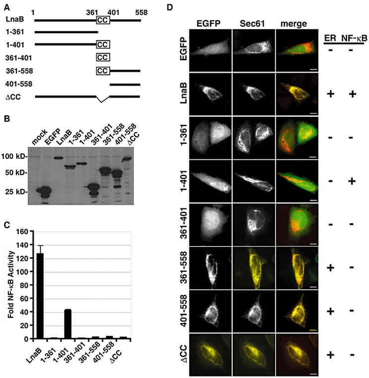
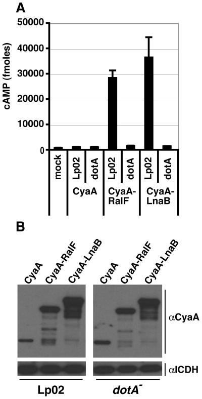
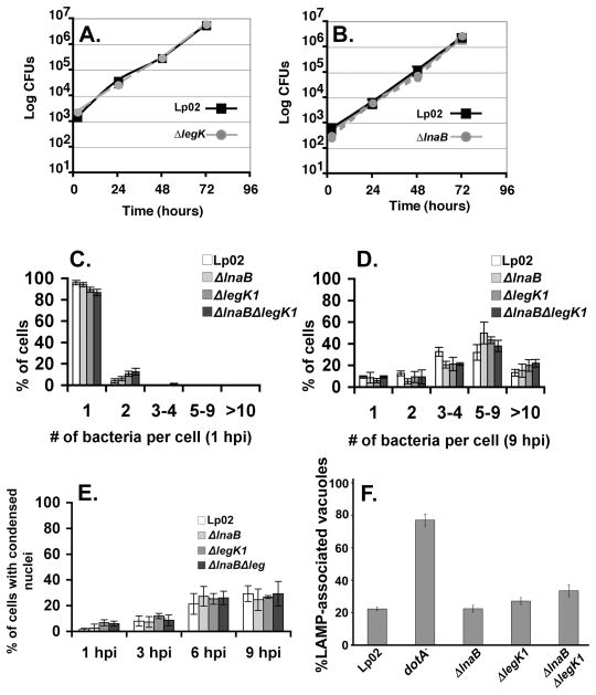
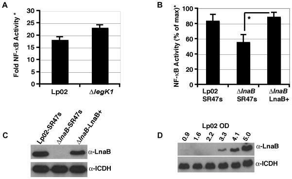
Legionella pneumophila possesses a large arsenal of type IV translocated substrates. Over 100 such proteins have been identified, but the functions of most are unknown. Previous studies have demonstrated that L. pneumophila activates NF-kappaB, a master transcriptional regulator of the mammalian innate immune response. Activation of NF-kappaB is dependent on the Legionella Icm/Dot type IV protein translocation system, consistent with the possibility that translocated bacterial proteins contribute to this response. To test this hypothesis, an expression library of 159 known and putative translocated substrates was created to evaluate whether ectopic production of a single L. pneumophila protein could activate NF-kappaB in mammalian cells. Expression of two of these proteins, LnaB (Legionella NF-kappaB activator B) and LegK1, resulted in approximately 150-fold induction of NF-kappaB activity in HEK293T cells, levels similar to the strong induction that occurs with ectopic expression of the known activator Nod1. LnaB is a substrate of the Icm/Dot system, and in the absence of this protein, a partial reduction of NF-kappaB activation in host cells occurs after challenge by post-exponential phase bacteria. These data indicate that LnaB is an Icm/Dot substrate that contributes to NF-kappaB activation during L. pneumophila infection in host cells.


Figures

Figure 1
Figure 1. L. pneumophila induced NF-κB activation is not dependent on Rip2
BM macrophages from C57Bl/6J (B6) and Rip2 knockout (Rip2KO) mice were challenged with L. pneumophila strain Lp02 ΔflaA and dot ΔflaA at MOI 1 for 7 hours. Fixed cells were stained for NF-κB, probed with anti- L. pneumophila antibody and the percent of cells harboring bacteria with detectable nuclear NF-κB staining were scored. Data represents the mean ± SE from three independent experiments, with 300 cells scored. * T-test p-value ≤ 0.01.
Figure 2
Figure 2. NF-κB activation by L. pneumophila in HEK293T cells is dependent on the Icm/Dot translocation system and independent of flagellin
(A) Time course of NF-κB activation at 2 (black bars), 4 (white bars), 6 (gray bars), or 10 (dark gray bars) hours post infection (hpi) with wild-type L. pneumophila (Lp02) or a Icm/Dot mutant (dotA) at the MOI indicated. (B) Dose dependent activation of NF-κB in response to Lp02, dotA, or a flagellin mutant (ΔflaA). MOI is indicated on the x-axis. Luminescence was assayed after 8 hours post infection (hpi). ND: not determined. HEK293T cells were transfected with the NFκB-luciferase reporter plasmid, and then challenged with the L. pneumophila strains indicated. Fold NF-κB activity is expressed as the light units of samples relative to transfected cells incubated in absence of bacteria. (C) High efficiency L. pneumophila uptake into HEK293T cells requires flagellin. Uptake was measured at 2 hpi by lysing cells and enumerating CFUs. Data represent the mean and SE of triplicate samples from a representative experiment. Experiments were repeated in duplicate.
Figure 3
Figure 3. Candidate IDTS can activate NF-κB
(A). Varied expression levels of L. pneumophila IDTS expressed in 293T cells. Protein expression levels of the indicated GFP fusions that had enhanced NF-κB activation are displayed on immunoblots, probing with anti-GFP. To allow clearer comparisons of the relative increases in NFκB activity, IDTS are grouped according to their expression levels in HEK293T cells. (B) Low level activation of NF-κB by L. pneumophila IDTS. Legionella NF-κB activators fused to GFP are marked on the x-axis. Shown are those IDTS that show statistically significant activation of NF-κB (T-test p-value ≤ 0.05). Bars representing those IDTS expressed at high levels are marked in black. Medium and low expression levels are shown in grey and white, respectively. (C) High level NF-κB activation by two Legionella IDTS. HEK293T cells were co-transfected with 200 ng of pNFκB-luciferase reporter and either 50 ng of the control plasmid (pGFP) or the indicated pGFP fusions and luminescence was assayed at 24 hours. pGFP-CAT (chloramphenicol aminotransferase) served as a negative control and pGFP-Nod1 was used as a positive control for NF-κB activation. Fold NF-κB activity is the relative light units of pGFP fusion compared to the pGFP control. Data represents the mean ± SE from three independent experiments preformed in triplicate.
Figure 4
Figure 4. The ser/thr kinase activity of LegK1 is required for NF-κB activation
(A) Alignment of the predicted catalytic domain of L. pneumophila LegK1 with other ser/thr kinase domain containing proteins: Human MAPK10 (NP_002744), Chlamydomonas reinhardtii MAPKK (XP_001696437), Mycobacterium tuberculosis PknA (NP_214529) and PknB (NP_214528). The catalytic aspartate (shown in bold) is preceded by an arginine in all the ser/thr kinases. (B) Mutation of the putative LegK1 catalytic aspartate to alanine eliminates NF-κB activation. HEK293T cells were co-transfected with 200 ng per well of pNFκB-luciferase and either 50ng of pGFP fused to CAT (negative control), Nod1 (positive control), LegK1, or catalytically inactive mutant LegK1 D223A. Data represent the mean ± SE from triplicate samples of a representative experiment. The experiment was preformed in triplicate. (C) Western blot of the protein expression levels of GFP, GFP-Nod1, GFP-LegK1, and GFP-LegK1-D223A probed with anti-GFP antibodies. HEK293T cells were transfected for 48 hours with 200 ng of the indicated plasmids; the expected protein based on molecular weight are marked by a white dot.
Figure 5
Figure 5. The coiled-coil domain of LnaB is required for NF-κB activity
(A) Schematic of EGFP-LnaB fusions. Full-length LnaB is 558 amino acids and contains a putative coiled-coil domain (CC) between amino acids 361-401. (B) Expression of EGFP fusions, 24 hours after transfection of HEK293T cells with the corresponding plasmids as detected by Western blot with anti-GFP. (C) HEK293T cells were co-transfected with 200 ng of pNFκB-luciferase and 50 ng of indicated EGFP fusion and luminescence was assayed after 24 hours. Fold NF-κB activity is the relative light units of pEGFP fusions compared to the pEGFP control. Data represents the mean ± SE from triplicate samples of a representative experiment. The experiment was preformed in triplicate. (D) Subcellular localization of indicated EGFP-LnaB fusions in HeLa cells expressing the ER marker Sec61 (red) and EGFP (green) in the merged image. Constructs that co-localized with Sec61 or activated NF-κB are marked with a +. Scale bar = 2μm.
Figure 6
Figure 6. LnaB is a substrate of the Icm/Dot translocation system
(A) U937 cells were challenged with the indicated L. pneumophila strains expressing CyaA (negative control), CyaA-RalF (positive control), CyaA-LnaB. After a 1 hour incubation, cAMP was extracted and relative cAMP levels were measured. Data represent the mean ± SE of triplicate samples from a representative experiment performed in triplicate. (B) Protein expression of CyaA, CyaA-RalF, CyaA-LnaB in the corresponding L. pneumophila strains prior to challenge of U937 cells. Isocitrate dehydrogenase (ICDH) was used as a loading control.
Figure 7
Figure 7. LnaB is dispensable for intracellular growth and protection from cell death
(A and B). Intracellular growth of L. pneumophila within A/J mouse BM macrophages. Displayed is the wild-type (Lp02) strain compared to ΔlegK1 (A) or ΔlnaB (B). At 2, 24, 48, and 72 hpi macrophages were lysed and colony forming units (CFUs) were enumerated (Experimental Procedures). Data represents the mean ± SE from three independent infections of a representative experiment preformed in duplicate. (C,D) Bacterial replication at 1 hpi and 9 hpi. The percentage of A/J mouse BM macrophages harboring the denoted number of bacteria is shown. The wild type (Lp02) was compared to ΔlnaB, ΔlegK1 and ΔlnaBΔlegK1 after infection of MOI=1. (E) Host cell survival. At the denoted time points post infection with wild type (Lp02), ΔlnaB, ΔlegK1 and ΔlnaBΔlegK1, condensed nuclei (cell death) were observed by Hoechst DNA staining and immunofluorescence microscopy. Average and SE of three cover slips of the respective experiments are displayed. (F). Intracellular targeting of the L. pneumophila replication vacuole in mutants defective for NF-κB activators. Bone marrow-derived macrophages from A/J mice were challenged with the noted L. pneumophila strains at MOI = 1.0 for one hour (Experimental Procedures). Cells were fixed, probed as described for L. pneumophila and late endocytic marker LAMP-1 (Experimental Procedures) and visualized by indirect immunofluorescence microscopy. Data are the mean ± SD of nine coverslips from three experiments for samples except ΔlegK1 which was the mean ± SD of 6 coverslips from two experiments.
Figure 8
Figure 8. LnaB is required for full activation of NF-κB
(A and B) HEK293T cells were transfected with the NF-κB-luciferase reporter plasmid and then challenged with the wild-type L. pneumophila (Lp02), the ΔlegK1 (A) or the ΔlnaB (B) strains at MOI = 1 for 7 hours. Fold NF-κB activity* is the relative light units of cells incubated with bacteria compared to cells incubated without bacteria. Samples were normalized to bacterial uptake at 2 hpi (Experimental Procedures). Data represents the mean ± SE of triplicate samples from a representative experiment performed in duplicate (A) or the mean ± SE from 4 independent experiments performed in triplicate (B) in which the maximum NF-κB activity was normalized to 100%. *T-test p-value < 0.05. (C) LnaB production in wild-type (Lp02::SR47s), ΔlnaB::SR47s, and ΔlnaB-LnaB+ complement strains. (D) LnaB expression is induced in post-exponential phase of growth. Lp02 was grown overnight to indicated A600 and immunoblotted with anti-LnaB or anti-ICDH as loading control.

Similar articles

Cited by

References

    1. Abu-Zant A, Jones S, Asare R, Suttles J, Price C, Graham J, Kwaik YA. Anti-apoptotic signalling by the Dot/Icm secretion system of L. pneumophila. Cell Microbiol. 2007;9:246–264. - PubMed
    1. Backert S, Ziska E, Brinkmann V, Zimny-Arndt U, Fauconnier A, Jungblut PR, et al. Translocation of the Helicobacter pylori CagA protein in gastric epithelial cells by a type IV secretion apparatus. Cell Microbiol. 2000;2:155–164. - PubMed
    1. Banga S, Gao P, Shen X, Fiscus V, Zong WX, Chen L, Luo ZQ. Legionella pneumophila inhibits macrophage apoptosis by targeting pro-death members of the Bcl2 protein family. Proc Natl Acad Sci U S A. 2007;104:5121–5126. - PMC - PubMed
    1. Bardill JP, Miller JL, Vogel JP. IcmS-dependent translocation of SdeA into macrophages by the Legionella pneumophila type IV secretion system. Mol Microbiol. 2005;56:90–103. - PubMed
    1. Bartfeld S, Engels C, Bauer B, Aurass P, Flieger A, Bruggemann H, Meyer TF. Temporal resolution of two-tracked NF-κB activation by Legionella pneumophila. Cell Microbiol. 2009;11:1638–1651. - PubMed

Publication types