

Mical links semaphorins to F-actin disassembly
- PMID: 20148037
- PMCID: PMC3215588
- DOI: 10.1038/nature08724
Mical links semaphorins to F-actin disassembly
Abstract
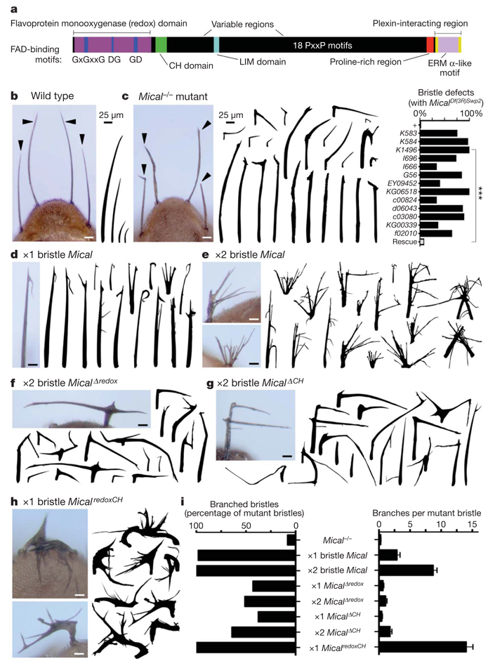
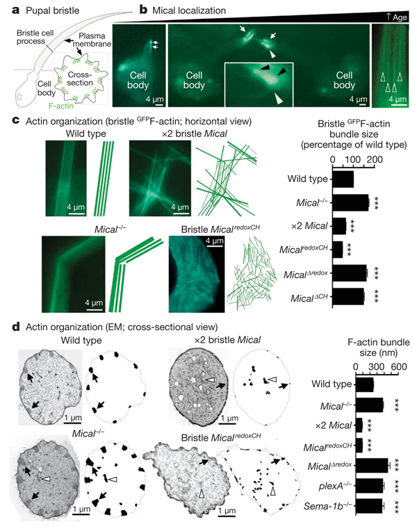
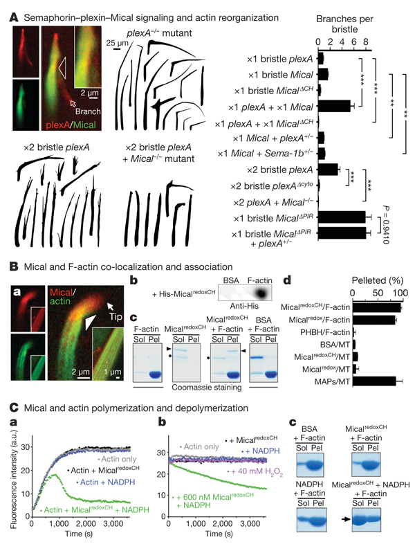
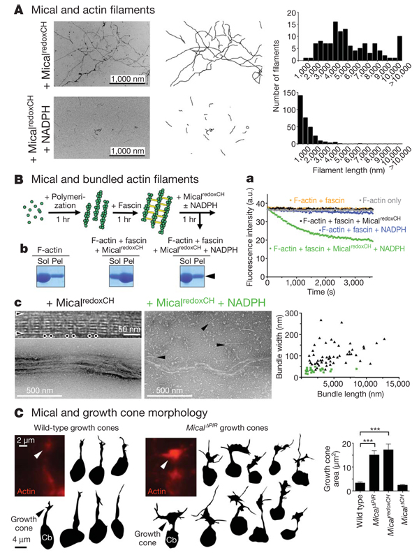
How instructive cues present on the cell surface have their precise effects on the actin cytoskeleton is poorly understood. Semaphorins are one of the largest families of these instructive cues and are widely studied for their effects on cell movement, navigation, angiogenesis, immunology and cancer. Semaphorins/collapsins were characterized in part on the basis of their ability to drastically alter actin cytoskeletal dynamics in neuronal processes, but despite considerable progress in the identification of semaphorin receptors and their signalling pathways, the molecules linking them to the precise control of cytoskeletal elements remain unknown. Recently, highly unusual proteins of the Mical family of enzymes have been found to associate with the cytoplasmic portion of plexins, which are large cell-surface semaphorin receptors, and to mediate axon guidance, synaptogenesis, dendritic pruning and other cell morphological changes. Mical enzymes perform reduction-oxidation (redox) enzymatic reactions and also contain domains found in proteins that regulate cell morphology. However, nothing is known of the role of Mical or its redox activity in mediating morphological changes. Here we report that Mical directly links semaphorins and their plexin receptors to the precise control of actin filament (F-actin) dynamics. We found that Mical is both necessary and sufficient for semaphorin-plexin-mediated F-actin reorganization in vivo. Likewise, we purified Mical protein and found that it directly binds F-actin and disassembles both individual and bundled actin filaments. We also found that Mical utilizes its redox activity to alter F-actin dynamics in vivo and in vitro, indicating a previously unknown role for specific redox signalling events in actin cytoskeletal regulation. Mical therefore is a novel F-actin-disassembly factor that provides a molecular conduit through which actin reorganization-a hallmark of cell morphological changes including axon navigation-can be precisely achieved spatiotemporally in response to semaphorins.
Figures
Similar articles
-
Extracellular inhibitors, repellents, and semaphorin/plexin/MICAL-mediated actin filament disassembly.Cytoskeleton (Hoboken). 2011 Aug;68(8):415-33. doi: 10.1002/cm.20527. Epub 2011 Aug 25. Cytoskeleton (Hoboken). 2011. PMID: 21800438 Free PMC article. Review.
-
Profilin and Mical combine to impair F-actin assembly and promote disassembly and remodeling.Nat Commun. 2021 Sep 20;12(1):5542. doi: 10.1038/s41467-021-25781-3. Nat Commun. 2021. PMID: 34545088 Free PMC article.
-
Amplification of F-Actin Disassembly and Cellular Repulsion by Growth Factor Signaling.Dev Cell. 2017 Jul 24;42(2):117-129.e8. doi: 10.1016/j.devcel.2017.06.007. Epub 2017 Jul 6. Dev Cell. 2017. PMID: 28689759 Free PMC article.
-
Direct redox regulation of F-actin assembly and disassembly by Mical.Science. 2011 Dec 23;334(6063):1710-3. doi: 10.1126/science.1211956. Epub 2011 Nov 24. Science. 2011. PMID: 22116028 Free PMC article.
-
Semaphorins: green light for redox signaling?Sci STKE. 2002 Oct 22;2002(155):pe44. doi: 10.1126/stke.2002.155.pe44. Sci STKE. 2002. PMID: 12393918 Review.
Cited by
-
14-3-3ε couples protein kinase A to semaphorin signaling and silences plexin RasGAP-mediated axonal repulsion.Neuron. 2012 Apr 12;74(1):108-21. doi: 10.1016/j.neuron.2011.12.034. Neuron. 2012. PMID: 22500634 Free PMC article.
-
Semaphorin3E-PlexinD1 signaling in coronary artery and lymphatic vessel development with clinical implications in myocardial recovery.iScience. 2021 Mar 15;24(4):102305. doi: 10.1016/j.isci.2021.102305. eCollection 2021 Apr 23. iScience. 2021. PMID: 33870127 Free PMC article.
-
Regulating small G protein signaling to coordinate axon adhesion and repulsion.Small GTPases. 2013 Jan-Mar;4(1):34-41. doi: 10.4161/sgtp.22765. Epub 2012 Dec 17. Small GTPases. 2013. PMID: 23247636 Free PMC article.
-
Redox Imbalance Associates with Clinical Worsening in Spinocerebellar Ataxia Type 2.Oxid Med Cell Longev. 2021 Feb 19;2021:9875639. doi: 10.1155/2021/9875639. eCollection 2021. Oxid Med Cell Longev. 2021. PMID: 33688396 Free PMC article.
-
MsrB1 and MICALs regulate actin assembly and macrophage function via reversible stereoselective methionine oxidation.Mol Cell. 2013 Aug 8;51(3):397-404. doi: 10.1016/j.molcel.2013.06.019. Epub 2013 Aug 1. Mol Cell. 2013. PMID: 23911929 Free PMC article.
References
-
- Tran TS, Kolodkin AL, Bharadwaj R. Semaphorin regulation of cellular morphology. Annu. Rev. Cell Dev. Biol. 2007;23:263–292. - PubMed
-
- Luo Y, Raible D, Raper JA. Collapsin: a protein in brain that induces the collapse and paralysis of neuronal growth cones. Cell. 1993;75:217–227. - PubMed
-
- Zhou Y, Gunput RA, Pasterkamp RJ. Semaphorin signaling: progress made and promises ahead. Trends Biochem. Sci. 2008;33:161–170. - PubMed
-
- Terman JR, Mao T, Pasterkamp RJ, Yu HH, Kolodkin AL. MICALs, a family of conserved flavoprotein oxidoreductases, function in plexin-mediated axonal repulsion. Cell. 2002;109:887–900. - PubMed
Publication types
MeSH terms
Substances
Grants and funding
LinkOut - more resources
Full Text Sources
Other Literature Sources
Molecular Biology Databases
