. 2010 May;84(9):4481-93.
doi: 10.1128/JVI.02141-09. Epub 2010 Feb 10.

The Elk-1 and serum response factor binding sites in the major immediate-early promoter of human cytomegalovirus are required for efficient viral replication in quiescent cells and compensate for inactivation of the NF-kappaB sites in proliferating cells

Affiliations

The Elk-1 and serum response factor binding sites in the major immediate-early promoter of human cytomegalovirus are required for efficient viral replication in quiescent cells and compensate for inactivation of the NF-kappaB sites in proliferating cells

Patrizia Caposio et al. J Virol. 2010 May.

Abstract

The major immediate-early promoter (MIEP) region of human cytomegalovirus (HCMV) plays a critical role in the regulation of lytic and latent infections by integrating multiple signals supplied by the infecting virus, the type and physiological state of the host cell, and its extracellular surroundings. The interaction of cellular transcription factors with their cognate binding sites, which are present at high densities within the enhancer upstream from the MIEP core promoter, regulate the rate of IE gene transcription and thus affect the outcome of HCMV infection. We have shown previously that the NF-kappaB binding sites within the MIEP enhancer and cellular NF-kappaB activity induced by HCMV infection are required for efficient MIEP activity and viral replication in quiescent cells (P. Caposio, A. Luganini, G. Hahn, S. Landolfo, and G. Gribaudo, Cell. Microbiol. 9:2040-2054, 2007). We now show that the inactivation of either the Elk-1 or serum response factor (SRF) binding site within the enhancer also reduces MIEP activation and viral replication of recombinant HCMV viruses in quiescent fibroblasts. In these cells, we show that the expression of either Elk-1 or SRF is required for optimal IE gene expression, and that the HCMV-stimulated activation of the MEK1/2-ERK1/2 signaling axis leads to Elk-1 transcriptional competency. Furthermore, the replication kinetics of recombinant viruses in which NF-kappaB, Elk-1, and SRF binding sites all are inactivated demonstrate that the higher levels of Elk-1 and SRF binding to MIEP in proliferating cells can compensate even for a lack of HCMV-induced NF-kappaB-mediated MIEP transactivation. These observations highlight the importance of the combination of different MIEP binding sites to optimize IE gene expression in cells in different physiological states.


Figures
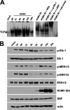
FIG. 1.
FIG. 1.
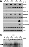
HCMV infection in quiescent cells induces TCF assembly and stimulates Elk-1 activation through the MAPK MEK1/2-ERK1/2 pathway. (A) HCMV infection rapidly induces TCF formation on the SEE of MIEP. HELF cells were growth arrested for 96 h in low-serum medium and then infected with HCMV AD169 (MOI of 5 PFU/cell) or mock infected. Nuclear extracts then were prepared at the indicated times p.i. and assayed for TCF complex formation by EMSA using the SEE probe. Autoradiographs demonstrate the nucleoprotein complex formed with the 32P-labeled wild-type MIEP SEE (from −518 to −547 with respect to the IE1/2 transcription start site). Supershift experiments were performed by adding 1 μg of rabbit polyclonal antibodies raised against SRF, 1 μg of a mouse monoclonal raised against p-Elk-1, or normal rabbit serum (NRS) as a control. An unlabeled 30-bp annealed SEE oligonucleotide was added as competitor DNA in a 300-fold molar excess above the level of the SEE probe. (B) Activation of MEK1/2, ERK1/2, and Elk-1 following HCMV infection in quiescent cells. Fibroblasts were grown to subconfluence, serum starved for 96 h, and then infected with HCMV AD169 (MOI, 5 PFU/cell). At the indicated times p.i., total protein cell extracts were prepared and analyzed by immunoblotting with rabbit anti-SRF, anti-Elk-1, anti-p-MEK1/2, anti-ERK1/2, or anti-p-ERK1/2 polyclonal antibody and with an anti-p-Elk-1 MAb. Actin immunodetected with a MAb served as an internal control.
FIG. 2.
FIG. 2.
Effects of the inhibition of HCMV-induced MEK1/2 activation on TCF formation and viral IE and E gene expression. (A) UO126 inhibits HCMV IE and E gene expression. Fibroblasts were grown to subconfluence and serum starved for 96 h, and then they were infected with HCMV AD169 (MOI, 5 PFU/cell). Where indicated, serum-starved HELFs were treated with U0126 (20 μM) for 1 h prior to infection. U0126 also was present during infection and subsequent incubation periods. At the indicated times p.i., total cell extracts were prepared and analyzed by immunoblotting with rabbit anti-SRF, anti-Elk-1, anti-p-MEK1/2, anti-ERK1/2, or anti-p-ERK1/2 polyclonal antibody and with anti-p-Elk-1, anti-IEA, or anti-UL44 MAb. Actin immunodetected with a MAb served as an internal control. (B) Virus-mediated TCF induction is reduced by UO126. HELF cells were growth arrested for 96 h in low-serum medium and then infected with HCMV AD169 (MOI of 5 PFU/cell) or mock infected. Where indicated, serum-starved HELFs were treated with U0126 (20 μM) for 1 h prior to infection. U0126 also was present during infection and subsequent incubation periods. Nuclear extracts then were prepared at the indicated times and assayed for TCF complex formation by EMSA analysis using the 32P-labeled wild-type MIEP SEE motif (from −518 to −547 with respect to the IE1/2 transcription start site).
FIG. 3.
FIG. 3.
Elk-1 silencing reduces MIEP activity in quiescent cells. (A) Inhibition of cellular Elk-1 protein expression by short hairpin RNAs (shRNAs). HELFs cells were transiently transfected with 5 μg of either a pRS shRNA expression plasmid specific for Elk-1 (shElk-1 A or shElk-1 B) or a pRS plasmid containing a noneffective shRNA cassette against GFP as a negative control for specific gene downregulation (NCS). Cells then were incubated in high- or low-serum medium for 72 h. Total cell extracts subsequently were prepared and analyzed by immunoblotting with rabbit anti-Elk-1 antibodies. The immunodetection of SRF served as a control for the specificity of shRNA-mediated Elk-1 silencing. NT, nontransfected HELF cells. (B) Silencing of cellular Elk-1 expression reduces HCMV IE1 and IE2 expression in quiescent cells. HELF cells were transiently transfected with 5 μg of either a pRS shRNA expression plasmid specific for Elk-1 (shElk-1 A or shElk-1 B) or a pRS plasmid containing a noneffective shRNA cassette against GFP (NCS) and then incubated in high- or low-serum medium for 72 h. Quiescent or proliferating shRNA Elk-1-expressing HELFs then were infected with HCMV AD169 (MOI of 3 PFU/cell). At 24 h p.i., total RNA was isolated and reverse transcribed. Real-time RT-PCR then was carried out with the appropriate IE1, IE2, and β-actin primers to quantify the expression levels of IE1 and IE2 mRNA. The results were analyzed using a standard-curve model. The levels of IE1 and IE2 mRNA were normalized to levels of endogenous β-actin mRNA. The data shown are the averages of three experiments ± standard errors of the means (error bars). NT, nontransfected HELF cells that were infected with HCMV as described above.
FIG. 4.
FIG. 4.
SRF expression is required for efficient IE gene expression in quiescent cells. (A) Silencing SRF protein expression by shRNA. HELFs were transiently transfected with 5 μg of either a pRS shRNA expression plasmid with a 29-nucleotide SRF-specific sequence insert (shSRF C or shSRF D) or a pRS plasmid containing a noneffective shRNA cassette against GFP as a negative control for specific gene downregulation (NCS) and then incubated in high- or low-serum medium for 72 h. Thereafter, total cell extracts were prepared and analyzed by immunoblotting with rabbit anti-SRF. The immunodetection of Elk-1 served as a control for the specificity of shRNA-mediated SRF silencing. NT, nontransfected HELF cells infected with HCMV as described above. (B) SRF expression is required for optimal IE1 and IE2 mRNA expression in quiescent cells. HELF cells were transiently transfected with 5 μg of either a pRS shRNA expression plasmid specific for SRF (shSRF C or shSRF D) or a pRS plasmid containing a noneffective shRNA cassette against GFP (NCS) and then incubated in high- or low-serum medium for 72 h. Quiescent or proliferating shRNA Elk-1-expressing HELFs then were infected with HCMV AD169 (MOI of 3 PFU/cell). At 24 h p.i., total RNA was isolated and reverse transcribed, and the levels of IE1 and IE2 mRNA were determined as described above. The data shown are the averages of two experiments ± standard errors of the means (error bars).
FIG. 5.
FIG. 5.
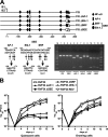
Mutagenesis of the MIEP SEE negatively affects HCMV replication in quiescent cells. (A) A schematic representation of the HCMV MIEP region and the mutations that were introduced to inactivate AP-1, Elk-1, SRF, and SEE (SRF and Elk-1) binding sites. The position of the AP-1 (at −168), Elk-1 (at −521), SRF (at −529), and SEE (between −521 and −539) sites are numbered with respect to the IE1/2 transcription start site. The point mutations and unique restriction sites introduced into each specific element are indicated below the wild-type sequence (−168 to −174, EcoRI; −521 to −528, EcoRI; −532 to −538, KpnI). To confirm the successful disruption of the AP-1, Elk-1, SRF, and SEE sites, the MIEP enhancer sequences between nucleotides −52498 and −53104 were PCR amplified from FIX (lanes 1 and 2), FIX ΔSEE REV (lanes 3 and 4), FIX ΔAP-1 (lanes 5 and 6), FIX ΔElk-1 (lanes 7 and 8), FIX ΔSRF (lanes 9 and 10), and FIX ΔSEE (lanes 11 and 12) reconstituted viruses (RVs). The amplified products were restriction digested with either EcoRI (lanes 1, 3, 5, 7, 9, and 11) or KpnI (lanes 2, 4, 6, 8, 10, and 12), and the DNA fragments were separated by gel electrophoresis. (B) Growth kinetics of ΔAP-1, ΔElk-1, ΔSRF, and ΔSEE viruses in growing and quiescent cells. Growth-arrested or proliferating HELF cells were infected with the parental RVFIX, RVFIX ΔAP-1, RVFIX ΔElk-1, RVFIX ΔSRF, RVFIX ΔSEE, or RVFIX SEE REV (MOI of 0.1 PFU/cell). The extent of viral replication was measured at the indicated days p.i. by titrating the infectivity of cell suspension supernatants on HELFs using the IE antigen indirect immunoperoxidase staining technique (13). The data shown are the averages of three experiments ± standard errors of the means (error bars).
FIG. 6.
FIG. 6.
MIEP SEE binding site is required for efficient viral IE gene expression in quiescent cells. Growing or quiescent HELFs were infected with the parental RVFIX or RVFIX ΔSEE (MOI of 0.1 PFU/cell). Total RNA was isolated at the indicated time p.i. and reverse transcribed. Real-time RT-PCR was carried out with the appropriate IE1, IE2, and β-actin primers to quantify the expression levels of IE1 and IE2 mRNA. The results then were analyzed using a standard-curve model, and the levels of IE1 and IE2 mRNA were normalized to levels of endogenous β-actin mRNA. The data shown are the averages of three experiments ± standard errors of the means (error bars). The value at each time point then was normalized to the value observed with cells infected for 12 h, which was set at 1.
FIG. 7.
FIG. 7.
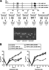
SEE of HCMV MIEP compensates for the lack of NF-κB-mediated MIEP transactivation in proliferating cells. (A) A schematic representation of the HCMV MIEP region and the mutations that were introduced to inactivate the AP-1 binding site, SEE (SRF and Elk-1 binding sites), and the four NF-κB binding sites. The AP-1, SEE, and NF-κB sites were defined with respect to the IE1/2 transcription start site. The point mutations and unique restriction sites introduced into each specific element are indicated below the wild-type sequence (−98 to −103, StuI; −161 to −166, KpnI; −168 to −174, EcoRI; −265 to −270 and −412 to −421, BglII; −522 to −528, EcoRI; and −532 to −538, KpnI). To confirm the successful disruption of AP-1, SEE, and all four NF-κB sites, the MIEP enhancer sequences between nucleotides −52498 and −53104 were PCR amplified from FIX Δ4NK-κB (lanes 1, 2, 3, and 4), FIX Δ4NK-κB-AP-1 (lanes 5, 6, 7, and 8), and FIX Δ4NK-κB-ΔSEE (lanes 9, 10, 11, and 12) reconstituted viruses (RVs). The amplified products were restriction digested using EcoRI (lanes 1, 5, and 9), KpnI (lanes 2, 6 and 10), StuI (lanes 3, 7 and 11), or BglII (lanes 4, 8 and 12), and the DNA fragments were separated by gel electrophoresis. (B) The NF-κB and SEE binding sites of MIEP both are required for optimal viral replication in quiescent cells. Growth-arrested or proliferating HELF cells were infected with the parental RVFIX, RVFIX Δ4NF-κB, RVFIX Δ4NF-κB-AP-1, RVFIX Δ4NF-κB-SEE, or RVFIX Δ4NF-κB-SEE REV (MOI of 0.1 PFU/cell). The extent of viral replication then was assessed at the indicated days p.i. by titrating the infectivity of the cell suspension supernatants on HELFs using the IE antigen indirect immunoperoxidase staining technique (13). The data shown are the averages of three experiments ± standard errors of the means (error bars).
FIG. 8.
FIG. 8.
Reduced replication rate of the RVFIX Δ4NF-κB-SEE virus in proliferating cells stems from reduced levels of IE gene expression. Growing or quiescent HELFs were infected with the parental RVFIX or RVFIX Δ4NF-κB-SEE (MOI of 0.1 PFU/cell). Total RNA was isolated at the indicated time p.i. and reverse transcribed. Real-time RT-PCR was carried out using the appropriate IE1, IE2, and β-actin primers to quantify the expression levels of IE1 and IE2 mRNA. The results then were analyzed using a standard-curve model, and the levels of IE1 and IE2 mRNA were normalized to the endogenous levels of β-actin mRNA. The data shown are the averages of three experiments ± standard errors of the means (error bars). The value at each time point then was normalized to the value observed with cells infected for 12 h, which was set at 1.

Similar articles

Cited by

References

    1. Benedict, C. A., A. Angulo, S. Ha, H. Huang, M. Messerle, C. F. Ware, and P. Ghazal. 2004. Neutrality of the canonical NF-κB pathway for human and murine cytomegalovirus transcription and replication in vitro. J. Virol. 78:741-750. - PMC - PubMed
    1. Bissinger, A. L., C. Sinzger, E. Kaiserling, and G. Jahn. 2002. Human cytomegalovirus as a direct pathogen: correlation of multiorgan involvement and cell distribution with clinical and pathological findings in a case of congenital inclusion disease. J. Med. Virol. 67:200-206. - PubMed
    1. Boldogh, I., S. AbuBakar, C. Z. Deng, and T. Albrecht. 1991. Transcriptional activation of cellular oncogenes fos, jun, and myc by human cytomegalovirus. J. Virol. 65:1568-1571. - PMC - PubMed
    1. Boldogh, I., M. P. Fons, and T. Albrecht. 1993. Increased levels of sequence-specific DNA-binding proteins in human cytomegalovirus infected cells. Biochem. Biophys. Res. Commun. 197:1505-1510. - PubMed
    1. Britt, W. 2008. Manifestations of human cytomegalovirus infection: proposed mechanisms of acute and chronic disease. Curr. Top. Microbiol. Immunol. 325:417-470. - PubMed

Publication types