. 2010 Mar;137(5):705-14.
doi: 10.1242/dev.039404.

Transcriptional control of stem cell maintenance in the Drosophila intestine

Affiliations

Transcriptional control of stem cell maintenance in the Drosophila intestine

Allison J Bardin et al. Development. 2010 Mar.

Abstract

Adult stem cells maintain tissue homeostasis by controlling the proper balance of stem cell self-renewal and differentiation. The adult midgut of Drosophila contains multipotent intestinal stem cells (ISCs) that self-renew and produce differentiated progeny. Control of ISC identity and maintenance is poorly understood. Here we find that transcriptional repression of Notch target genes by a Hairless-Suppressor of Hairless complex is required for ISC maintenance, and identify genes of the Enhancer of split complex [E(spl)-C] as the major targets of this repression. In addition, we find that the bHLH transcription factor Daughterless is essential to maintain ISC identity and that bHLH binding sites promote ISC-specific enhancer activity. We propose that Daughterless-dependent bHLH activity is important for the ISC fate and that E(spl)-C factors inhibit this activity to promote differentiation.


Figures
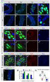
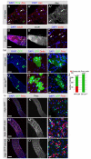
Fig. 1.
Fig. 1.
Hairless is required for ISC self-renewal. The growth of positively marked clones (GFP, green) as well as the expression of Delta (red) and Pros (blue) were monitored over time in the posterior midgut (nuclear DAPI, blue). (A-H) The growth of positively marked wild-type (A-D) and HE31 mutant (E-H) clones was monitored over a 2-week period at 4, 10 and 14 days (d) after heat shock (AHS). (D,H) High-magnification views at 14 days. (I-J″′) Whereas wild-type control clones (I-I″′) usually contained 1-2 Delta+ small cells and 0-2 Pros+ cells, HE31 clones (J-J″′) lacked Delta+ small cells and Pros+ cells. Note that Delta expression was detected in some Pros+ cells in wild-type midguts (see I′). (K-K″′) Overexpression of Hairless in MARCM clones under the control of tub-GAL4 over a 6-day period resulted in ectopic Delta+ ISC-like cells and ectopic Pros+ cells. (L) numb2 mutant ISCs proliferated into large clones by 10 days AHS. (M) Analysis of the size of wild-type and HE31clones at 6 days AHS. For each category of clone size, the number of clones is given as the percentage of the total number of clones (wild-type, n=215 clones; HE31, n=334 clones). The frequency of single-cell clones, corresponding to transient clones and non-proliferative ISC clones, was significantly increased upon loss of Hairless activity, whereas large HE31 mutant clones (6 cells or more) were rare compared with wild-type clones (see Materials and methods for statistics). (N) The ISC and its progeny. The ISC produces an ISC and an enteroblast (EB) upon division. The EB further differentiates into either an enteroendocrine cell (ee) expressing the transcription factor Pros or a large polyploid enterocyte (EC). Scale bars: 100 μm in A-C,E-G,L; 10 μm, in D,H,I-K.
Fig. 2.
Fig. 2.
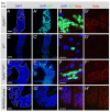
Su(H) is required for the Hairless ISC maintenance defect. Clone growth (GFP, green; DAPI, blue) and Delta expression (red) were examined in clones produced using a two heat-shock protocol. Low (A,A′,C,C′,E,E′,G,G′) and high (B,B′,D,D′,F,F′,H,H′) magnification views of representative midguts are shown. (A-B′) Su(H)Δ47 clones (outlined in A) contained ISC-like cells expressing Delta. (C-D′) HE31 clones failed to proliferate and did not express Delta. (E-F′) HE31 positively marked clones within Su(H)Δ47 unmarked clones (outlined in E) proliferated and contained Delta-expressing ISC-like cells. (G-H′) Negative control clones. The potential loss of GAL80 expression independent of a recombination event was monitored in Su(H)Δ47 single-mutant clones (outlined in G) in y w P[hs-FLP] P[pTub-GAL4] P[UAS-nlsGFP]; FRT40A P[l(2)35Bg] Su(H)Δ47/FRT40A P[pTub-GAL80]; FRT82B P[pTub-GAL80] / MKRS flies. This genotype produced virtually no GFP+ cells, indicating that the spontaneous loss of GAL80 expression cannot account for the phenotype seen in E-F′. Scale bars: 100 μm in A,A′,C,C′,E,E′,G,G′; 10 μm in B,B′,D,D′,F,F′,H,H′.
Fig. 3.
Fig. 3.
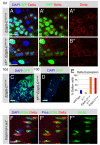
Deletion of the E(spl)-C suppresses the Hairless ISC maintenance defect. (A-B″) Both E(spl)-Cmδ-m6 (A-A″) and E(spl)-Cmδ-m6 HE31 (B-B″) positively marked mutant clones (GFP, green; nuclear DAPI, blue) grew and contained many small Delta+ (red) ISC-like cells at 6 days AHS. (C,D) E(spl)-Cmδ-m6 clones (C) were larger than E(spl)-Cmδ-m6 HE31 clones (D) at 10 days AHS. (E) Analysis of the percentage of Delta+ cells in wild-type, HE31, E(spl)-Cmδ-m6 and E(spl)-Cmδ-m6 HE31 clones at 10 days AHS. The difference between HE31 and either E(spl)-Cmδ-m6 or E(spl)-Cmδ-m6 HE31 was statistically significant, whereas the difference between E(spl)-Cmδ-m6 and E(spl)-Cmδ-m6 HE31 was not (see Materials and methods for statistics). (F-F″) E(spl)-mβ1.5-lacZ expression (β-galactosidase, green) was high in presumptive EBs adjacent to ISCs (Delta, red) that exhibited low E(spl)-mβ1.5-lacZ expression. Only basal nuclei are shown in this confocal section (Pros and DAPI, blue). Scale bars: 10 μm in A-B″, F-F″; 100 μm in C,D.
Fig. 4.
Fig. 4.
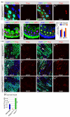
E(spl)-C genes regulate enteroendocrine differentiation. (A-A″′) The expression of Pros as a marker for ee fate and polyploidy (DAPI, blue) as a marker for EC fate were assessed in E(spl)-C and neur mutant clones (GFP, green). EC nuclei were detected in apical planes (A). A schematic of cells in apical/basal planes is shown in Fig. S3B in the supplementary material. Apical (A,A′) and basal (A″,A″′) views are shown of a 6-day AHS E(spl)-Cmδ-m6 mutant clone. E(spl)-Cmδ-m6 mutant clones produced many ISC-like cells expressing Delta (red in A,A″) but not Pros (red in A′,A″′). (B-C′) At 6 days, E(spl)-Cmδ-m6 mutant clones (GFP, green in C′) had an increase in small nuclei (DAPI, blue) but overall tissue architecture (phalloidin, green in B,C and blue in C′; Pdm1, red) was similar to the wild type (B). (D) The density and specification of large (>7 μm) polyploid ECs was unaffected by the deletion of the E(spl)-C, although a slightly higher number of cells expressing Pdm1 was present. Error bars represent s.d. from the mean. (E-E″′) Apical and basal low-magnification views of a field of 14-day E(spl)-Cmδ-m6 mutant cells. Polyploid ECs were properly specified (E), whereas Pros was not expressed in E(spl)-Cmδ-m6 mutant cells (E-E″′; an exceptional Pros+ cell is indicated in E″ by an arrow). (F-F″′) Apical and basal low-magnification views of a field with 14-day neurIF65 mutant cells. As previously noted (Ohlstein and Spradling, 2007), neurIF65 mutant clones contain few polyploid ECs (arrow in F) but many ectopic Pros+ ee-like cells (F-F″′). (G-G″′) Apical and basal low-magnification views of a field with 14-day neurIF65 E(spl)-Cmδ-m6 mutant clones. Many Pros+ ee-like cells were detected (G′,G″′). (H) Quantification of the number of Pros+ ee cells. E(spl)-CΔmδ-m6 mutant clones lack Pros+ cells (0.5%, n=622 cells) compared with neurIF65 mutant (76%, n=327 cells) and wild-type (6%, n=294 cells) clones. See Materials and methods for statistics. Scale bars: 10 μm.
Fig. 5.
Fig. 5.
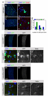
daughterless is required for ISC identity. (A-D) Clone growth (nuclear GFP, green; DAPI, blue) and expression of Delta (red) were examined in wild-type (A,B) and da10 (C,D) clones at 6 days AHS. da10 mutant clones (C,D) did not contain Delta+ cells, failed to grow and were composed of single or pairs of differentiated EC cells. (E) Quantification of clone size [number of cells in wild-type (blue, n=139) and da10 mutant (green, n=62) clones] at 6 days AHS. For each clone size category, the number of clones is given as the percentage of the total number of clones. The frequency of single-cell clones, corresponding to transient clones and non-proliferative ISC clones, was significantly increased upon loss of da activity, whereas large da10 mutant clones (6 cells or more) were not seen (see Materials and methods for statistics). (F-G″) The role of Da-binding motifs was assessed in the context of a mira-promoter-GFP transgene (mira-prom-GFP) that was specifically expressed in ISCs (nuclear GFP, green in G and white in G′; Delta, red in G and white in G″; DAPI, blue). A dividing ISC is marked by an asterisk in G. Pairs of GFP+ cells were also seen, probably owing to inheritance of GFP by ISC progeny cells. (H-I″) Mutation of the seven E-box motifs in the mira-promoter-GFP transgene (miraΔEbox-prom-GFP) largely abolished nuclear GFP expression (compare nuclear signals in G′ and I′). Scale bars: 100 μm in A,C,F,F′,H,H′; 10 μm in B,D,G-G″,I-I″.
Fig. 6.
Fig. 6.
achaete-scute complex genes are dispensable for ISC fate, but act in enteroendocrine fate. (A-A″) Expression of the bHLH protein Asense (Ase, red in A,A′, white in A″) was specifically detected in a subset of Pros+ cells (arrows; DAPI, blue; Pros, green). (B-C) scute RNA (red) was detected by fluorescent in situ hybridization in small nuclei cells both within and outside (arrows in C) of the Su(H) mutant clone area (identified by DAPI staining and outlined in B). (D-J) Df(1)scB57 mutant clones (G-I) grew similarly to wild-type control clones (D-F) and contained Delta+ ISCs as well as polyploid ECs (Delta, red in E,H; DAPI, blue; GFP, green). However, Df(1)scB57 mutant clones did not contain Pros+ cells (red). (J) Quantification revealed that one-third of wild-type clones at 10 days AHS contained at least one Pros+ cell (red bar, 12/32 clones), whereas Df(1)scB57 (0/37) did not contain Pros+ cells (P=0.00003, Fisher's exact test; see Materials and methods for statistics). (K-P) Expression of scute (M-N) and asense (O-P) in ISCs and EBs of adult flies using esgGAL4Gal80ts produced ectopic Pros+ ee cells, as compared with control flies (K-L; GFP, green, Pros, white or red; DAPI, blue). Scale bars: 100 μm in B,B′,D,G,K,K′,M,M′,O,O′; 10 μm in A-A″,C,E,F,H,I,L,N,P.
Fig. 7.
Fig. 7.
Model for ISC maintenance. We propose that Hairless prevents ISC loss by repressing expression of Notch target genes, including the E(spl)-C genes. We further propose that Da-dependent bHLH activity promotes ISC identity, including the ability to self-renew and to express Delta. Delta, in turn, activates Notch in the adjacent EB (Micchelli and Perrimon, 2006; Ohlstein and Spradling, 2006; Ohlstein and Spradling, 2007), releasing the intracellular domain of Notch (NICD). We speculate that, in response to Notch activation, the E(spl)-bHLH repressors downregulate Da-dependent bHLH activity in EBs as described in other systems (reviewed by Kageyama et al., 2007; Alifragis et al., 1997; Gigliani et al., 1996; Heitzler et al., 1996; Oellers et al., 1994), thereby shutting off ISC identity and promoting differentiation. The solid lines represent interactions for which we provide evidence, whereas the dashed line represents a proposed mechanism based on interaction data from other systems.

Similar articles

Cited by

References

    1. Acar M., Jafar-Nejad H., Giagtzoglou N., Yallampalli S., David G., He Y., Delidakis C., Bellen H. J. (2006). Senseless physically interacts with proneural proteins and functions as a transcriptional co-activator. Development 133, 1979-1989 - PubMed
    1. Alifragis P., Poortinga G., Parkhurst S. M., Delidakis C. (1997). A network of interacting transcriptional regulators involved in Drosophila neural fate specification revealed by the yeast two-hybrid system. Proc. Natl. Acad. Sci. USA 94, 13099-13104 - PMC - PubMed
    1. Amcheslavsky A., Jiang J., Ip Y. T. (2009). Tissue damage-induced intestinal stem cell division in Drosophila. Cell Stem Cell 4, 49-61 - PMC - PubMed
    1. Bailey A. M., Posakony J. W. (1995). Suppressor of hairless directly activates transcription of enhancer of split complex genes in response to Notch receptor activity. Genes Dev. 9, 2609-2622 - PubMed
    1. Bang A. G., Posakony J. W. (1992). The Drosophila gene Hairless encodes a novel basic protein that controls alternative cell fates in adult sensory organ development. Genes Dev. 6, 1752-1769 - PubMed

Publication types

MeSH terms

Substances