. 2010 Feb 5;6(2):117-29.
doi: 10.1016/j.stem.2009.12.015.

Sprouty1 regulates reversible quiescence of a self-renewing adult muscle stem cell pool during regeneration

Affiliations

Sprouty1 regulates reversible quiescence of a self-renewing adult muscle stem cell pool during regeneration

Kelly L Shea et al. Cell Stem Cell. .

Abstract

Satellite cells are skeletal muscle stem cells capable of self-renewal and differentiation after transplantation, but whether they contribute to endogenous muscle fiber repair has been unclear. The transcription factor Pax7 marks satellite cells and is critical for establishing the adult satellite cell pool. By using a lineage tracing approach, we show that after injury, quiescent adult Pax7(+) cells enter the cell cycle; a subpopulation returns to quiescence to replenish the satellite cell compartment, while others contribute to muscle fiber formation. We demonstrate that Sprouty1 (Spry1), a receptor tyrosine kinase signaling inhibitor, is expressed in quiescent Pax7(+) satellite cells in uninjured muscle, downregulated in proliferating myogenic cells after injury, and reinduced as Pax7(+) cells re-enter quiescence. We show that Spry1 is required for the return to quiescence and homeostasis of the satellite cell pool during repair. Our results therefore define a role for Spry1 in adult muscle stem cell biology and tissue repair.


Figures

Figure 1
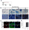
Figure 1. The pool of quiescent Pax7 satellite cells returns to homeostasis during adult muscle regeneration
(A) Transverse sections of uninjured (0 days after injury) and regenerating (4, 12, and 50 days after injury) TA muscle contain quiescent (MyoD-, Ki67-) (green arrowhead) and cycling (MyoD+, Ki67+; yellow arrowhead) Pax7+ cells in sublaminar location (laminin+; magenta). Differentiating myogenic cells (red arrowhead) could be observed in regenerating muscle underneath the basal lamina. (B) Total number of Pax7+ cells (left) and quiescent (Ki67-; MyoD-) Pax7+ cells (right) per muscle section from 4-6 mice per time point (mean ± sem; p<0.05). (C) BrdU+/Pax7+ cells in transverse sections from fully regenerated muscle from BrdU-treated mice. Top panel: Pax7 (red) and DAPI (blue) staining, Bottom panel: BrdU (green) and DAPI staining. Regenerated muscle fibers are distinguished with DAPI+ central nuclei (white arrowhead). The contour of muscle fiber is denoted by white line. Scale bar; 40 μm (A), 80 μm (C).
Figure 2
Figure 2. Pax7-derived satellite cells are capable of self-renewal and differentiation
(A) The cartoon depicts the tamoxifen (TM) injection scheme for lineage tracing muscle satellite cells. Pax7+ satellite cells in the Pax7-CreERtm;R26R-lacZ mouse were permanently labeled by IP administration of TM 14 days prior to muscle injury. (B) Transverse sections were collected from uninjured and regenerating muscle and stained with X-gal. Five days after injury non centrally-nucleated fibers in regenerating muscle had no detectable X-gal reactivity (*). After 50 days of regeneration, X-gal+ mononucleated cells were observed in the satellite cell position of regenerated muscle fibers (black arrows). (C) Single fibers from uninjured and regenerated Pax7-CreERtm;R26R-lacZ muscle stained with Pax7 (red), β-gal (green) and DAPI (blue). Regenerated muscle fibers are characterized by DAPI+ ‘central-myonuclear chains’. (D) The percentage of β-gal+/Pax7+ satellite cells expressing on single fibers isolated from uninjured and regenerated muscle fibers in the presence (+) or absence (-) of Cre or TM. Data is presented as mean ±sem. n=4-6 mice. (p<0.05). Scale bars in (B) are 60 μm (left) and 30 μm (right) and 40 μm in (C).
Figure 3
Figure 3. Sprouty1 is a marker of quiescent Pax7+ satellite cells
(A) Relative Spry1 transcript levels assessed by real-time qRT-PCR from FACS-purified myogenic cells (See Figure S3 for FACS sorting and Ct values) isolated from uninjured and regenerating muscle. (B) Transverse sections and single muscle fibers from uninjured muscles of Spry1lacZ/+ mice were stained with X-gal (upper panels). X-gal+ mononucleated cells (blue; black arrows) were observed in the satellite cell position in muscle sections (left) and single fibers (right). Single fibers from Spry1lacZ/+ mice were stained with anti-β-gal (green) and Pax7 (red) (middle row; white arrows) or β-gal (green) and MyoD (red) (bottom row; white arrowhead) after 0 hours or 36 hours in culture. Inset show β-gal+/Pax7+ cell (middle row) and β-gal-/ MyoD+/ cell (lower row). (C) Muscles from Spry1lacZ+/- were injured and left to regenerate from 4 to 50 days or remained uninjured. Muscle sections stained with anti-Pax7 and β-gal is shown from uninjured and regenerating muscle. Arrows show Pax7+/β-gal+ cells (at 0 and 30 days after injury) and Pax7+/β-gal- cells (at 12 days after injury). (D) Quantitation of (C), showing the percentage of β-gal+/Pax7+ cells in muscle sections. Data were presented as mean ± sem. n=4-6 mice. Scale bar in (B) is 80 μm and (C) is 40 μm.
Figure 4
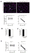
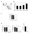
Figure 4. Sprouty1 is required for restoring the muscle stem cell pool during regeneration
(A) Mice were injected with TM to permanently activate Cre in Pax7-CreERtm;Spry1flox/flox; R26R-lacZ (SC-Null) and Pax7-CreERtm;Spry1WT/WT;R26R-lacZ (Control) adult mice. 14 days after TM treatment TA/EDL muscles from one leg were injured and left to regenerate for 50 days. Muscle sections stained with anti-Pax7, MyoD, Ki67, laminin and DAPI (Upper panels is shown from Control and SC-Null regenerated muscle; white arrow heads denote sub-laminar Pax7 cells). Graphs (lower panels) present the average number of Pax7+ cells in sub-laminar position. The bar between each point defines each animal. (p<0.01). (B) Single muscle fibers from (A) were stained with anti-Pax7 and β-gal to quantify the percentage of Pax7+ cells expressing β-gal driven from the Pax7-Cre reporter. (C) BrdU was administered in (A) during first 10 days after muscle injury. Muscle sections were stained with anti-BrdU, Pax7 and laminin. Histogram shows the percentage of BrdU+/Pax7+ cells satellite cells after 50 days of regeneration. (D) Muscle sections from Spry1+/+ and Spry1lacZ/lacZ were stained with anti-Pax7, MyoD, Ki67 and laminin (see Figure 4A). Graphs show the average number of Pax7+ cells in sub-laminar position in uninjured and regenerated muscle sections of individual mice. The bar between each point defines each animal. Scale bar; 40 μm.
Figure 5
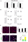
Figure 5. Disruption of Spry1 in Pax7 cells affects apoptosis but not differentiation of myogenic cells during regeneration
(A) Quantitative analysis of regenerating muscle (13 days after injury) from TM-treated Pax7-CreERtm;Spry1WT/WT (Control) and Pax7-CreER;Spry1flox/flox (SC-Null) mice. Graph shows the average number of quiescent (MyoD-, Ki67-) (left panel), cycling (MyoD+, Ki67+) (middle panel) Pax7+ cells and differentiated myogenic cells (Myogenin+) (right panel) in serial sections of TA muscle. Cell counts are expressed relative to Control muscle. Data were averaged (n=3 mice per group) and expressed as mean ±sem. (p<0.05). (B) Quantitative analysis of uninjured and regenerated muscle from TM-treated Control and SC-Null mice. Graph shows the average muscle fiber cross-sectional area (top panels) and the total number of muscle fibers (bottom panels) of uninjured and regenerated muscle in serial sections from the mid-belly of TA muscle. (C) TM treated Control (Pax7-CreERtm;Spry1WT/WT) and SC-Null (Pax7-CreERtm;Spry1flox/flox) muscle at 50 days after injury stained with laminin and DAPI (left panels). Histogram shows the average muscle fiber size in uninjured and regenerated muscle (n=4 mice per group) and expressed as mean ±sem. (D) Control and SC-Null muscles described in (C), were stained for TUNEL (green), MyoD (red), laminin (magenta) and DAPI (left panels). TUNEL+ cells are located sub- and peri-laminar position (white arrowheads). Inset: TUNEL+/MyoD+ nucleus (yellow) located underneath the basal lamina of a regenerating muscle fiber (white arrowhead). Histogram (right) shows the number of TUNEL+, MyoD+ (yellow) and MyoD- (green) cells per muscle section. Data were averaged (4 mice per group) and expressed as mean ±sem. (p<0.05). Scale bar; 80 μm.
Figure 6
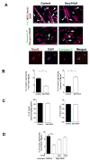
Figure 6. Sprouty1 regulates reversible quiescence of a subset of satellite cell progenitors
(A) Primary myoblasts from Spry1WT/WT;R26R-lacZ and Spry1flox/flox:R26R-lacZ mice were incubated with Cre adenovirus (Control and Spry1Null respectively) and switched to low mitogen media. Cultures were stained with markers to determine quiescence (top panel; Pax7+, MyoD-, Ki67-), differentiation (middle panel; Myogenin+) and apoptosis (middle panel; activated Caspase-3). White arrowheads show quiescent cells; white arrows show differentiated (top panels) and apoptotic (middle panels) cells. Lower panel shows activated Caspase-3 staining in mononucleated MyoD+ cells. (B) Histograms show that the percentage of quiescent Pax7+ cells (left), percentage of apoptotic cells (right) and (C) percentage of differentiated myogenic cells (left) and ability to fuse (right). (D) MEK inhibitor, U0126 (3 and 10 μM) or diluent alone (DMSO (-)) was added to Spry1flox/flox (Spry1Null) and Spry1WT/WT (Control) reserve cell cultures for 2 days. Histogram shows the percentage of quiescent (Pax7+, MyoD-, Ki67-) cells. Scale bar; 40 μm.
Figure 7
Figure 7. Return to quiescence and self-renewal is temporally instructed in a subpopulation of cycling Pax7 satellite cells
(A) Cartoon depicting the TM strategy to induce Cre activation in Control (Pax7-CreERtm;Spry1WT/WT) and SC-Null (Pax7-CreERtm;Spry1flox/flox) mice 14 days prior to injury) or at distinct phases of muscle regeneration. Muscles were analyzed 50 days after muscle injury. (B) Muscle sections from experiments depicted in (A) were stained with anti-Pax7, MyoD, Ki67, laminin and DAPI. Histogram shows the average number of Pax7+ cells per muscle section in regenerated SC-Null muscle expressed relative to the number of Pax7+ cells in the uninjured contra-lateral muscle. Data were averaged over a minimum of 4 mice per group and expressed as mean ± sem. (C) Muscles from Spry1+/+ and Spry1lacZ/lacZ adult mice were injured and left to regenerate for 13 days. Mice were treated with U0126 or vehicle (DMSO) via IP injection 10 and 11 days after injury. Histograms show the number of quiescent Pax7+ satellite cells located underneath the basal lamina of muscle sections. (D) Mice were subjected to two rounds of injury (1° and 2°) and 50 days of repair. Fifty days after the second injury muscle was analyzed for the number of Pax7+ cells in satellite cell position in Spry1+/+ and Spry1lacZ/lacZ (left panel) and TM-treated SC-Null and littermate controls (right panel). (**,p<0.01). (E) The average muscle fiber size from Control and SC-Null muscles from (D).

Similar articles

Cited by

References

    1. Abou-Khalil R, Le GF, Pallafacchina G, Valable S, Authier FJ, Rudnicki MA, Gherardi RK, Germain S, Chretien F, Sotiropoulos A, Lafuste P, Montarras D, Chazaud B. Autocrine and paracrine angiopoietin 1/Tie-2 signaling promotes muscle satellite cell self-renewal. Cell Stem Cell. 2009;5:298–309. - PMC - PubMed
    1. Allen RE, Dodson MV, Luiten LS. Regulation of skeletal muscle satellite cell proliferation by bovine pituitary fibroblast growth factor. Exp Cell Res. 1984;152:154–160. - PubMed
    1. Basson MA, Akbulut S, Watson-Johnson J, Simon R, Carroll TJ, Shakya R, Gross I, Martin GR, Lufkin T, McMahon AP, Wilson PD, Costantini FD, Mason IJ, Licht JD. Sprouty1 is a critical regulator of GDNF/RET-mediated kidney induction. Dev Cell. 2005;8:229–239. - PubMed
    1. Bockhold KJ, Rosenblatt JD, Partridge TA. Aging normal and dystrophic mouse muscle: analysis of myogenicity in cultures of living single fibers. Muscle Nerve. 1998;21:173–183. - PubMed
    1. Brack AS, Conboy IM, Conboy MJ, Shen J, Rando TA. A temporal switch from notch to Wnt signaling in muscle stem cells is necessary for normal adult myogenesis. Cell Stem Cell. 2008;2:50–59. - PubMed

Publication types

MeSH terms