. 2010 Feb 8;188(3):401-13.
doi: 10.1083/jcb.200907018.

Synaptobrevin N-terminally bound to syntaxin-SNAP-25 defines the primed vesicle state in regulated exocytosis

Affiliations

Synaptobrevin N-terminally bound to syntaxin-SNAP-25 defines the primed vesicle state in regulated exocytosis

Alexander M Walter et al. J Cell Biol. .

Abstract

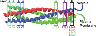
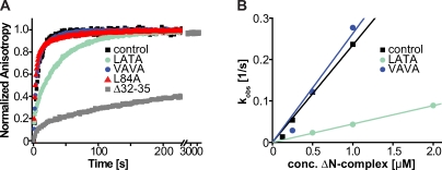
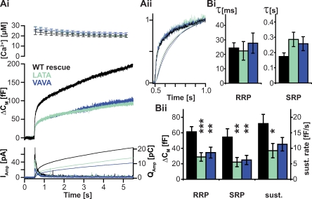
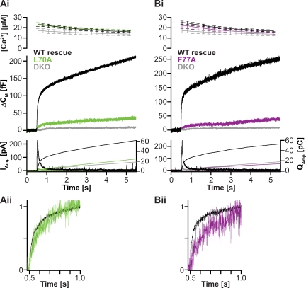
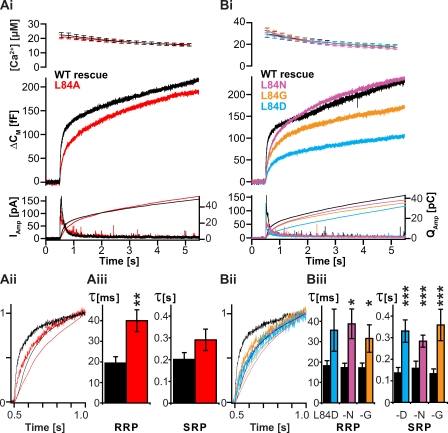
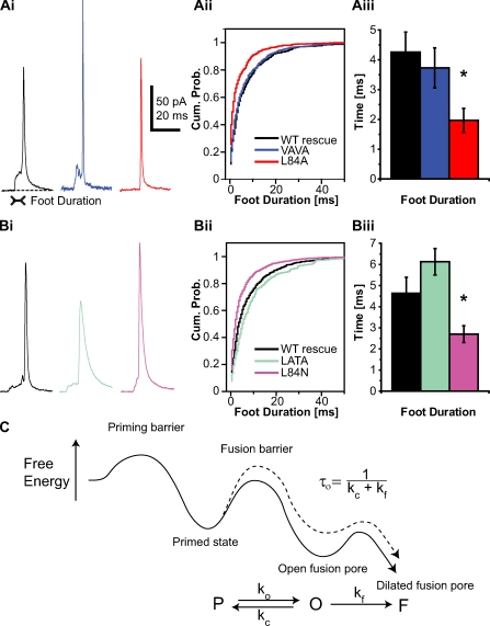
Rapid neurotransmitter release depends on the ability to arrest the SNAP receptor (SNARE)-dependent exocytosis pathway at an intermediate "cocked" state, from which fusion can be triggered by Ca(2+). It is not clear whether this state includes assembly of synaptobrevin (the vesicle membrane SNARE) to the syntaxin-SNAP-25 (target membrane SNAREs) acceptor complex or whether the reaction is arrested upstream of that step. In this study, by a combination of in vitro biophysical measurements and time-resolved exocytosis measurements in adrenal chromaffin cells, we find that mutations of the N-terminal interaction layers of the SNARE bundle inhibit assembly in vitro and vesicle priming in vivo without detectable changes in triggering speed or fusion pore properties. In contrast, mutations in the last C-terminal layer decrease triggering speed and fusion pore duration. Between the two domains, we identify a region exquisitely sensitive to mutation, possibly constituting a switch. Our data are consistent with a model in which the N terminus of the SNARE complex assembles during vesicle priming, followed by Ca(2+)-triggered C-terminal assembly and membrane fusion.


Figures

Figure 1.
Figure 1.
Overview of mutations introduced into syb-2 in the present investigation. All targeted sites are layer residues facing the inside of the complex (Fasshauer et al., 1998) either at the N-terminal end (left) or at the C-terminal end (right) of the complex. In the LATA mutant, aa 32 (leucine) and aa 35 (threonine) corresponding to layers −7 and −6 were mutated to alanine. The VAVA mutant bears mutations in layers −5 and −4, where valines (residues 39 and 42) were mutated to alanines. In the C-terminal half of the complex, leucine-70 was mutated to alanine (L70A) and phenylalanine was mutated to alanine (F77A) in layers 4 and 6, respectively. In layer 8, leucine was substituted by alanine (L84A), arginine (L84N), glycine (L84G), or aspartate (L84D).
Figure 2.
Figure 2.
In vitro binding of syb-2 to a syntaxin–SNAP-25 acceptor complex. (A) Fluorescence anisotropy measurements of labeled syb-2 mutants and WT protein binding to the ΔN complex. Comparison of two N-terminal mutants (LATA and VAVA) with WT syb-2 (control) and a C-terminal mutant (L84A) is shown. An N-terminally truncated syb-2 (Δ32–35, deletion of aa 32–35) displays greatly decreased binding kinetics (note the axis break on the abscissa; 100 nM synaptobrevin was added to 500 nM ΔN complex). (B) A plot of the observed binding rates of the synaptobrevin WT protein (control trace) and the LATA and VAVA mutants against the different concentrations (conc.) of the acceptor ΔN complex allows calculation of the association rate (slope of linear fits; values given in Biophysical characterization of mutants).
Figure 3.
Figure 3.
N-terminal mutants of syb-2 reduce pool size and sustained component when expressed in adrenal chromaffin cells. The N-terminal mutants of syb-2 are shown in Fig. 1. (Ai) N-terminal SNARE destabilization shows a reduced secretory response in exocytosis evoked by Ca2+ uncaging. (top) Mean ± SEM of intracellular Ca2+ concentration after UV-induced Ca2+ uncaging at time = 0.5 s. (middle) Mean capacitance increase. (bottom) Mean amperometric current (thick traces; left ordinate) and amperometric charge (thin traces; right ordinate). All cells were from double syb-2/cellubrevin knockout mice; control cells were infected with the WT protein (WT rescue). (Aii) A detailed view of normalized traces in Ai shows that the kinetics of release are unaffected (normalized capacitance, thick traces; and normalized amperometric charge, thin traces). (Bi and Bii) Kinetic analysis reveals significant reduction of pool sizes in LATA and VAVA mutants but no effect on rate of release. Fitting of exponentials to capacitance responses was used to quantify parameters reflecting time constants (Bi) and sizes (Bii) of the RRP and SRP. The sustained component (sust.) was found as the slope of the capacitance trace between 1.5 and 5.5 s. Mean ± SEM is shown (WT rescue, n = 21 cells; LATA, n = 20 cells; and VAVA, n = 29 cells; and *, P < 0.05; **, P < 0.01; and ***, P < 0.001 in t test).
Figure 4.
Figure 4.
Mutations in layers 4 and 6 severely affect transmitter release. The mutations in layers 4 and 6 are shown in Fig. 1. (Ai and Bi) Mean secretory responses in Ca2+-triggered release are dramatically reduced in the L70A (Ai) and the F77A (Bi) mutant. For comparison, traces from double knockout cells (DKO) are included. Double knockout traces are pooled from an independent dataset and were not recorded in parallel to L70A and F77A measurements. Mean ± SEM of Ca2+ levels is shown. (Aii and Bii) Detailed view of normalized traces in Ai and Bi. (A: WT rescue, n = 23; L70A, n = 15; double knockout, n = 21; B: WT rescue, n = 8; F77A, n = 13; double knockout, n = 21.)
Figure 5.
Figure 5.
C-terminal mutants of syb-2 cause a slowdown of transmitter release. The C-terminal mutants of syb-2 are shown in Fig. 1. (Ai and Bi) Mean secretion in flash-evoked transmitter release is slowed down. (Aii and Bii) Detailed views of normalized traces in Ai and Bi show that the kinetics of release are slowed down in all cases. (Aiii and Biii) Time constants of the RRP and SRP obtained by fitting of exponentials show slowdown of release in the L84A mutant (Aiii: control, n = 19; and L84A, n = 25) and L84N, L84G, and L84D mutations (Biii: control, n > 15; L84D, n = 9/14; L84G, n = 13/17; and L84N, n = 15/19 for RRP/SRP time constants; and *, P < 0.05; **, P < 0.01; and ***, P < 0.001 in t test). (Ai, Aiii, Bi, and Biii) Mean ± SEM is shown.
Figure 6.
Figure 6.
Fusion pore stability is affected by C- but not N-terminal destabilization of the SNARE complex. (Ai and Bi) Examples of spikes recorded by means of carbon fiber amperometry. (Aii and Bii) Mean of cumulative probability (Cum. Prob.) distributions of the prespike foot duration shows a shift to shorter feet for C-terminal synaptobrevin mutants (L84A in Aii and L84N in Bii). (Aiii and Biii) The mean foot duration (mean of cell medians) of C-terminal mutants is significantly reduced. Mean ± SEM is shown (A: WT rescue, n = 16 cells; VAVA, n = 19 cells; and L84A, n = 19 cells; and B: control, n = 14 cells; LATA, n = 15 cells; and L84N, n = 19 cells; and *, P < 0.05 in one-way ANOVA after log transformation yielded normally distributed homoscedastic data). (C) Proposed energy landscape for fusion (line), including barriers for vesicle priming, fusion triggering, and fusion pore expansion. The kinetic scheme comprises rate constants for pore formation (ko), pore closure (kc), and pore dilation (kf). In this model, the lifetime of the pore (τo) is defined by the inverse sum of the rate constants kc and kf. The dashed line shows the proposed energy landscape after C-terminal mutation in syb-2.

Similar articles

Cited by

References

    1. An S.J., Almers W. 2004. Tracking SNARE complex formation in live endocrine cells. Science. 306:1042–1046 10.1126/science.1102559 - DOI - PubMed
    1. Antonin W., Fasshauer D., Becker S., Jahn R., Schneider T.R. 2002. Crystal structure of the endosomal SNARE complex reveals common structural principles of all SNAREs. Nat. Struct. Biol. 9:107–111 10.1038/nsb746 - DOI - PubMed
    1. Ashery U., Betz A., Xu T., Brose N., Rettig J. 1999. An efficient method for infection of adrenal chromaffin cells using the Semliki Forest virus gene expression system. Eur. J. Cell Biol. 78:525–532 - PubMed
    1. Bai J., Wang C.T., Richards D.A., Jackson M.B., Chapman E.R. 2004. Fusion pore dynamics are regulated by synaptotagmin*t-SNARE interactions. Neuron. 41:929–942 10.1016/S0896-6273(04)00117-5 - DOI - PubMed
    1. Baumert M., Maycox P.R., Navone F., De Camilli P., Jahn R. 1989. Synaptobrevin: an integral membrane protein of 18,000 daltons present in small synaptic vesicles of rat brain. EMBO J. 8:379–384 - PMC - PubMed

Publication types

LinkOut - more resources