. 2010 Feb 12;86(2):148-60.
doi: 10.1016/j.ajhg.2010.01.016. Epub 2010 Feb 4.

Mutations in Grxcr1 are the basis for inner ear dysfunction in the pirouette mouse

Affiliations

Mutations in Grxcr1 are the basis for inner ear dysfunction in the pirouette mouse

Hana Odeh et al. Am J Hum Genet. .

Abstract

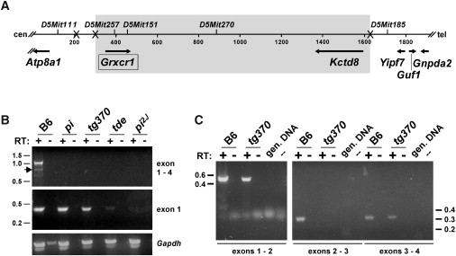
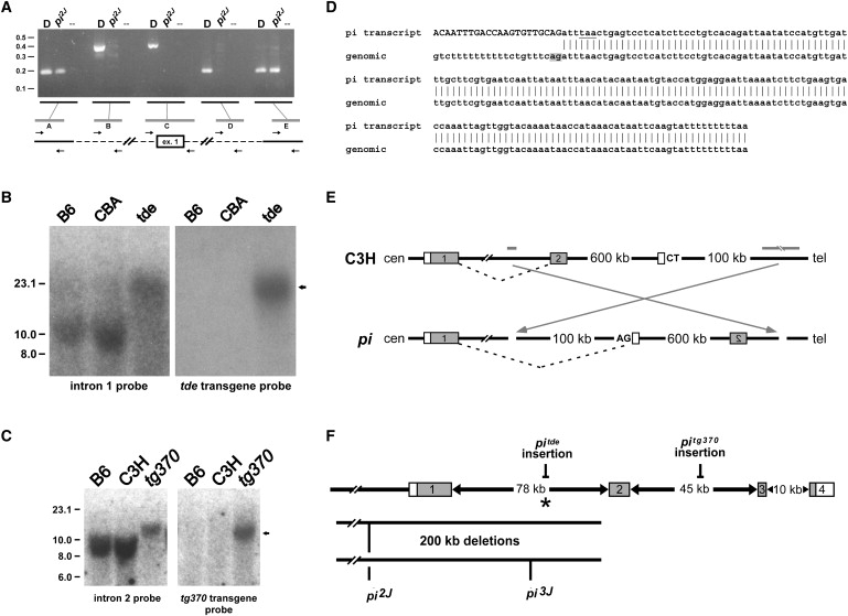
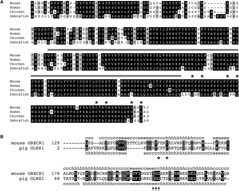
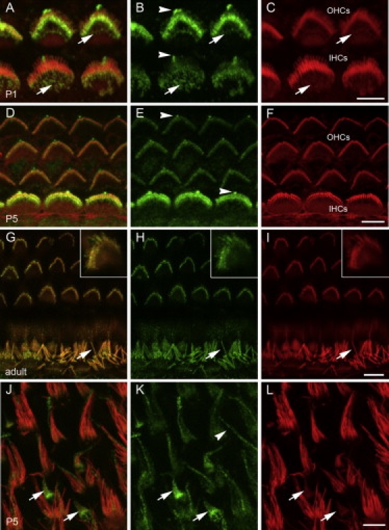
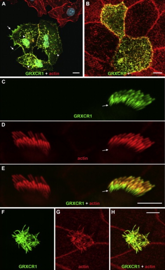
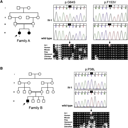
Recessive mutations at the mouse pirouette (pi) locus result in hearing loss and vestibular dysfunction due to neuroepithelial defects in the inner ear. Using a positional cloning strategy, we have identified mutations in the gene Grxcr1 (glutaredoxin cysteine-rich 1) in five independent allelic strains of pirouette mice. We also provide sequence data of GRXCR1 from humans with profound hearing loss suggesting that pirouette is a model for studying the mechanism of nonsyndromic deafness DFNB25. Grxcr1 encodes a 290 amino acid protein that contains a region of similarity to glutaredoxin proteins and a cysteine-rich region at its C terminus. Grxcr1 is expressed in sensory epithelia of the inner ear, and its encoded protein is localized along the length of stereocilia, the actin-filament-rich mechanosensory structures at the apical surface of auditory and vestibular hair cells. The precise architecture of hair cell stereocilia is essential for normal hearing. Loss of function of Grxcr1 in homozygous pirouette mice results in abnormally thin and slightly shortened stereocilia. When overexpressed in transfected cells, GRXCR1 localizes along the length of actin-filament-rich structures at the dorsal-apical surface and induces structures with greater actin filament content and/or increased lengths in a subset of cells. Our results suggest that deafness in pirouette mutants is associated with loss of GRXCR1 function in modulating actin cytoskeletal architecture in the developing stereocilia of sensory hair cells.


Figures

Figure 1
Figure 1
Grxcr1 Cochlear Expression Is Altered in the Pirouette Alleles (A) Combined genetic and sequence-based map is shown, with Xs indicating the relative positions of recombination breakpoints detected in a (pi × CAST/EiJ) F1 intercross. Map length is indicated in 200 kb increments. The gray box represents the nonrecombinant region that is ≤ 0.2 cM in size (95% confidence interval). In addition to the two genes located within the nonrecombinant region (Grxcr1 and Kctd8), four genes are located in the immediate flanking regions: Atp8a1 (U75321),Yipf7 (AF217188),Guf1 (AK084627), and Gnpda2 (AK016785). The predicted transcriptional orientation of each gene is indicated by an arrow. Centromeric (cen) and telomeric (tel) orientation of the chromosome is indicated. (B) RT-PCR products were amplified from cochlear RNA of C57BL/6J (B6) control mice and from homozygous pirouette mutants with the use of Grxcr1 primers complementary to sequences in exon1 and exon 4 (top panel) or within exon 1 (middle panel) and with the use of control Gapdh primers (bottom panel). Sequence analysis indicated that a minor product amplified from B6 RNA represents an alternatively spliced transcript lacking exon 2 (arrow). Positions of molecular-size standards are indicated at left in kilobases. (C) RT-PCR products were amplified from cochlear RNA of C57BL/6J (B6) control mice and from homozygous pitg370 mutants with the use of Grxcr1 primers complementary to sequences in exons 1 and 2 (left), in exons 2 and 3 (middle), and in exons 3 and 4 (right). Control PCR reactions that lacked template (–) were also performed, along with reactions with genomic DNA used as template. RT indicates the presence or absence of reverse transcriptase in corresponding reactions.
Figure 2
Figure 2
Genomic Alterations within Grxcr1 Are Present in Each of the Pirouette Alleles (A) PCR products were amplified from genomic DNA of control DBA/2J (“D”) and homozygous pi2J mice, with the use of primer pairs (“A”–“E”) designed from sequences upstream of exon 1, immediately flanking exon 1, and within intron 1. The distance between pairs A and E in normal genomic DNA is approximately 200 kb and represents the maximum size of a deletion in the mutant. The distance between pairs B and D is approximately 175 kb and represents the minimal size of the deletion. The deletion breakpoint in intron 1 is located within the 9 kb region between pairs D (no amplification in pi2J DNA) and E (positive for amplification in both control and pi2J DNA), approximately 3 kb upstream of exon 2. Positions of molecular-size standards are indicated at left in kilobases. (B) Southern blots containing EcoRV-digested genomic DNA from control (C57BL/6J, B6; CBA/CaJ, CBA) and homozygous pitde mice were hybridized with a probe derived from genomic sequences 32 kb upstream of exon 2 (left panel), stripped, and rehybridized with a probe complementary to the tde transgene construct (right panel). The arrow indicates common-sized 20 kb fragments that hybridize with both probes only in pitde DNA, consistent with insertion of the tde transgene into this region of intron 1. (C) Southern blots containing EcoRI-digested genomic DNA from control (C57BL/6J, B6; C3He/FeJ, C3H) and homozygous pitg370 mice were hybridized with a probe derived from genomic sequences 25 kb downstream of exon 2 (left panel), stripped, and rehybridized with a probe complementary to the tg370 transgene construct (right panel). The arrow indicates common-sized 13 kb fragments that hybridize with both probes only in pitg370 DNA, consistent with insertion of the tg370 transgene into this region of intron 2. (D) The sequence of a hybrid Grxcr1 cochlear transcript detected in affected pi mice. The top sequence is from a 3′ RACE product amplified with the use of nested primers derived from exon 1 of Grxcr1. The upper-case nucleotides indicate identity to the 3′ end of exon 1. The underlined nucleotides indicate a stop codon that would result in a truncated GRXCR1 protein. Vertical bars represent identity to genomic sequence located 600 kb telomeric of Grxcr1 (bottom, in telomere-to-centromere orientation). The gray box denotes a cryptic splice acceptor signal; an adjacent polypyrimidine tract is also present. (E) Structure of a predicted chromosomal inversion in pi. Gray bars represent the relative position in the WT background strain C3H of putative inversion breakpoints located in intron 1 of Grxcr1 and in a region approximately 700 kb telomeric (see Figure S3). Gray boxes indicate the first two exons of Grxcr1. The open boxes represent the cryptic exon detected by 3′ RACE. An inversion in the pi allele (arrows) would place the cryptic exon in the correct orientation for inclusion in the hybrid transcript with exon 1 of Grxcr1, with the use of the AG splice acceptor signal shown. (F) Summary of the Grxcr1 mutations in each of the pirouette alleles. The asterisk indicates the position of the putative centromeric inversion breakpoint in the original pi allele.
Figure 3
Figure 3
Grxcr1 Encodes a Potential “Redox” Protein (A) The predicted amino acid sequence of mouse GRXCR1 is indicated with one-letter abbreviations and aligned with orthologs from human (NP_001073945), chicken (ENSGALT00000037467), and zebrafish (ENSDARP00000102510). Black or gray shading indicates positions at which residues from at least three species are identical or biochemically similar, respectively. The region of similarity with glutaredoxin proteins is indicated with a gray bar, and conserved cysteines in the C terminus are marked with asterisks. (B) Alignment of the central region of mouse GRXCR1 with pig glutaredoxin (Protein Data Bank no. d1kte). The paired catalytic Cys residues found in many glutaredoxins are indicated with asterisks. Residues predicted to be required for contact with glutathione are indicated with arrows. Secondary structure information: c, random coil; h, alpha helix; e, beta sheet.
Figure 4
Figure 4
GRXCR1 Is Localized to Stereocilia on the Apical Surface of Sensory Hair Cells in the Inner Ear Inner ear tissues from WT C57BL/6J mice at three different time points (P1, P5, and adult) were incubated with antiserum raised against GRXCR1 (green) and rhodamine-phalloidin (red). Panels at left are merged images. (A–F) The major site of GRXCR1 immunoreactivity in the cochlea at P1 (A–C) and P5 (D–F) was sensory hair cells and their stereocilia bundles, with apparently higher levels of GRXCR1 in OHC stereocilia at P1 (A and B) and in IHC stereocilia at P5 (D and E). In P1 mice, GRXCR1 immunoreactivity was prominent in each row of stereocilia within bundles, including immature, shorter stereocilia that have lower relative levels of filamentous actin revealed by rhodamine-phalloidin staining (A–C). GRXCR1 immunoreactivity was also noted in apical microvilli of sensory cells at P1 (arrows, A–C) and in kinocilia at PI and P5 (arrowheads, B and E). (G–I) In the adult, GRXCR1 was localized throughout the length of stereocilia of OHCs (inserts) and IHCs (arrows). (J–L) GRXCR1 is localized along the length of vestibular hair cell stereocilia of the utricular macula. In vestibular epithelia, GRXCR1 staining was most prominent in immature stereocilia bundles (arrows). Immunoreactivity was also present in the kinocilia of vestibular sensory cells (arrowhead, K). Scale bars represent 5 μm.
Figure 5
Figure 5
GRXCR1 Protein Localizes to Actin-Filament Bundles in Transfected Cells (A) In COS-7 fibroblast cells transfected with Grxcr1-CFP, GRXCR1 (green) colocalizes with actin filaments (red) within filopodia-like structures at the cortical (arrows) and dorsal (arrowheads) surfaces of transfected cells. GRXCR1 did not colocalize significantly with actin stress fibers (data not shown). The number and actin filament content of dorsal projections in the GRXCR1-positive cells are increased relative to that of neighboring untransfected cells. (B) GRXCR1-GFP colocalizes with actin filaments in the apical microvilli of transfected CL4 epithelial cells but does not appreciably alter microvilli dimensions. (C–E) In transfected cochlear explants from WT mice, GRXCR1-GFP localized along the entire length of stereocilia of an inner hair cell, including the immature shorter row (arrows), which have lower actin filament content than that of longer rows. Stereocilia dimensions in GRXCR1-positive stereocilia bundles appeared similar to those of a neighboring untransfected hair cell (D, left). (F–H) GRXCR1-GFP also colocalizes with actin filaments in the apical microvilli of nonsensory epithelial cells in transfected explant cultures. The GRXCR1-positive microvilli were substantially longer than those of neighboring untransfected cells. Scale bars represent 10 μm (A) and 5 μm (B, E, and H).
Figure 6
Figure 6
Missense Variants in Human GRXCR1 Are Associated with Congenital Nonsyndromic Hearing Loss (A and B) Consanguineous pedigrees, sequence chromatograms of probands (arrows), and partial alignments of GRXCR1 encoded by genomic sequences from probands and controls and from other vertebrate species are shown for the double homozygous variants p.G64S (c.190G>A) and p.F153V (c.457T>G) (A) and for the single variant p.P38L (c.113C>T) (B).

Similar articles

Cited by

References

    1. Petit C., Richardson G.P. Linking genes underlying deafness to hair-bundle development and function. Nat. Neurosci. 2009;12:703–710. - PMC - PubMed
    1. Lim D.J., Anniko M. Developmental morphology of the mouse inner ear. A scanning electron microscopic observation. Acta Otolaryngol. 1985;422(Supplement):1–69. - PubMed
    1. Goodyear R.J., Marcotti W., Kros C.J., Richardson G.P. Development and properties of stereociliary link types in hair cells of the mouse cochlea. J. Comp. Neurol. 2005;485:75–85. - PubMed
    1. Gillespie P.G., Müller U. Mechanotransduction by hair cells: models, molecules, and mechanisms. Cell. 2009;139:33–44. - PMC - PubMed
    1. Flock A., Cheung H.C. Actin filaments in sensory hairs of inner ear receptor cells. J. Cell Biol. 1977;75:339–343. - PMC - PubMed

Publication types

MeSH terms