

A possible neuroprotective action of a vinylic telluride against Mn-induced neurotoxicity
- PMID: 20133376
- PMCID: PMC2855358
- DOI: 10.1093/toxsci/kfq036
A possible neuroprotective action of a vinylic telluride against Mn-induced neurotoxicity
Abstract
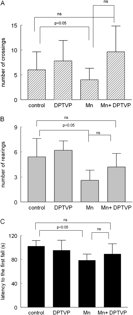
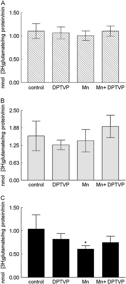
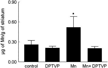
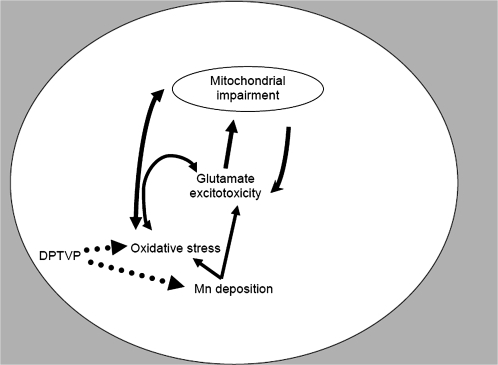
Manganese (Mn) is a metal required by biological systems. However, environmental or occupational exposure to high levels of Mn can produce a neurological disorder called manganism, which has similarities to Parkinson's disease. Diethyl-2-phenyl-2-tellurophenyl vinylphosphonate (DPTVP) is an organotellurium compound with a high antioxidant activity, especially in the brain. The present study was designed to investigate the effects of long-term low-dose exposure to Mn in drinking water on behavioral and biochemical parameters in rats and to determine the effectiveness of vinylic telluride in attenuating the effects of Mn. After 4 months of treatment with MnCl(2) (13.7 mg/kg), rats exhibited clear signs of neurobehavioral toxicity, including a decrease in the number of rearings in the open field and altered motor performance in rotarod. The administration of DPTVP (0.150 micromol/kg, ip, 2 weeks) improved the motor performance of Mn-treated rats, indicating that the compound could be reverting Mn neurotoxicity. Ex vivo, we observed that Mn concentrations in the Mn-treated group were highest in the striatum, consistent with a statistically significant decrease in mitochondrial viability and [(3)H]glutamate uptake, and increased lipid peroxidation. Mn levels in the hippocampus and cortex were indistinguishable from controls, and no significant differences were noted in the ex vivo assays in these areas. Treatment with DPTVP fully reversed the biochemical parameters altered by Mn. Furthermore, DPTVP treatment was also associated with a reduction in striatal Mn levels. Our results demonstrate that DPTVP has neuroprotective activity against Mn-induced neurotoxicity, which may be attributed to its antioxidant activity and/or its effect on striatal Mn transport.
Figures


Similar articles
-
Manganese-exposed developing rats display motor deficits and striatal oxidative stress that are reversed by Trolox.Arch Toxicol. 2013 Jul;87(7):1231-44. doi: 10.1007/s00204-013-1017-5. Epub 2013 Feb 6. Arch Toxicol. 2013. PMID: 23385959 Free PMC article.
-
Short-term manganese inhalation decreases brain dopamine transporter levels without disrupting motor skills in rats.J Toxicol Sci. 2016;41(3):391-402. doi: 10.2131/jts.41.391. J Toxicol Sci. 2016. PMID: 27193731
-
Neurotoxicity of manganese chloride in neonatal and adult CD rats following subchronic (21-day) high-dose oral exposure.J Appl Toxicol. 2000 May-Jun;20(3):179-87. doi: 10.1002/(sici)1099-1263(200005/06)20:3<179::aid-jat631>3.0.co;2-c. J Appl Toxicol. 2000. PMID: 10797470
-
"Manganese-induced neurotoxicity: a review of its behavioral consequences and neuroprotective strategies".BMC Pharmacol Toxicol. 2016 Nov 4;17(1):57. doi: 10.1186/s40360-016-0099-0. BMC Pharmacol Toxicol. 2016. PMID: 27814772 Free PMC article. Review.
-
Manganese-Induced Parkinsonism and Parkinson's Disease: Shared and Distinguishable Features.Int J Environ Res Public Health. 2015 Jul 6;12(7):7519-40. doi: 10.3390/ijerph120707519. Int J Environ Res Public Health. 2015. PMID: 26154659 Free PMC article. Review.
Cited by
-
Scientific opinion on the tolerable upper intake level for manganese.EFSA J. 2023 Dec 8;21(12):e8413. doi: 10.2903/j.efsa.2023.8413. eCollection 2023 Dec. EFSA J. 2023. PMID: 38075631 Free PMC article.
-
Parthenolide ameliorates 3-nitropropionic acid-induced Huntington's disease-like aberrations via modulating NLRP3 inflammasome, reducing microglial activation and inducing astrocyte shifting.Mol Med. 2024 Sep 26;30(1):158. doi: 10.1186/s10020-024-00917-5. Mol Med. 2024. PMID: 39327568 Free PMC article.
-
Development and In Vivo Assessment of 4-Phenyltellanyl-7-chloroquinoline-loaded Polymeric Nanocapsules in Alzheimer's Disease Models.Brain Sci. 2023 Jun 28;13(7):999. doi: 10.3390/brainsci13070999. Brain Sci. 2023. PMID: 37508931 Free PMC article.
-
Manganese-induced sex-specific gut microbiome perturbations in C57BL/6 mice.Toxicol Appl Pharmacol. 2017 Sep 15;331:142-153. doi: 10.1016/j.taap.2017.06.008. Epub 2017 Jun 10. Toxicol Appl Pharmacol. 2017. PMID: 28610994 Free PMC article.
-
Metals, oxidative stress and neurodegeneration: a focus on iron, manganese and mercury.Neurochem Int. 2013 Apr;62(5):575-94. doi: 10.1016/j.neuint.2012.12.006. Epub 2012 Dec 21. Neurochem Int. 2013. PMID: 23266600 Free PMC article. Review.
References
-
- Aschner M, Erikson KM, Dorman DC. Manganese dosimetry: species differences and implications for neurotoxicity. Crit. Rev. Toxicol. 2005;35:1–32. - PubMed
-
- Aschner M, Gannon M, Kimelberg HK. Manganese uptake and efflux in cultured rat astrocytes. J. Neurochem. 1992;58:730–735. - PubMed
-
- Avila DS, Gubert P, Dalla Corte CL, Alves D, Nogueira CW, Rocha JB, Soares FA. A biochemical and toxicological study with diethyl 2-phenyl-2-tellurophenyl vinylphosphonate in a sub-chronic intraperitoneal treatment in mice. Life Sci. 2007;80:1865–1872. - PubMed
Publication types
MeSH terms
Substances
Grants and funding
LinkOut - more resources
Full Text Sources
Medical
