. 2010 Jan 20;5(1):e8794.
doi: 10.1371/journal.pone.0008794.

A role for non-covalent SUMO interaction motifs in Pc2/CBX4 E3 activity

Affiliations

A role for non-covalent SUMO interaction motifs in Pc2/CBX4 E3 activity

Jacqueline C Merrill et al. PLoS One. .

Abstract

Background: Modification of proteins by the small ubiquitin like modifier (SUMO) is an essential process in mammalian cells. SUMO is covalently attached to lysines in target proteins via an enzymatic cascade which consists of E1 and E2, SUMO activating and conjugating enzymes. There is also a variable requirement for non-enzymatic E3 adapter like proteins, which can increase the efficiency and specificity of the sumoylation process. In addition to covalent attachment of SUMO to target proteins, specific non-covalent SUMO interaction motifs (SIMs) that are generally short hydrophobic peptide motifs have been identified.

Methodology/principal findings: Intriguingly, consensus SIMs are present in most SUMO E3s, including the polycomb protein, Pc2/Cbx4. However, a role for SIMs in SUMO E3 activity remains to be shown. We show that Pc2 contains two functional SIMs, both of which contribute to full E3 activity in mammalian cells, and are also required for sumoylation of Pc2 itself. Pc2 forms distinct sub-nuclear foci, termed polycomb bodies, and can recruit partner proteins, such as the corepressor CtBP. We demonstrate that mutation of the SIMs in Pc2 prevents Pc2-dependent CtBP sumoylation, and decreases enrichment of SUMO1 and SUMO2 at polycomb foci. Furthermore, mutational analysis of both SUMO1 and SUMO2 reveals that the SIM-interacting residues of both SUMO isoforms are required for Pc2-mediated sumoylation and localization to polycomb foci.

Conclusions/significance: This work provides the first clear evidence for a role for SIMs in SUMO E3 activity.


Conflict of interest statement

Competing Interests: The authors have declared that no competing interests exist.

Figures

Figure 1
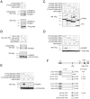
Figure 1. Pc2 contains two potential SIMs.
A) Human Pc2 is shown schematically, with the location of the CtBP-interaction motif (PIDLR in Pc2), the major sumoylation site in Pc2 (VKPE) and the two consensus SIMs. CD: chromodomain, His: poly-histidine stretch, C-box: carboxyl-terminal homology box. B) Alignments of the conserved regions surrounding SIM1 and SIM2 are shown, together with the SIM consensus sequences. Amino acid number are from human Pc2, sequences are from human, mouse, chicken, Xenopus laevis and tropicalis, zebra fish and Tetraodon nigroviridis. The PIDLR, adjacent to SIM2, is indicated by a line above the sequence. Identity (black) and similarity (gray) in at least 5/7 sequences is shown. C) An alignment of known SIMs (protein names to the left) is shown. The SIM in each is boxed, and acidic residues within 10 amino-acids of the SIM are shaded. D) His6-YFP fusion proteins encoding amino acids 250–560 of Pc2 (either wild type, or lacking SIM1, SIM2 or both, as indicated) were incubated with GST fusions to SUMO1 or SUMO2 bound to glutathione agarose, and interacting proteins were identified by western blot. A portion of the input proteins was analyzed by western blot (below), with either a His6 antibody for the Pc2 fusions, or a GST antibody for the SUMO fusions. E) COS1 cells were transfected with the indicated expression constructs and lysates analyzed for CtBP and sumoylated CtBP. F) The interaction of wild type or SIM1 mutant Pc2 with CtBP was determined by co-immunoprecipitation from COS1 cells transfected as indicated. Proteins were precipitated on anti-Flag agarose and analyzed by T7 western blot. Expression in the lysates was monitored by direct western (lower panels). The sequence around SIM1 and of the two Pc2 SIM1 mutants are shown below. The positions of molecular weight markers are indicated.
Figure 2
Figure 2. Identification of a minimal functional domain around SIM1.
A) COS1 cells were transfected with Flag-tagged Pc2(2-290), or a version with the VKPE sequence added after amino acid 290, and analyzed by cobalt affinity and Flag western blot (above) or by direct western on the lysates (below). The positions of the unmodified Pc2 and the sumoylated forms are shown). B) Cells were analyzed as in A, using a series of fusions of the NLS and sumoylation site from IκBα, with Myc epitope-tagged pyruvate kinase. C) A series of Pc2 deletion mutants was expressed in COS1 cells with or without co-expressed His-tagged SUMO1, and analyzed by direct Flag western blot, as in A. For each pair of lanes, the Pc2-VKPE fusion is indicated with an arrow, and the major sumoylated with an arrowhead. In lanes 1, 5 and 7, modification with endogenous SUMO is also detected. D and E) Lysates from transfected COS1 cells were analyzed as in C, for a series of Pc2(2-290)-based mutants (D), or Pc2(236-290)-based mutants (E), as indicated. F) The Pc2 constructs used in this figure are shown schematically, together with a summary of their sumoylation status in the presence of co-expressed SUMO1. The positions of molecular weight markers are indicated.
Figure 3
Figure 3. Mutational analysis of the SIM1 surroundings.
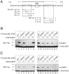
A) The minimal sequence surrounding SIM1 shown to have E3 activity is shown. The line and dashed lines above represent the regions of highest similarity across species (see Figure 1). The alanine mutants analyzed are shown below, together with a summary of their activity. B and C) COS1 cells were transfected with a series of Flag-Pc2(2-290)-VKPE expression constructs, in which pairs of conserved residues surrounding the SIM were altered to alanines. Constructs were expressed with or without co-expressed His-tagged SUMO1 (B) or SUMO2 (C). Lysates were analyzed by direct Flag western blot to detect unmodified and sumoylated Pc2 constructs. The positions of molecular weight markers are indicated.
Figure 4
Figure 4. Pc2 SIMs are required for interaction with SUMO3.
A) A series of Pc2 expression constructs with either SIM1, SIM2 or both deleted (ΔS1, ΔS2, ΔS1/2, respectively) was created, and tested for interaction with CtBP by coimmunoprecipitation from transfected COS1 cells, as in Figure 1. The CtBP interaction mutant (ΔPIDLR) was included as a control. Co-precipitating CtBP is indicated by an arrow, the upper band is the heavy chain. Expression in the lysates is shown in the lower panels. B and C) COS1 cells were transfected with the indicated Flag-Pc2 expression constructs together with a vector encoding a GFP-tagged non-conjugatable mutant of SUMO3 (GFP-nc-SUMO3). Proteins were precipitated on anti-Flag agarose and analyzed by western blot with a GFP antibody. Expression in the lysates is shown below. Arrows indicate co-precipitating proteins. D) Lysates from COS1 cells transfected with the indicated Pc2 constructs were incubated with glutathione agarose to which recombinant bacterially expressed GST-SUMO1 or GST-SUMO2 had been pre-bound. Bound proteins were analyzed by western blot for Flag-Pc2 constructs. A portion of the lysate was analyzed in parallel (input), and the GST-SUMO fusions were visualized by Coomassie blue staining the lower part of the gel. E) The Pc2 expression constructs analyzed are shown schematically. The positions of molecular weight markers are indicated.
Figure 5
Figure 5. SIM1 and SIM2 contribute to E3 activity.
A) Sumoylation of Pc2 and Pc2 mutants (upper panel) was tested by direct western blot in lysates from transfected COS1 cell either without or with coexpressed His6-SUMO1. Lower panel shows sumoylation of CtBP, analyzed by direct western blot of COS1 cell lysates, transfected with the series of Pc2 constructs. B) Sumoylation of Pc2 (above) and CtBP (below) was analyzed as in A, except that all Pc2 expression constructs are all in the context of the 2-531 deletion mutant of Pc2, which lacks the carboxyl-terminal domain (C-box) and is delocalized from polycomb foci. C) Sumoylation of Pc2 (upper panel) and CtBP (lower panel) was analyzed by direct western blot as in A and B. Only the series using Pc2 and mutants in the context of amino-acids 2-531 is shown, as these mutations did not affect the full length protein. D) Sumoylation of Pc2 (upper panel) and CtBP (lower panel) in the presence of either coexpressed SUMO1 or SUMO2, as indicated, was analyzed by direct western blot. Wild type Pc2 and SIM deletion mutants in the context of full length Pc2 are shown. The positions of molecular weight markers are indicated.
Figure 6
Figure 6. Sub-cellular localization of SUMO and Pc2 SIM mutants.
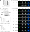
A) COS1 cells were transfected with the indicated eYFP-tagged Pc2 expression constructs with eCFP-tagged CtBP and visualized by live cell fluorescence microscopy. Cells with Pc2 foci were identified and scored for colocalization of eCFP-CtBP. The percentage of cells with colocalization is plotted for each Pc2 mutant. B) Examples of CtBP and Pc2 colocalization are shown. Separate YFP and CFP images (false colored to green and white for maximal contrast) are shown, together with a merged image and the corresponding Hoechst stain. C) Modification of Pc2 and CtBP was analyzed by direct western blot of COS1 cell lysates transfected with the indicated wild type or non-conjugatable (ΔGG) versions of SUMO1 and SUMO2. The positions of molecular weight markers are indicated. D and E) The proportion of cells in which the indicated monomeric Cherry (mC-) SUMO1 (D) and SUMO2 (E) fusions co-localized with the indicated eYFP-Pc2 constructs is shown. The SUMO expression constructs are either wild type, ΔGG (as in panel C), or are the SUMO1(FV) and SUMO2(QFI) SIM interaction mutants shown schematically in Figure 7D. F) Representative live cell fluorescent images showing localization of a selection of Pc2 and SUMO expression constructs are shown. The coexpressed eYFP-Pc2 and mC-SUMO constructs are indicated to the left, and individual eYFP (in green) and mCherry (in white) images are shown, together with the merged image and the Hoechst stain.
Figure 7
Figure 7. Mutational analysis of SUMO1 and SUMO2.
A) Modification of wild type Flag-tagged Pc2 by the indicated His6-tagged SUMO variants in transfected COS1 cells was analyzed by direct western blot. B and C) Modification of CtBP by the indicated SUMO variants alone or coexpressed with Ubc9 or Pc2 was analyzed as in A. Expression of the Flag-tagged Pc2 and Ubc9 is shown in the lower panels. D) Partial amino acid sequences of SUMO1 and SUMO2 are shown. Numbers indicate amino acid numbers. The two arrows indicate the positions of β strands 1 and 2, and the cylinder the first α helix. The residues in each SUMO isoform which were altered to alanines to create the FV and QFI mutants are shown. The positions of molecular weight markers are indicated.

Similar articles

Cited by

References

    1. Nacerddine K, Lehembre F, Bhaumik M, Artus J, Cohen-Tannoudji M, et al. The SUMO pathway is essential for nuclear integrity and chromosome segregation in mice. Dev Cell. 2005;9:769–779. - PubMed
    1. Geiss-Friedlander R, Melchior F. Concepts in sumoylation: a decade on. Nat Rev Mol Cell Biol. 2007;8:947–956. - PubMed
    1. Gill G. Something about SUMO inhibits transcription. Curr Opin Genet Dev. 2005;15:536–541. - PubMed
    1. Johnson ES. Protein modification by SUMO. Annu Rev Biochem. 2004;73:355–382. - PubMed
    1. Hay RT. Protein modification by SUMO. Trends Biochem Sci. 2001;26:332–333. - PubMed

Publication types