

The role of primary cilia in neuronal function
- PMID: 20097287
- PMCID: PMC2953617
- DOI: 10.1016/j.nbd.2009.12.022
The role of primary cilia in neuronal function
Abstract
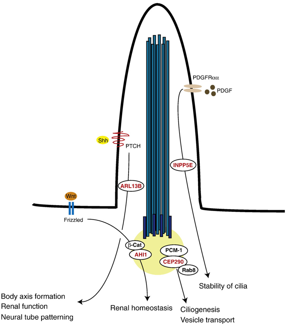
The "ciliopathies" are a newly defined group of disorders characterized by defects in the structure or function of the cellular primary cilium. Patients with these disorders display variably expressive fibrocystic renal disease, retinal blindness, polydactyly, obesity, and brain dysgenesis as well as neurocognitive impairments. Joubert syndrome is a ciliopathy defined by cerebellar vermis hypoplasia, oculomotor apraxia, intermittent hyperventilation, and mental retardation. Recent evidence suggests important roles for the primary cilium in mediating a host of extracellular signaling events such as morphogen, mitogen, homeostatic and polarity signals. Based upon the clinical features of ciliopathies and cilia mediated signaling pathways, the data support a role for the primary cilium in modulating neurogenesis, cell polarity, axonal guidance and possibly adult neuronal function.
Copyright 2009 Elsevier Inc. All rights reserved.
Figures

Similar articles
-
The ciliopathies in neuronal development: a clinical approach to investigation of Joubert syndrome and Joubert syndrome-related disorders.Dev Med Child Neurol. 2011 Sep;53(9):793-798. doi: 10.1111/j.1469-8749.2011.04021.x. Epub 2011 Jun 17. Dev Med Child Neurol. 2011. PMID: 21679365 Free PMC article. Review.
-
Joubert syndrome: insights into brain development, cilium biology, and complex disease.Semin Pediatr Neurol. 2009 Sep;16(3):143-54. doi: 10.1016/j.spen.2009.06.002. Semin Pediatr Neurol. 2009. PMID: 19778711 Free PMC article. Review.
-
Neuronal ciliary signaling in homeostasis and disease.Cell Mol Life Sci. 2010 Oct;67(19):3287-97. doi: 10.1007/s00018-010-0425-4. Epub 2010 Jun 11. Cell Mol Life Sci. 2010. PMID: 20544253 Free PMC article. Review.
-
Mutations in ARMC9, which Encodes a Basal Body Protein, Cause Joubert Syndrome in Humans and Ciliopathy Phenotypes in Zebrafish.Am J Hum Genet. 2017 Jul 6;101(1):23-36. doi: 10.1016/j.ajhg.2017.05.010. Epub 2017 Jun 15. Am J Hum Genet. 2017. PMID: 28625504 Free PMC article.
-
TALPID3 in Joubert syndrome and related ciliopathy disorders.Curr Opin Genet Dev. 2019 Jun;56:41-48. doi: 10.1016/j.gde.2019.06.010. Epub 2019 Jul 19. Curr Opin Genet Dev. 2019. PMID: 31326647 Review.
Cited by
-
Neuronal and astrocytic primary cilia in the mature brain.Pharmacol Res. 2018 Nov;137:114-121. doi: 10.1016/j.phrs.2018.10.002. Epub 2018 Oct 4. Pharmacol Res. 2018. PMID: 30291873 Free PMC article. Review.
-
The DNA methylome in panic disorder: a case-control and longitudinal psychotherapy-epigenetic study.Transl Psychiatry. 2019 Nov 21;9(1):314. doi: 10.1038/s41398-019-0648-6. Transl Psychiatry. 2019. PMID: 31754096 Free PMC article.
-
Type 3 adenylyl cyclase: a key enzyme mediating the cAMP signaling in neuronal cilia.Int J Physiol Pathophysiol Pharmacol. 2016 Sep 30;8(3):95-108. eCollection 2016. Int J Physiol Pathophysiol Pharmacol. 2016. PMID: 27785336 Free PMC article. Review.
-
Primary Cilia Structure Is Prolonged in Enteric Neurons of 5xFAD Alzheimer's Disease Model Mice.Int J Mol Sci. 2021 Dec 17;22(24):13564. doi: 10.3390/ijms222413564. Int J Mol Sci. 2021. PMID: 34948356 Free PMC article.
-
Primary Cilia as a Possible Link between Left-Right Asymmetry and Neurodevelopmental Diseases.Genes (Basel). 2017 Jan 25;8(2):48. doi: 10.3390/genes8020048. Genes (Basel). 2017. PMID: 28125008 Free PMC article. Review.
References
-
- Adams NA, et al. The retinal ciliopathies. Ophthalmic Genet. 2007;28:113–125. - PubMed
-
- Ahdab-Barmada M, Claassen D. A distinctive triad of malformations of the central nervous system in the Meckel-Gruber syndrome. J. Neuropathol. Exp. Neurol. 1990;49:610–620. - PubMed
-
- Andersen JS, et al. Proteomic characterization of the human centrosome by protein correlation profiling. Nature. 2003;426:570–574. - PubMed
-
- Avidor-Reiss T, et al. Decoding cilia function: defining specialized genes required for compartmentalized cilia biogenesis. Cell. 2004;117:527–539. - PubMed
-
- Badano JL, et al. The ciliopathies: an emerging class of human genetic disorders. Annu. Rev. Genomics Hum. Genet. 2006;7:125–148. - PubMed
Publication types
MeSH terms
Grants and funding
LinkOut - more resources
Full Text Sources
