

Review
doi: 10.1038/nrc2791.
Instructive role of the vascular niche in promoting tumour growth and tissue repair by angiocrine factors
Affiliations
- PMID: 20094048
- PMCID: PMC2944775
- DOI: 10.1038/nrc2791
Item in Clipboard
Review
Instructive role of the vascular niche in promoting tumour growth and tissue repair by angiocrine factors
Nat Rev Cancer.
2010 Feb.
Abstract
The precise mechanisms whereby anti-angiogenesis therapy blocks tumour growth or causes vascular toxicity are unknown. We propose that endothelial cells establish a vascular niche that promotes tumour growth and tissue repair not only by delivering nutrients and O2 but also through an 'angiocrine' mechanism by producing stem and progenitor cell-active trophogens. Identification of endothelial-derived instructive angiocrine factors will allow direct tumour targeting, while diminishing the unwanted side effects associated with the use of anti-angiogenic agents.
Figures
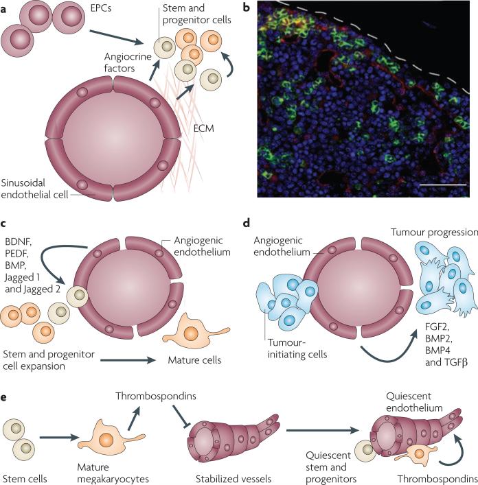
a | By expressing angiocrine factors and producing extracellular matrix (ECM), endothelial cells and endothelial progenitor cells (EPCs) establish a microenvironment, referred to as the vascular niche, which supports the expansion of normal and malignant stem and progenitor cells. b | Establishment of the vascular niche by the bone marrow sinusoidal endothelial cells. The confocal image shows a cross-section of the bone marrow taken from transgenic Notch reporter mice 7 days after sublethal irradiation with 6.5 Gy. Regenerating Notch-activated green fluorescent protein (GFP)+ haematopoietic stem and progenitor cells (green fluorescence) could be detected in the proximity of VE-cadherin+ Notch ligand+ sinusoidal endothelial cells (red staining). Therefore, angiocrine factors, such as Notch ligands, reconstitute haematopoiesis. c | Vascular endothelial growth factor A (VEGFA)- or fibroblast growth factor 2 (FGF2)-activated endothelial cells promote the proliferation and lineage-specific differentiation of normal cells by release of angiocrine factors (FGFs, BMPs, jagged 1 and jagged 2) and direct cellular contact. d | Identical pathways to those described in (c) affect malignant stem and progenitor cells. e | The mature progeny of stem cells, such as megakaryocytes, enforce a quiescent state in the endothelial cells by inhibiting their angiogenic activity through the release of anti-angiogenic factors, such as thrombospondins, which in turn promotes stem cell quiescence. BDNF, bone-derived neurotrophic factor; BMP, bone morphogenetic protein; PEDF, pigment epithelium-derived factor; TGFβ, transforming growth factor-β. Bar represents 50 μm.
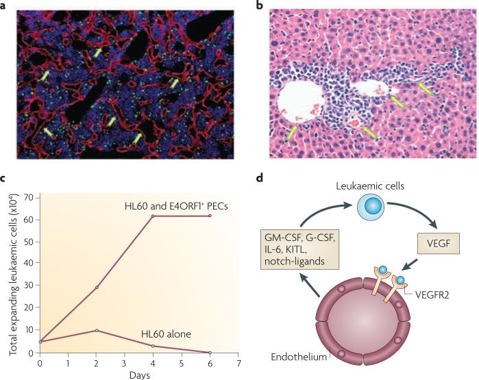
The prototypical angiogenic factor vascular endothelial growth factor A (VEGFA) released by leukaemic cells (green fluorescent protein+ cells in (a) and infiltrating cells in (b)) activates the tyrosine kinase VEGF receptor 2 (VEGFR2) expressed on the endothelial cells and this promotes the proliferation of VE-cadherin+ vessels in the bone marrow (a, arrows) and liver (b, arrows). In vitro E4ORF1+ primary endothelial cells (PECs) support the long term expansion of HL60 leukaemic cells in serum- and cytokine-free conditions (c), and in the absence of the E4ORF1+ primary endothelial cells HL60 leukaemic cells undergo cell death (c). The mechanism by which endothelial cells support the proliferation of the leukaemic cells is through the VEGFA–VEGFR2-mediated upregulation of the pro-leukaemic factors (d) leading to the uncontrolled proliferation of the leukaemic cells. G-CSF, granulocyte-colony stimulating factor; GM-CSF, granulocyte-macrophage-colony stimulating factor; IL-6, interleukin-6; KITL, KIT ligand. Part (c) of this figure is reproduced from ref . .
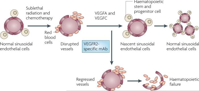
Timely regeneration of the sinusoidal endothelial cells supports regeneration of the haematopoietic stem and progenitor cells, guaranteeing prompt reconstitution of haematopoiesis after treatment with myeloablative chemotherapy and irradiation. As such, bone marrow sinusoidal endothelial cells establish a vascular niche that, through the release of angiocrine factors, promotes the reconstitution of the haematopoietic stem and progenitor cells. VEGF, vascular endothelial growth factor; VEGFR2, VEGF receptor 2.
Similar articles
-
Beyond Angiogenesis: Exploiting Angiocrine Factors to Restrict Tumor Progression and Metastasis.Cancer Res. 2020 Feb 15;80(4):659-662. doi: 10.1158/0008-5472.CAN-19-3351. Epub 2019 Dec 12. Cancer Res. 2020. PMID: 31831463
-
Tumours can adapt to anti-angiogenic therapy depending on the stromal context: lessons from endothelial cell biology.Eur J Cell Biol. 2006 Feb;85(2):61-8. doi: 10.1016/j.ejcb.2005.10.003. Epub 2005 Nov 11. Eur J Cell Biol. 2006. PMID: 16439306 Review.
-
Controlling escape from angiogenesis inhibitors.Nat Rev Cancer. 2012 Oct;12(10):699-709. doi: 10.1038/nrc3366. Nat Rev Cancer. 2012. PMID: 23001349 Free PMC article. Review.
-
Two-domain vascular disruptive agents in cancer therapy.Curr Cancer Drug Targets. 2004 Sep;4(6):501-9. doi: 10.2174/1568009043332826. Curr Cancer Drug Targets. 2004. PMID: 15379635 Review.
-
Recent advances in angiogenesis, anti-angiogenesis and vascular targeting.Trends Pharmacol Sci. 2002 Dec;23(12):576-82. doi: 10.1016/s0165-6147(02)02109-0. Trends Pharmacol Sci. 2002. PMID: 12457776 Review.
Cited by
-
The perivascular niche regulates breast tumour dormancy.Nat Cell Biol. 2013 Jul;15(7):807-17. doi: 10.1038/ncb2767. Epub 2013 Jun 2. Nat Cell Biol. 2013. PMID: 23728425 Free PMC article.
-
EphA3 CAR T cells are effective against glioblastoma in preclinical models.J Immunother Cancer. 2024 Aug 7;12(8):e009403. doi: 10.1136/jitc-2024-009403. J Immunother Cancer. 2024. PMID: 39111832 Free PMC article.
-
The cancer stem cell niche: how essential is the niche in regulating stemness of tumor cells?Cell Stem Cell. 2015 Mar 5;16(3):225-38. doi: 10.1016/j.stem.2015.02.015. Cell Stem Cell. 2015. PMID: 25748930 Free PMC article. Review.
-
Infantile Hemangioma Originates From A Dysregulated But Not Fully Transformed Multipotent Stem Cell.Sci Rep. 2016 Oct 27;6:35811. doi: 10.1038/srep35811. Sci Rep. 2016. PMID: 27786256 Free PMC article.
-
Endothelial cells promote the colorectal cancer stem cell phenotype through a soluble form of Jagged-1.Cancer Cell. 2013 Feb 11;23(2):171-85. doi: 10.1016/j.ccr.2012.12.021. Epub 2013 Jan 31. Cancer Cell. 2013. PMID: 23375636 Free PMC article.
References
-
- Folkman J. Angiogenesis: an organizing principle for drug discovery? Nature Rev. Drug Discov. 2007;6:273–286. - PubMed
-
- Ferrara N, Hillan KJ, Gerber HP, Novotny W. Discovery and development of bevacizumab, an anti-VEGF antibody for treating cancer. Nature Rev. Drug Discov. 2004;3:391–400. - PubMed
-
- Carmeliet P, Jain RK. Angiogenesis in cancer and other diseases. Nature. 2000;407:249–257. - PubMed
Publication types
MeSH terms
Substances
Grants and funding
- P01 HL059312-090006/HL/NHLBI NIH HHS/United States
- R01 HL061849-04/HL/NHLBI NIH HHS/United States
- R01 HL097797-01/HL/NHLBI NIH HHS/United States
- R01 HL061849-03S1/HL/NHLBI NIH HHS/United States
- U01 HL066952-040002/HL/NHLBI NIH HHS/United States
- P01 HL059312-080006/HL/NHLBI NIH HHS/United States
- R01 HL075234/HL/NHLBI NIH HHS/United States
- R21 HL083222-01/HL/NHLBI NIH HHS/United States
- R01 HL075234-04/HL/NHLBI NIH HHS/United States
- U01 HL066952/HL/NHLBI NIH HHS/United States
- P01 HL067839-020004/HL/NHLBI NIH HHS/United States
- P01 HL072942-010004/HL/NHLBI NIH HHS/United States
- R01 HL097797-03/HL/NHLBI NIH HHS/United States
- P50 HL084936/HL/NHLBI NIH HHS/United States
- R01 HL058707-04/HL/NHLBI NIH HHS/United States
- P01 HL059312-100006/HL/NHLBI NIH HHS/United States
- P01 HL072942/HL/NHLBI NIH HHS/United States
- U01 HL066952-030002/HL/NHLBI NIH HHS/United States
- P50 HL084936-010003/HL/NHLBI NIH HHS/United States
- R21 HL083222-02/HL/NHLBI NIH HHS/United States
- P50 HL084936-030003/HL/NHLBI NIH HHS/United States
- P01 HL067839/HL/NHLBI NIH HHS/United States
- R01 HL075234-03/HL/NHLBI NIH HHS/United States
- U01 HL066952-020002/HL/NHLBI NIH HHS/United States
- R01 HL058707-03/HL/NHLBI NIH HHS/United States
- P01 HL059312-060006/HL/NHLBI NIH HHS/United States
- R01 HL097797-02/HL/NHLBI NIH HHS/United States
- R01 HL097797/HL/NHLBI NIH HHS/United States
- P50 HL084936-020003/HL/NHLBI NIH HHS/United States
- P01 HL059312/HL/NHLBI NIH HHS/United States
- R01 HL061849-03/HL/NHLBI NIH HHS/United States
- R01 HL061849/HL/NHLBI NIH HHS/United States
- R01 HL075234-02/HL/NHLBI NIH HHS/United States
- R01 HL061849-05/HL/NHLBI NIH HHS/United States
- P01 HL067839-050004/HL/NHLBI NIH HHS/United States
- P01 HL067839-030004/HL/NHLBI NIH HHS/United States
- P01 HL059312-070006/HL/NHLBI NIH HHS/United States
- P50 HL084936-040003/HL/NHLBI NIH HHS/United States
- P01 HL067839-010004/HL/NHLBI NIH HHS/United States
- R01 HL075234-01/HL/NHLBI NIH HHS/United States
- R01 HL061849-02/HL/NHLBI NIH HHS/United States
- U01 HL066952-050002/HL/NHLBI NIH HHS/United States
- U01 HL066952-010002/HL/NHLBI NIH HHS/United States
- HHMI/Howard Hughes Medical Institute/United States
- P01 HL067839-040004/HL/NHLBI NIH HHS/United States
- R21 HL083222/HL/NHLBI NIH HHS/United States
LinkOut - more resources
Full Text Sources
Other Literature Sources
