. 2010 Jan 15;6(1):e1000721.
doi: 10.1371/journal.ppat.1000721.

Marburg virus evades interferon responses by a mechanism distinct from ebola virus

Affiliations

Marburg virus evades interferon responses by a mechanism distinct from ebola virus

Charalampos Valmas et al. PLoS Pathog. .

Abstract

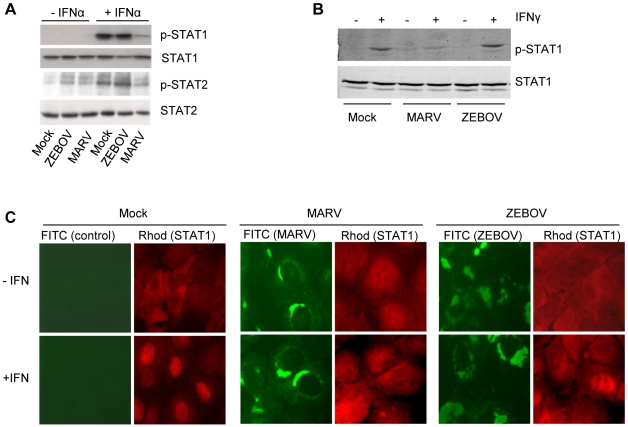
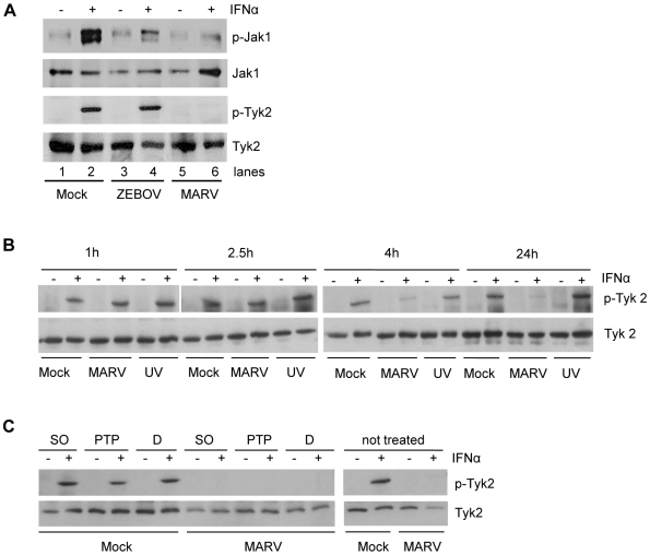
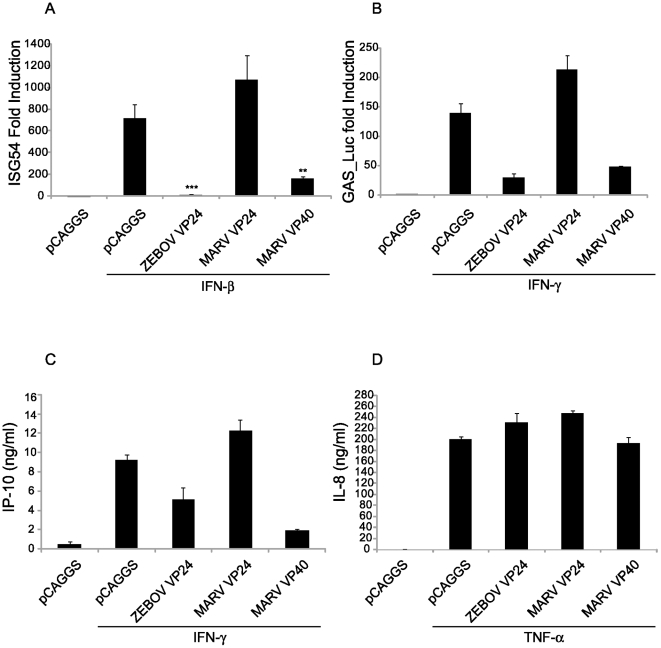
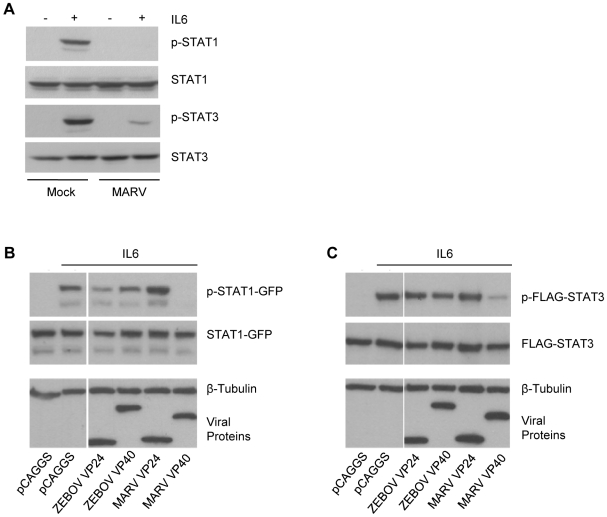
Previous studies have demonstrated that Marburg viruses (MARV) and Ebola viruses (EBOV) inhibit interferon (IFN)-alpha/beta signaling but utilize different mechanisms. EBOV inhibits IFN signaling via its VP24 protein which blocks the nuclear accumulation of tyrosine phosphorylated STAT1. In contrast, MARV infection inhibits IFNalpha/beta induced tyrosine phosphorylation of STAT1 and STAT2. MARV infection is now demonstrated to inhibit not only IFNalpha/beta but also IFNgamma-induced STAT phosphorylation and to inhibit the IFNalpha/beta and IFNgamma-induced tyrosine phosphorylation of upstream Janus (Jak) family kinases. Surprisingly, the MARV matrix protein VP40, not the MARV VP24 protein, has been identified to antagonize Jak and STAT tyrosine phosphorylation, to inhibit IFNalpha/beta or IFNgamma-induced gene expression and to inhibit the induction of an antiviral state by IFNalpha/beta. Global loss of STAT and Jak tyrosine phosphorylation in response to both IFNalpha/beta and IFNgamma is reminiscent of the phenotype seen in Jak1-null cells. Consistent with this model, MARV infection and MARV VP40 expression also inhibit the Jak1-dependent, IL-6-induced tyrosine phosphorylation of STAT1 and STAT3. Finally, expression of MARV VP40 is able to prevent the tyrosine phosphorylation of Jak1, STAT1, STAT2 or STAT3 which occurs following over-expression of the Jak1 kinase. In contrast, MARV VP40 does not detectably inhibit the tyrosine phosphorylation of STAT2 or Tyk2 when Tyk2 is over-expressed. Mutation of the VP40 late domain, essential for efficient VP40 budding, has no detectable impact on inhibition of IFN signaling. This study shows that MARV inhibits IFN signaling by a mechanism different from that employed by the related EBOV. It identifies a novel function for the MARV VP40 protein and suggests that MARV may globally inhibit Jak1-dependent cytokine signaling.


Conflict of interest statement

The authors have declared that no competing interests exist.

Figures

Figure 1
Figure 1. MARV infection prevents IFN-mediated phosphorylation and nuclear translocation of STAT proteins.
Huh-7 cells were infected with MARV or ZEBOV at an MOI of 5 or mock-infected (Mock). At 24 h p.i., cells were either treated with 100 IU/ml of IFNα-2b (A) or 10 IU/ml of IFNγ (B) for 30 min where indicated. Cell lysates were analyzed by western blotting using antibodies directed against total and phosphorylated forms of STAT1 (A and B) or STAT2 (A). (C) Huh-7 cells were infected with MARV or ZEBOV at an MOI of 5 or left uninfected. At 24 h p.i., the cells were stimulated with 1000 IU/ml of IFNα-2b for 45 min, fixed with 4% paraformaldehyde, and stained with antibody directed against STAT1, MARV, or ZEBOV as indicated.
Figure 2
Figure 2. IFNα-induced tyrosine phosphorylation of Janus kinases is inhibited in MARV- but not in EBOV-infected cells.
This inhibition occurs early in infection and is insensitive to PTP inhibitors. (A) Huh-7 cells were infected with MARV or ZEBOV at an MOI of 5 or left uninfected. At 24 h p.i., cells were treated with 1000 IU/ml of IFNα for 20 min where indicated. Cell lysates were analyzed by western blotting using antibodies directed against total protein and phosphorylated forms of Jak1 and Tyk2. (B) Huh-7 cells were infected with MARV or UV-inactivated MARV at an MOI of 5 or left uninfected. 20 min before lysis (24 h p.i.), cells were treated with 2000 IU/ml of IFNα and harvested at the indicated time points. Cell lysates were analyzed by western blotting using antibodies directed against total protein and phosphorylated forms of Tyk2. (C) Huh-7 cells were infected with MARV at an MOI of 5. The cells were treated with DMSO (D) or the phosphatase inhibitors sodium orthovanadate (SO) or PTP1B inhibitor (PTP) prior to IFN treatment (24 h p.i., 2000 IU/ml IFNα for 20 min). Cell lysates were analyzed by western blotting using antibodies directed against total protein and phosphorylated forms of Tyk2. Note that the cuts in the films excised samples irrelevant to this study.
Figure 3
Figure 3. MARV VP40 acts as an IFN antagonist.
(A) Vero cells were transfected with 1 µg empty vector (pCAGGS) or the indicated expression plasmids. 24 h post transfection (p.t.) cells were treated with 1000 IU/ml of IFNβ for 24 h and infected with NDV GFP. At 16 h p.i., green fluorescence (indicating viral replication) was visualized and photographed with a fluorescence microscope. (B) Huh-7 cells were transfected with 2 µg of the indicated plasmids expressing the indicated viral proteins from rabies virus, ZEBOV and MARV. At 24 h p.t., the cells were stimulated with 2000 IU/ml of IFNα-2b for 45 min, fixed with 4% paraformaldehyde, and stained with anti-STAT2 antibody and antibodies to detect viral proteins.
Figure 4
Figure 4. MARV VP40 inhibits IFN-induced STAT and Jak phosphorylation.
(A) STAT1 or STAT2 (1 µg) fused to a C-terminal GFP was co-expressed in Huh-7 cells with 2 µg of the indicated expression plasmids, treated with 1000 IU/ml of universal IFNα/β for 30 min. Cells were lysed and assayed by western blot for tyrosine phosphorylated STAT1 (p-STAT1-GFP), STAT2 (p-STAT2-GFP), as well as for total expression levels of over-expressed proteins. (B) Huh-7 cells were transfected with 2 µg of the indicated expression plasmids and 1 µg of a plasmid expressing STAT1 fused to GFP at the C-terminus (STAT1-GFP). 24h p.t., cells were treated with 1000 IU/ml of IFNγ for 30 min and lysed. Lysates were analyzed by western blot for phosphorylation of STAT1 and total levels of STAT1 as well as for expression of the tagged proteins. (C and D) 293T cells were transfected with 2 µg of the indicated expression plasmids, treated with 1000 IU/ml of IFNα/β (C) or IFNγ (D) for 30 min, lysed and subjected to western blot analysis for detection of phosphorylated and total Jak1 (C and D), Tyk2 (C) or Jak2 (D). Note that the cuts in the films excised samples irrelevant to this study. All the presented data for a given protein is from the same gel and the same exposure.
Figure 5
Figure 5. MARV VP40 inhibits ISRE- and GAS-induced gene expression.
(A) MARV VP40 inhibits type I IFN-induced gene expression. 293T cells were co-transfected with a construct expressing the CAT gene driven by an ISG54 promoter along with a constitutively expressed Renilla luciferase gene and 1 µg of plasmids that express the indicated viral proteins and 24h.p.t. treated with 1000 IU/ml IFNα/β for 18 h and assayed for CAT and luciferase activities. The IFN-induced CAT activity was normalized to Renilla luciferase activity. Presented is the mean fold induction of 3 experiments compared to the untreated negative control, error bars represent the standard deviations and the asterisks the p-values (**p-val = 0.016; ***p = 0.0006). Lysates were analyzed for viral protein expression (data not shown). (B) MARV VP40 inhibits IFNγ dependent gene expression. Huh-7 cells were co-transfected with the IFNγ inducible firefly luciferase reporter plasmid pGAS-Luc along with a constitutively expressed Renilla luciferase gene and 0.6 µg of the indicated expression plasmids. Cells were treated with a 1000 IU/ml of IFNγ for 18 h and assayed for dual luciferase activities. The IFN-induced firefly luciferase activity was normalized to Renilla luciferase activity. The bars represent the mean fold induction of 3 experiments compared to the untreated negative control, and the error bars represent the standard deviations. (C) MARV VP40 inhibits the IFNγ dependent IP-10 production. 2×10⁵ HUVECs were transfected with 2 µg of the indicated expression plasmids. Cells were treated with 100 IU/ml IFNγ for 24 h; and supernatants were collected, cleared by centrifugation and analyzed by ELISA for IP-10. (D) MARV VP40 does not inhibit TNFα-induced IL-8 production. HUVECs were transfected as in (C) and treated with 50 ng/ml of TNFα for 24 h. Supernatants were collected, cleared by centrifugation and analyzed by ELISA for IL-8 concentrations.
Figure 6
Figure 6. MARV inhibits IL-6 signaling.
(A) Huh-7 cells were infected with MARV at an MOI of 5 or left uninfected. At 24 h p.i., cells were treated with 50 ng/ml IL-6 for 30 min where indicated. Cell lysates were analyzed by western blotting using antibodies directed against total protein and phosphorylated forms of STAT1 and STAT3. (B and C) Huh-7 cells were co-transfected with 2 µg of the indicated viral protein expression plasmids and either 1 µg of plasmids encoding STAT1-GFP (B) or FLAG-STAT3 (C). The samples were analyzed as described in (A). Note that for panels B and C, the cuts in the films excised samples irrelevant to this study. All the presented data for a given protein is from the same gel and the same exposure.
Figure 7
Figure 7. Jak1 phosphorylation is inhibited by MARV VP40.
(A and C) 1 µg of HA-tagged human Jak1 (A) or Tyk2 (C) expression plasmid was transfected into Huh-7 cells along with 2 µg of empty vector (pCAGGS) or the indicated viral protein expression plasmids and 0.5 µg plasmid encoding STAT2 fused to GFP. Cells were lysed and subjected to western blot analysis using total and phospho-specific antibodies against Jak1 (A), Tyk2 (C), and STAT2 or GFP. Anti-β-tubulin was used as a loading control and anti-Flag to detect expression of viral proteins. (B) 1 µg of human Jak1 plasmid was transfected along with 2 µg of empty vector (pCAGGS) or the indicated viral protein expression plasmids. Cells were lysed, subjected to SDS-PAGE and analyzed with total and phospho-specific antibodies against Jak1, STAT1 and STAT3. Anti-β-tubulin was used as a loading control and anti-Flag to detect expression of viral proteins. (D) Two-fold dilutions of HA-Tyk2 expression plasmid starting at 1 µg were transfected in Huh-7 cells with either 2 µg of empty plasmid (pCAGGS) or 2 µg of expression plasmid for MARV VP40. Levels of phospho-Tyk2 and phospho-STAT1 as well as total Tyk2 and STAT1 were assayed by western blot. Anti-β-tubulin and anti-Flag antibodies were used as indicated. Note that for panels A and B, the cuts in the films excised samples irrelevant to this study. All the presented data for a given protein is from the same gel and the same exposure.
Figure 8
Figure 8. MARV VP40 inhibition of IFN signaling does not require an intact late domain.
(A) M40-AAAA buds less efficiently than wild type MARV VP40. A MARV VP40 late domain mutant (M40-AAAA) was made using site directed mutagenesis. M40-AAAA, wild type MARV VP40, ZEBOV VP40 or GFP (2 µg) was expressed in 293T cells. 48 h later, supernatants were harvested and virus-like particles (VLPs) were purified through a sucrose cushion. Cells were lysed and examined together with the VLPs for protein expression levels (WCE: whole cell extract). (B) MARV VP40 does not require the late domain to inhibit the phosphorylation of STAT1. Huh-7 cells were transfected with 1 µg STAT1-GFP expression plasmid along with 2 µg plasmid DNA encoding Flag-tagged versions of viral proteins from LGTV, ZEBOV and MARV or the late domain mutant M40-AAAA and treated with 1000 IU/ml of IFNα/β for 30 min. Lysates were analyzed for phosphorylation of STAT1 and for total expression levels of all over-expressed proteins. β-tubulin expression was assessed as indicated. All the presented data for a given protein is from the same gel and the same exposure. (C) MARV VP40 does not need the late domain to inhibit the IFNα/β-dependent induction of ISG54-Luc. 293T cells were co-transfected with a construct expressing the luciferase gene under control of an ISG54 promoter along with a constitutively expressed Renilla luciferase reporter gene and 1 µg empty vector (pCAGGS) or expression plasmids expressing wild-type MARV VP40 or M40-AAAA. Cells were treated with 1000 IU/ml IFNα/β for 18 h and assayed for firefly and Renilla luciferase activities. IFN-induced firefly luciferase activity was normalized to Renilla luciferase activity. The bars represent the mean fold induction of 3 experiments compared to the untreated negative control and error bars represent the standard deviations.

Similar articles

Cited by

References

    1. Bray M, Geisbert TW. Ebola virus: the role of macrophages and dendritic cells in the pathogenesis of Ebola hemorrhagic fever. Int J Biochem Cell Biol. 2005;37:1560–1566. - PubMed
    1. Tough DF. Type I interferon as a link between innate and adaptive immunity through dendritic cell stimulation. Leuk Lymphoma. 2004;45:257–264. - PubMed
    1. Biron CA, Cousens LP, Ruzek MC, Su HC, Salazar-Mather TP. Early cytokine responses to viral infections and their roles in shaping endogenous cellular immunity. Adv Exp Med Biol. 1998;452:143–149. - PubMed
    1. Platanias LC. Mechanisms of type-I- and type-II-interferon-mediated signalling. Nat Rev Immunol. 2005;5:375–386. - PubMed
    1. Muller M, Briscoe J, Laxton C, Guschin D, Ziemiecki A, et al. The protein tyrosine kinase JAK1 complements defects in interferon-alpha/beta and -gamma signal transduction. Nature. 1993;366:129–135. - PubMed

Publication types

MeSH terms