. 2010 Mar 1;588(Pt 5):831-46.
doi: 10.1113/jphysiol.2009.180570. Epub 2010 Jan 18.

Glutamate-mediated astrocyte-to-neuron signalling in the rat dorsal horn

Affiliations

Glutamate-mediated astrocyte-to-neuron signalling in the rat dorsal horn

Rita Bardoni et al. J Physiol. .

Abstract

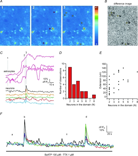
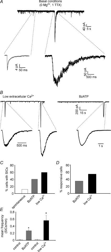
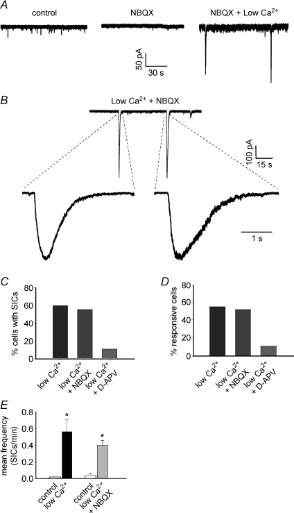
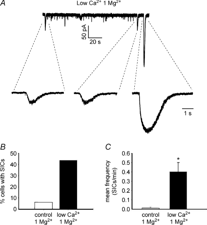
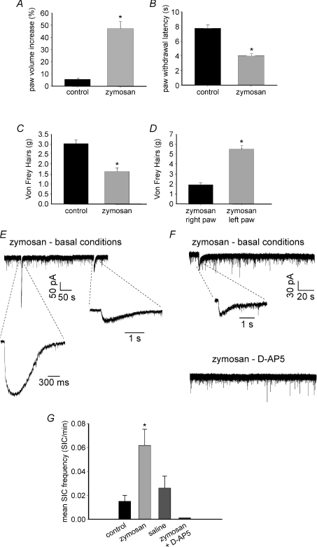
By releasing neuroactive agents, including proinflammatory cytokines, prostaglandins and neurotrophins, microglia and astrocytes are proposed to be involved in nociceptive transmission, especially in conditions of persistent, pathological pain. The specific action on dorsal horn neurons of agents released from astrocytes, such as glutamate, has been, however, poorly investigated. By using patch-clamp and confocal microscope calcium imaging techniques in rat spinal cord slices, we monitored the activity of dorsal horn lamina II neurons following astrocyte activation. Results obtained revealed that stimuli that triggered Ca(2+) elevations in astrocytes, such as the purinergic receptor agonist BzATP and low extracellular Ca(2+), induce in lamina II neurons slow inward currents (SICs). Similarly to SICs triggered by astrocytic glutamate in neurons from other central nervous system regions, these currents (i) are insensitive to tetrodotoxin (TTX), (ii) are blocked by the NMDA receptor (NMDAR) antagonist d-AP5, (iii) lack an AMPA component, and (iv) have slow rise and decay times. Ca(2+) imaging also revealed that astrocytic glutamate evokes NMDAR-mediated episodes of synchronous activity in groups of substantia gelatinosa neurons. Importantly, in a model of peripheral inflammation, the development of thermal hyperalgesia and mechanical allodynia was accompanied by a significant increase of spontaneous SICs in dorsal horn neurons. The NMDAR-mediated astrocyte-to-neuron signalling thus represents a novel pathway that may contribute to the control of central sensitization in pathological pain.


Figures

Figure 4
Figure 4. Ca2+ elevation in astrocytes triggers domain Ca2+ responses in lamina II neurons of spinal cord slices
A, sequence of dorsal horn pseudocolour images after incubation with Oregon Green BAPTA 1-AM illustrating the Ca2+ signal at rest (1) and during BzATP application (2 and 3) in constant presence of TTX. The BzATP-induced Ca2+ increase in astrocytes (2, red arrowheads) is followed by a domain response in five neurons (3, yellow arrowheads). B, difference image obtained by subtracting the image of the neuronal domain Ca2+ response (3) from the image captured before the domain response (2). C, time course of the BzATP-mediated Ca2+ signal change in three astrocytes (labelled in panel A2) and the following highly synchronous Ca2+ elevation in five neurons belonging to the domain response. Numbers 1, 2 and 3 refer to the corresponding panels in A. D, histogram showing the number of neurons composing a domain response following stimulation with BzATP, PGE2 or low Ca2+ (n= 14). E, maximal spatial extension of domains in relation to the number of neurons in the domain. F, time course of the BzATP-mediated Ca2+ signal change in five neurons: BzATP evokes two successive, similar domain responses from the same neurons (b and d).
Figure 1
Figure 1. Slow inward currents (SICs) recorded from lamina II neurons in basal conditions and after astrocyte stimulation
A, representative patch-clamp recordings showing several miniature EPSCs (mEPSCs), mediated by both AMPA and NMDA receptors, and a single slow inward current. Insets: expanded mEPSC (left) and SIC (right). The SIC exhibits slower kinetics and larger amplitude than the miniature synaptic current. The decay phase of the mEPSC is interpolated by a double exponential function (red line), while the SIC is fitted by a single exponential. B, recordings of SICs from two different lamina II neurons, in ‘low extracellular Ca2+’ (left) and in the presence of the purinergic receptor agonist benzoylbenzoyl-ATP (BzATP, 100 μm, right). C, percentage of cells exhibiting at least one SIC in the different conditions, during 5 min recording. D, percentage of responsive cells, i.e. cells where the number of SICs during the stimulus is higher than in control. E, the application of BzATP or low extracellular Ca2+ causes a significant increase of SIC mean frequency in the responsive neurons (Wilcoxon's signed rank test, P < 0.01 for both groups; low Ca2+, n= 11; BzATP, n= 13).
Figure 2
Figure 2. SICs are mediated by NMDA receptors, while AMPA receptors are not involved
A, effects of NBQX application (10 μm) on synaptic activity (mEPSCs) and appearance of SICs in a lamina II neuron. Control conditions are represented by zero extracellular Mg2+ and 1 μm TTX. Application of NBQX blocks the AMPA mediated mEPSCs, while two SICs are still apparent in low extracellular Ca2++ NBQX. B, examples of SICs recorded in low Ca2+ and NBQX from another lamina II neuron. Again, blocking non-NMDA receptors does not prevent the appearance of SICs. C and D, application of NBQX in low Ca2+ does not alter the percentage of cells with SICs or the percentage of responsive neurons, while addition of 100 μm d-AP5 inhibits almost completely the appearance of SICs (only 1 SIC observed in 9 recorded neurons). E, a significant increase of SIC frequency during low Ca2+ can be observed also in the presence of NBQX (paired t test, P < 0.01 and n= 14). Frequencies of SICs in low Ca2+ and low Ca2++ NBQX are not significantly different (Mann–Whitney U test, P > 0.05).
Figure 3
Figure 3. SICs can be observed also in the presence of extracellular magnesium
A, recording of SICs from a lamina II neuron, during low extracellular Ca2+ and in the presence of 1 mm Mg2+. Three SICs can be observed in these conditions. B and C, lowering Ca2+ concentration in 1 mm Mg2+ still causes an increase in the percentage of cells exhibiting at least one SIC in 5 min recording (B) and a significant enhancement of SIC frequency (C, Wilcoxon's signed rank test, P < 0.01, n= 13).
Figure 5
Figure 5. Effects of intraplantar injection of zymosan on nociceptive behaviour and induction of SICs
AD, behavioural tests. A, zymosan (30 μl, 35–40 mg ml−1) causes a significant increase of paw volume (oedema), compared with control animals injected with the same volume of saline (Mann–Whitney U test, P < 0.05; mean baseline paw volume ranged from 281 to 301 μl in both groups). B, zymosan induces thermal hyperalgesia evaluated as a significant decrease of paw withdrawal latency to a thermal stimulus during the plantar test (Mann–Whitney U test, P < 0.01 for both sets of data). C, mechanical allodynia assessed using von Frey filaments on the right paw of animals receiving zymosan or saline (Mann–Whitney U test, P < 0.01). Zymosan causes a significant decrease of paw withdrawal threshold. D, mechanical allodynia tested by performing the von Frey test on the right (injected with zymosan) and left paw (non treated) of each animal. The injected paw exhibits a significant decrease of withdrawal threshold compared to the untreated one (Mann–Whitney U test, P < 0.01). Data are expressed as means ±s.e.m. of 5–6 rats for each group. EG, patch-clamp recordings of SICs in lamina II neurons, obtained from non-treated rats (control) and animals injected with zymosan or saline. Recordings were performed at −60 mV in basal conditions (i.e. zero extracellular Mg2+ and 1 μm TTX). E, representative traces of SICs recorded from an inflamed animal, in basal conditions. F, traces recorded in a different lamina II neuron, from a treated animal, showing the presence of a spontaneous SIC in control, abolished by the subsequent application of 100 μm d-AP5. G, mean frequency of SICs determined from all cells obtained from the 3 animal groups. Zymosan causes a significant increase of SIC frequency compared to control and saline (P < 0.001 and P < 0.05, respectively), while no significant difference is observed between control and saline (Kruskal–Wallis test followed by Dunn's method for multiple comparisons). The application of 100 μm d-APV during recording from treated animals completely abolishes SICs (control: n= 50; zymosan: n= 47; saline: n= 46, zymosan +d-APV: n= 10).

Similar articles

Cited by

References

    1. Ahmed Z, Lewis CA, Faber DS. Glutamate stimulates release of Ca2+ from internal stores in astroglia. Brain Research. 1990;516:165–169. - PubMed
    1. Aimar P, Pasti L, Carmignoto G, Merighi A. Nitric oxide-producing islet cells modulate the release of sensory neuropeptides in the rat substantia gelatinosa. J Neurosci. 1998;18:10375–10388. - PMC - PubMed
    1. Angulo MC, Kozlov AS, Charpak S, Audinat E. Glutamate released from glial cells synchronizes neuronal activity in the hippocampus. J Neurosci. 2004;24:6920–6927. - PMC - PubMed
    1. Araque A, Sanzgiri RP, Parpura V, Haydon PG. Calcium elevation in astrocytes causes an NMDA receptor-dependent increase in the frequency of miniature synaptic currents in cultured hippocampal neurons. J Neurosci. 1998;18:6822–6829. - PMC - PubMed
    1. Bezzi P, Carmignoto G, Pasti L, Vesce S, Rossi D, Rizzini BL, Pozzan T, Volterra A. Prostaglandins stimulate calcium-dependent glutamate release in astrocytes. Nature. 1998;391:281–285. - PubMed

Publication types

LinkOut - more resources