. 2010 Jan 14;463(7278):232-6.
doi: 10.1038/nature08673.

Long-term potentiation depends on release of D-serine from astrocytes

Affiliations

Long-term potentiation depends on release of D-serine from astrocytes

Christian Henneberger et al. Nature. .

Abstract

Long-term potentiation (LTP) of synaptic transmission provides an experimental model for studying mechanisms of memory. The classical form of LTP relies on N-methyl-D-aspartate receptors (NMDARs), and it has been shown that astroglia can regulate their activation through Ca(2+)-dependent release of the NMDAR co-agonist D-serine. Release of D-serine from glia enables LTP in cultures and explains a correlation between glial coverage of synapses and LTP in the supraoptic nucleus. However, increases in Ca(2+) concentration in astroglia can also release other signalling molecules, most prominently glutamate, ATP and tumour necrosis factor-alpha, whereas neurons themselves can synthesize and supply D-serine. Furthermore, loading an astrocyte with exogenous Ca(2+) buffers does not suppress LTP in hippocampal area CA1 (refs 14-16), and the physiological relevance of experiments in cultures or strong exogenous stimuli applied to astrocytes has been questioned. The involvement of glia in LTP induction therefore remains controversial. Here we show that clamping internal Ca(2+) in individual CA1 astrocytes blocks LTP induction at nearby excitatory synapses by decreasing the occupancy of the NMDAR co-agonist sites. This LTP blockade can be reversed by exogenous D-serine or glycine, whereas depletion of D-serine or disruption of exocytosis in an individual astrocyte blocks local LTP. We therefore demonstrate that Ca(2+)-dependent release of D-serine from an astrocyte controls NMDAR-dependent plasticity in many thousands of excitatory synapses nearby.


Figures
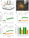
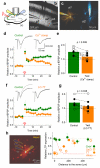
Figure 1
Figure 1. Clamping astrocytic Ca2+ blocks LTP at nearby synapses in a D-serine-dependent manner
a, Experimental arrangement: ip, intracellular patch-pipette; ep, extracellular pipette; green line, Schaffer collaterals. b, A typical patched astrocyte (Alexa Fluor 594, ~120 μm stack fusion); ep and ip as in a; bv, blood vessel; escape of Alexa to neighbouring cells can be seen; inset: DIC at lower magnification. c, LTP of AMPAR fEPSPs in control conditions, with (n = 23, green) or without (n = 29, grey) astrocyte patched. Arrow, HFS onset; traces, characteristic AMPAR fEPSPs before (black) and after (grey) LTP induction; notations apply in c-e. d, Clamping astrocytic Ca2+ (0.2 mM OGB-1, 0.45 mM EGTA, 0.14 mM CaCl2) abolishes local LTP (n = 19, orange) whereas 10 μM D-serine rescues it (n = 10, green). e, LTP in 10 μM D-serine (163 ± 12%, n = 8, green; no astrocyte patched) is no different from that in control (Fig. 1c) suggesting saturation of either the NMDAR co-agonist site or the downstream induction mechanism. 50 μM APV completely blocks LTP (n = 12, orange). f, Summary for experiments in c-e, as indicated. Bars, mean ± s.e.m for fEPSPs measured 25-30 min post-HFS relative to baseline. ***, p < 0.005 (one-population t-test); +++, p < 0.002 (two-population t-test).
Figure 2
Figure 2. Activation of the NMDAR co-agonist site is astrocyte- and use-dependent
a, NMDAR fEPSP slope (Fig. 1a arrangement) monitored during the transition from cell-attached to whole-cell mode, as indicated. Grey circles, control (n = 6; Methods); orange, Ca2+ clamp (n = 7 throughout, n = 13 before D-serine application); green, Ca2+ clamp with 10 μM D-serine in bath (n = 7). Segments, averaging epochs; traces, examples (also Supplementary Fig. 4). b, Summary of experiments shown in a. Bars, mean ± s.e.m. (applies throughout); numerals, epochs in a. ***, p < 0.0001 (one-population t-test); +++, p < 0.005 (two-population t-tests). c, The effect of D-serine on NMDAR EPSCs before and after LTP induction. Green circles, the amplitude of AMPAR EPSCs (n = 6; left panel; Vm = −70 mV) and, subsequently, NMDAR EPSCs in the same cell post-LTP (right panel; 10 μM NBQX, Vm = −10 mV); grey segment, D-serine application. Open circles (n = 5; top axis, right panel), NMDAR EPSC amplitudes in no-LTP conditions. Traces: upper-left, characteristic AMPAR EPSCs in control (black) and post-LTP (grey); lower-right, NMDAR EPSCs before (black) and after (grey) application of D-serine, in control and post-LTP, as indicated. d, 10 pulses at 50 Hz transiently potentiates synaptic NMDAR responses in a glycine- and glia- dependent manner. Traces: examples of single-stimulus EPSCs including a prominent NMDAR-mediated component (Vm = +40 mV; no AMPAR blockade was used to ensure pharmacological continuity with LTP induction protocols) in baseline conditions (left; grey, individual traces; black, average), 20 seconds (middle), and 2 min after the stimulus train (right), in control (upper row) and in 0.1 mM glycine (lower row). Bar graph: average ratio (post-train EPSC potentiation in glycine) / (post-train EPSC potentiation in control) for test experiments illustrated by traces (green; average change of the NMDAR-mediated response expressed as the area under the EPSC curve over the 100-300 ms post-peak interval: −23 ± 8%, p = 0.032, n = 6), and also for control experiments (orange) including AMPAR EPSCs (amplitude change 5 ± 9%; p = 0.41, n = 4) and NMDAR EPSCs in FAC (15 ± 14%; p = 0.32, n = 6), as indicated (examples in Supplementary Fig. 6); circles, individual experiments.
Figure 3
Figure 3. LTP expression depends on the occupancy of the NMDAR co-agonist sites controlled by D-serine synthesis in a nearby astrocyte
a, LTP of AMPAR fEPSPs in control conditions (n = 10; green circles) is abolished by either 50 μM APV (open circles, n = 12) or 750 nM DCKA (n = 9; orange; dose-response curve in Supplementary Fig. 8). Traces, characteristic responses in control (black) and following LTP induction (grey). b, Incubation with 5 mM FAC for >50 min blocks LTP (n = 16, orange circles) whereas 10 μM D-serine rescues it (n = 16; green circles). Notation is as in a. c, Summary of experiments depicted in a-b. Bars, mean ± s.e.m. (applies throughout); *, p = 0.0127; **, p = 0.0014; ***, p < 0.001 (two-population t-test). d, 400 μM intra-astrocyte HOAsp does not suppress LTP induction (potentiation is 50 ± 15%, n = 6, p = 0.021; arrangement as in Fig. 1a); traces, average fEPSPs before (black) and after (grey) application of HFS, one-cell example; time scale applies to d-e. e, Intra-astrocyte HOAsp blocks induction of LTP (fEPSP change: +12 ± 11%, n = 7, p = 0.32; orange) following depletion of D-serine using HFS in APV; arrows, HFS onset. Omitting HOAsp robustly induces LTP (52 ± 11%, n = 6, p = 0.0052; green). HOAsp was unlikely to affect glutamate metabolism because no rundown of glutamatergic responses was observed. f, Summary of experiments shown in d-e. * (left), p = 0.021; **, p = 0.0052 (one-population t-test); * (right), p = 0.024 (two-population t-test).
Figure 4
Figure 4. Individual astrocytes influence LTP induction mainly at nearby synapses
a, Experimental arrangement; notations as in Fig. 1a. b-c, An experiment seen in DIC (b) and in Alexa channel (c); a1 and a2, two patched astrocytes (λx2P = 800 nm, ~150 μm z-stack; false colours: sequential staining of a1 and a2 followed by subtraction of a1 image from combined a1+a2 image, Methods); st, stimulating electrode. d, Ca2+ clamp in test astrocyte suppresses local LTP, but not LTP near the neighbouring control astrocyte. Graph, a-fEPSP amplitudes (mean ± s.e.m.) recorded from the control (green) and test (orange) astrocyte (n = 9). Traces, respective characteristic a-fEPSPs (Supplementary Fig. 11) recorded before (black) and after (grey) LTP induction. Incomplete blockade of early potentiation near the test cell was likely due to delayed equilibration between the two dialysing pipettes. e, Summary of experiments in d; connected circles, recorded astrocyte pairs; black and hollow circles, experiments in which the test cell was, respectively, closer to and further away from the stimulating electrode (which might bias LTP expression); p-values, paired t-test (n = 9). f-g, Experiments similar to those shown in d-e, but with the test astrocyte loaded with the LC-TT (n = 8). Other notation is as in d-e. Slow equilibration of high molecular weight LC-TT might explain incomplete block of early potentiation near the test cell. h, Left panel, experimental arrangement (as in Fig. 1a-b): dotted cones, depiction of the extracellular pipette (ep). Graph: circles, relative potentiation (± s.e.m.) of AMPAR fEPSPs at different distances from the patched soma in control (n = 34; green), Ca2+-clamp (n = 48; open orange) and LC-TT (n = 15, filled orange) experiments. Dashed red line, the average emission intensity profile of Alexa (whole-cell loaded at 40 μM) escaping to neighbouring cells (n = 215 astrocytes imaged in eight 3-D stacks; details in Supplementary Fig. 13).

Comment in

Similar articles

Cited by

References

    1. Bliss TVP, Collingridge GL. A Synaptic Model of Memory - Long-Term Potentiation in the Hippocampus. Nature. 1993;361:31–39. - PubMed
    1. Schell MJ, Molliver ME, Snyder SH. D-serine, an endogenous synaptic modulator: localization to astrocytes and glutamate-stimulated release. Proc Natl Acad Sci U S A. 1995;92:3948–52. - PMC - PubMed
    1. Mothet JP, et al. D-serine is an endogenous ligand for the glycine site of the N-methyl-D-aspartate receptor. Proc Natl Acad Sci U S A. 2000;97:4926–31. - PMC - PubMed
    1. Panatier A, et al. Glia-derived D-serine controls NMDA receptor activity and synaptic memory. Cell. 2006;125:775–84. - PubMed
    1. Yang Y, et al. Contribution of astrocytes to hippocampal long-term potentiation through release of D-serine. Proc Natl Acad Sci U S A. 2003;100:15194–9. - PMC - PubMed

REFERENCES (METHODS)

    1. Bergles DE, Jahr CE. Glial contribution to glutamate uptake at Schaffer collateral-commissural synapses in the hippocampus. J Neurosci. 1998;18:7709–7716. - PMC - PubMed
    1. Volterra A, Meldolesi J. Astrocytes, from brain glue to communication elements: the revolution continues. Nat Rev Neurosci. 2005;6:626–40. - PubMed
    1. Xu T, Binz T, Niemann H, Neher E. Multiple kinetic components of exocytosis distinguished by neurotoxin sensitivity. Nature Neurosci. 1998;1:192–200. - PubMed
    1. Szerb JC, Issekutz B. Increase in the stimulation-induced overflow of glutamate by fluoroacetate, a selective inhibitor of the glial tricarboxylic cycle. Brain Res. 1987;410:116–20. - PubMed
    1. Rusakov DA, Fine A. Extracellular Ca2+ depletion contributes to fast activity-dependent modulation of synaptic transmission in the brain. Neuron. 2003;37:287–297. - PMC - PubMed

Publication types

MeSH terms