. 2009 Dec 24;139(7):1315-26.
doi: 10.1016/j.cell.2009.11.025.

Tumor self-seeding by circulating cancer cells

Affiliations

Tumor self-seeding by circulating cancer cells

Mi-Young Kim et al. Cell. .

Abstract

Cancer cells that leave the primary tumor can seed metastases in distant organs, and it is thought that this is a unidirectional process. Here we show that circulating tumor cells (CTCs) can also colonize their tumors of origin, in a process that we call "tumor self-seeding." Self-seeding of breast cancer, colon cancer, and melanoma tumors in mice is preferentially mediated by aggressive CTCs, including those with bone, lung, or brain-metastatic tropism. We find that the tumor-derived cytokines IL-6 and IL-8 act as CTC attractants whereas MMP1/collagenase-1 and the actin cytoskeleton component fascin-1 are mediators of CTC infiltration into mammary tumors. We show that self-seeding can accelerate tumor growth, angiogenesis, and stromal recruitment through seed-derived factors including the chemokine CXCL1. Tumor self-seeding could explain the relationships between anaplasia, tumor size, vascularity and prognosis, and local recurrence seeded by disseminated cells following ostensibly complete tumor excision.


Figures

Figure 1
Figure 1. Seeding of established tumors by CTCs
(A) A diagram of contralateral-seeding experiment. Unlabeled and GFP/luciferase-expressing breast cancer cells were injected into contralateral No. 2 mammary glands as a “recipient tumor” and a “donor tumor”, respectively. (B) BLI of recipient tumors extracted from mice bearing the indicated GFP/luciferase-expressing donor tumors. Color-range bars: photon flux. LM2: a lung metastatic derivative of MDA231. MCF7-BoM2: a bone-metastatic derivative of MCF7, CN34-BrM2: a brain-metastatic derivative of pleural effusion CN34, PyMT: cells derived from mammary tumors developed in MMTV-PyMT transgenic mice. (C) BLI of tumor-free and tumor-bearing mammary glands from mice bearing GFP/luciferase-expressing donor tumors. n=9–18. (D) Frozen sections of seeded MDA231-LM2 tumors were visualized by fluorescence microscopy. An entire tumor section and a higher-magnification image (×10) of a selected field are shown. (E) A contralateral-seeding experiment was performed with RFP- and GFP-expressing MDA231-LM2 cells. Frozen sections from RFP-labeled tumors were visualized under confocal microscopy at ×20. (F) A diagram to test mammary tumor seeding from lung metastases. GFP/luciferase-expressing MDA231-LM2 cells were injected intravenously. Once lung metastases were established, unlabeled MDA231 cells were injected into a mammary gland No. 2. (G) Left: burden of CTCs derived from lung metastases in mice described in panel F. Relative levels of CTC were plotted against the luminescent signals of recipient tumors. Right: BLI of three representative recipient tumors (i, ii and iii) identified in the graph.
Figure 2
Figure 2. Preferential tumor seeding by metastatic cell progenies
(A) Comparison of seeding activity of highly and poorly metastatic cells. Contralateral-seeding experiments were performed with the GFP/luciferase-expressing parental cancer cell lines, or with their lung metastatic (SW620-LM1, MDA231-LM2) or bone metastatic derivatives (A375-BoM2). Relative luminescent signals are plotted. (B) A schematic diagram of isolation of seeder cell populations. For details see Experimental Procedures. (C) Comparative tumor-seeding ability of in vivo-selected seeder cells (MDA231-S1a and S1b) and parental MDA231 cells in contralateral-seeding experiments as described in Figure1A. n= 6–8. (D) Comparative tumor-seeding ability between parental and in vivo-selected seeder cells (MDA231-S1 and A375-S1) from the circulation as described in Figure 3A. n= 8–10. (E) The gene expression profiles of parental MDA231, various metastatic derivatives and the seeder lines S1a and S1b were scored with gene expression classifiers for metastasis to lung (LMS), bone (BoMS) or brain (BrMS). Raw scores were scaled between 0 and 100. (F) Left: Parental or MDA231 S1a cells were inoculated intravenously into mice. Lung colonization was measured by BLI and quantified. n=6–7. Right: representative histological staining (H&E) of lung sections from the experiment on the left. (G, H) Cells were inoculated into the cardiac left ventricle. Bone colonization was measured by BLI and quantified; n=12. Brains were extracted and colonization was measured by BLI; n=5–7. Error bars in all cases represent SEM and p values were based on two-tailed Mann-Whitney test; for details see Experimental Procedures.
Figure 3
Figure 3. Tumor attraction and infiltration functions
(A) Unlabeled MDA231 cells were injected into a mammary gland No. 2. When tumors became palpable, LacZ/GFP/luciferase-expressing MDA231-LM2 cells were introduced into the circulation by intracardiac injection. (B) BLI of mice with seeded and unseeded tumors. Arrow, recipient tumor. (C) Comparative tumor-seeding ability of MDA231 and MDA231-LM2 cells from the circulation. Luminescent signals from recipient tumors at the indicated time points are shown. (D) Luminescent signals of recipient tumors from mice injected with indicated cell lines were quantified 10 (MDA-231) and 5 (A375) days after injection. n=6–10. (E) A diagram summarizing two functions involved in tumor self-seeding. Error bars in all cases represent SEM and p values were based on two-tailed Mann-Whitney test.
Figure 4
Figure 4. Tumor-derived mediators of cancer cell attraction
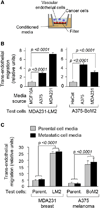
(A) Schematic diagram of the in vitro trans-endothelial migration assay. Conditioned media were placed in the bottom well. Test cells were plated in the top trans-well chamber. A confluent monolayer of human endothelial cells was present as indicated. Quantification was performed as described in the experimental procedures. (B) Trans-endothelial migration of LM2 and A375-BoM2 cells in the presence of media conditioned by the indicated cell lines. n= 60–150. (C) Trans-migration activity of tumor cells under indicated conditions was expressed as a fold difference relative to migration of parental MDA231 or A375 cells in the presence of media from non-tumorigenic cells (MCF10A conditioned media for MDA231; HaCat conditioned media for A375). n= 40–70. Error bars in all cases represent SEM and p values were based on two-tailed Mann-Whitney test.
Figure 5
Figure 5. Mediators of tumor infiltration by cancer cells
(A) Cytokine antibody arrays (Raybiotech) probed with MCF10A, MDA231, and MDA231-LM2 conditioned media. The position of IL-6 and IL-8 are indicated. For additional annotation see Figure S2. (B) Relative trans-well migration activity (without endothelial cell layer) of the indicated cell lines, with or without IL-6 or IL-8. n=89–110. (C) Relative trans-endothelial migration activity of indicated cells with or without IL-6 or IL-8. n=84–124. (D) Relative seeding actvity of MDA231-S1a cells expressing the indicated shRNA from the circulation. n=5–10. (E) Trans-endothelial migration activity of indicated cell lines. n= 30–45. (F) Relative seeding activity of MDA231-S1a cells expressing indicated shRNA from the circulation. n=5–8; error bars, maximum and minimum values. (G) Lung colonization activity of MDA231-S1a cells expressing indicated shRNAs. n=6–7. Error bars in all cases represent SEM and p values were based on two-tailed Mann-Whitney test unless indicated otherwise.
Figure 6
Figure 6. Promotion of tumor growth and stroma recruitment by self-seeding
(A) Tumor volumes of MDA231-LM2-seeded and unseeded tumors at the indicated time points. n= 13–14. (B, C) β-galactosidase staining of a typical tumor seeded with LacZ+ MDA231-LM2 cells. An entire tumor section (B) and a ×40 magnification (C) are shown. (D) CD31 staining of sections from unseeded and seeded tumors. (E) CD45 staining of sections from unseeded and seeded tumors. (F) Anti-neutrophil staining of sections from unseeded and seeded tumors. (G) CD68 staining of sections from unseeded and seeded tumors. (H) Seeding experiments from the circulation were performed with 4T1 in syngeneic mice and with A375-BoM2 cells. Recipient tumors were analyzed by FACS. The percentage of stromal cells positively stained for the indicated markers are shown. P values were calculated based on two-tailed student’s t-test. (I) Seeding experiments from the circulation were performed as described in Figure 3A with MDA231-S1a cells expressing indicated shRNAs. CD45 staining was performed on recipient tumors 19 days after the injection. (J) Quantification of CD45 positive cells in tumors seeded by MDA231-S1a control and CXCL1 knockdown cells. n= 9. Error bars in all cases represent SEM and p values were based on two-tailed Mann-Whitney test unless indicated otherwise.

Comment in

Similar articles

Cited by

References

    1. Acosta JC, O'Loghlen A, Banito A, Guijarro MV, Augert A, Raguz S, Fumagalli M, Da Costa M, Brown C, Popov N, et al. Chemokine signaling via the CXCR2 receptor reinforces senescence. Cell. 2008;133:1006–1018. - PubMed
    1. Adams JC. Roles of fascin in cell adhesion and motility. Curr Opin Cell Biol. 2004;16:590–596. - PubMed
    1. Arihiro K, Oda H, Kaneko M, Inai K. Cytokines facilitate chemotactic motility of breast carcinoma cells. Breast Cancer. 2000;7:221–230. - PubMed
    1. Aslakson CJ, Miller FR. Selective events in the metastatic process defined by analysis of the sequential dissemination of subpopulations of a mouse mammary tumor. Cancer Res. 1992;52:1399–1405. - PubMed
    1. Balasubramanian S, Eckert RL. Keratinocyte proliferation, differentiation, and apoptosis--differential mechanisms of regulation by curcumin, EGCG and apigenin. Toxicol Appl Pharmacol. 2007;224:214–219. - PMC - PubMed

Publication types

Substances

Associated data