. 2010 Mar 12;285(11):8196-206.
doi: 10.1074/jbc.M109.025684. Epub 2010 Jan 8.

SPARC suppresses apoptosis of idiopathic pulmonary fibrosis fibroblasts through constitutive activation of beta-catenin

Affiliations

SPARC suppresses apoptosis of idiopathic pulmonary fibrosis fibroblasts through constitutive activation of beta-catenin

Wenteh Chang et al. J Biol Chem. .

Abstract

Idiopathic pulmonary fibrosis (IPF) is a poorly understood progressive disease characterized by the accumulation of scar tissue in the lung interstitium. A hallmark of the disease is areas of injury to type II alveolar epithelial cells with attendant accumulation of fibroblasts in areas called fibroblastic foci. In an effort to better characterize the lung fibroblast phenotype in IPF patients, we isolated fibroblasts from patients with IPF and looked for activation of signaling proteins, which could help explain the exaggerated fibrogenic response in IPF. We found that IPF fibroblasts constitutively expressed increased basal levels of SPARC, plasminogen activator inhibitor-1 (PAI-1), and active beta-catenin compared with control cells. Control of basal PAI-1 expression in IPF fibroblasts was regulated by SPARC-mediated activation of Akt, leading to inhibition of glycogen synthase kinase-3beta and activation of beta-catenin. Additionally, IPF fibroblasts (but not control fibroblasts) were resistant to plasminogen-induced apoptosis and were sensitized to plasminogen-mediated apoptosis by inhibition of SPARC or beta-catenin. These findings uncover a newly discovered regulatory pathway in IPF fibroblasts that is characterized by elevated SPARC, giving rise to activated beta-catenin, which regulates expression of downstream genes, such as PAI-1, and confers an apoptosis-resistant phenotype. Disruption of this pathway may represent a novel therapeutic target in IPF.


Figures

FIGURE 1.
FIGURE 1.
α-SMA and SPARC are highly expressed in IPF fibroblasts. A, Western blot analysis of lysate from control (n = 4) and IPF (n = 4) fibroblast cultures showing expression of α-SMA and SPARC, with α-tubulin as a loading control. B, quantitation of α-SMA and SPARC in control (n = 4) and IPF (n = 4) fibroblasts.
FIGURE 2.
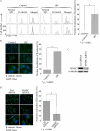
FIGURE 2.
Increased active β-catenin in IPF fibroblasts and its regulation by SPARC. A, quantitation of nuclear, i.e. active, β-catenin in control and IPF fibroblast cultures by FACS analysis with anti-β-catenin antibody. Results are shown from two control and two IPF samples. Statistical analysis of IPF (n = 4) and control (n = 4) fibroblast cultures was performed using Student's t test and is presented as the mean ± S.D. in the right panel. FITC, fluorescein isothiocyanate. B, quantitation of nuclear localization of active β-catenin in control and IPF fibroblasts by immunostaining with β-catenin antibody and 4′,6-diamidino-2-phenylindole (DAPI) for nuclear staining. Representative control cells show peripheral cytoplasmic localization of β-catenin, whereas β-catenin is expressed in the nuclei of IPF cells. Results are shown in higher magnification (lower panels). The percentages of nuclear localized β-catenin in control (n = 4) and IPF (n = 4) fibroblast cultures are presented as the mean ± S.D. in the right panel. Statistical analysis was performed using Student's t test. C and D, SPARC regulates nuclear localization of β-catenin. Following lentiviral transduction of control (Ctrl; scrambled) or SPARC shRNA, IPF fibroblasts were harvested for Western blot analysis with anti-SPARC antibody (C). The nuclear localization of active β-catenin in control or SPARC shRNA of four IPF samples was assessed as described for B. Representative results from two samples are shown in the left panel of D. In the right panel, the percentage of nuclear localized β-catenin in control shRNA (n = 4) versus SPARC shRNA in IPF fibroblasts is presented as the mean ± S.D. Statistical analysis was performed using Student's t test.
FIGURE 3.
FIGURE 3.
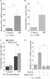
Elevated basal PAI-1 expression in IPF fibroblasts does not require the TGF-β1 receptor/ALK5. A, PAI-1 mRNA expression was measured by real-time qPCR in control and IPF cells after serum starvation. B, secreted PAI-1 was determined by enzyme-linked immunosorbent assay (average of triplicates) in serum-starved control or IPF cells. Data represent the mean ± S.D. (n = 4, control and IPF samples). Statistical analysis was performed using Student's t test. C, human lung fibroblasts from control (n = 4) and IPF (n = 4) cultures were incubated overnight with the TGF-β1 receptor/ALK5 inhibitor SB431542 at 10 μm. Secreted PAI-1 was determined by enzyme-linked immunosorbent assay as described for B. Data represent the mean ± S.D. Statistical analysis was performed using Student's t test. D, SB431542 suppressed the induction of PAI-1 mRNA expression by exogenous TGF-β1 as measured by real-time qPCR. Human control fibroblasts (n = 3) were preincubated with SB431542 at 10 μm for 1 h before the addition of TGF-β1 overnight at 2 ng/ml. The PAI-1 mRNA levels were measured by real-time qPCR. Results are shown as the mean ± S.D. with statistical analysis by Student's t test.
FIGURE 4.
FIGURE 4.
SPARC and β-catenin regulate expression of PAI-1. Down-regulation of SPARC or β-catenin suppressed PAI-1 in IPF fibroblasts. A, PAI-1 mRNA expression was assessed by real-time qPCR in IPF fibroblasts following transduction of control (Ctrl; scrambled), SPARC, or β-catenin (β-cat) shRNA. Data are from duplicate samples (mean ± S.D., n = 4). B, secreted PAI-1 in the culture medium from IPF fibroblasts was determined by enzyme-linked immunosorbent assay (done in triplicate), and data are the mean ± S.D. (n = 4). Statistical analysis was performed using Student's t test. C, Western blot analysis shows down-regulation of endogenous SPARC (upper panel) and β-catenin (lower panel) proteins in IPF cells following transduction of the respective shRNA constructs.
FIGURE 5.
FIGURE 5.
IPF fibroblasts are resistant to plasminogen-induced apoptosis. A, cell survival determined by the alamarBlue assay following overnight incubation with various concentrations of plasminogen is shown. Data are from experiments repeated twice with control (n = 4) and IPF (n = 4) fibroblast cultures and are shown as the mean ± S.D. Statistical analysis was performed using a one-way analysis of variance. B, Western blot analysis using an antibody specific for cleaved caspase-3 shows that plasminogen caused a concentration-dependent increase in cleaved caspase-3 in control cells. Results are representative of three independent experiments from control cell lines (n = 4). In the lower panel, a significant increase in caspase-3 activity is shown in control fibroblasts (n = 4) treated with various concentrations of plasminogen for 24 h but not in IPF fibroblasts (n = 4). Data are presented as the mean ± S.D. from triplicates for each sample. Statistical analysis was performed using a one-way analysis of variance. C, SPARC and β-catenin regulate plasminogen-induced apoptosis. Endogenous SPARC or β-catenin in IPF cells was attenuated by transduction of SPARC or β-catenin (β-Cat) shRNA. Left panel, following the addition of plasminogen, an alamarBlue assay revealed a significant decrease in cell viability in SPARC or β-catenin shRNA cells but not in control (Ctrl) shRNA cells. Right panel, under the same treatment conditions, a significant increase in caspase-3 activity was observed following attenuation of SPARC or β-catenin. Data are the mean ± S.D. (n = 4). Statistical analysis was performed using Student's t test.
FIGURE 6.
FIGURE 6.
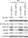
SPARC regulates β-catenin activity thorough Akt. Akt and GSK-3β activity was reduced in SPARC shRNA IPF fibroblasts with a coincident decrease in expression of PAI-1 and α-SMA. IPF fibroblast cultures (n = 4) were transduced with control (Ctrl) or SPARC shRNA, followed by Western blot analysis for the indicated proteins. Representative results from three independent experiments are shown.
FIGURE 7.
FIGURE 7.
Reduced nuclear β-catenin and PAI-1 expression in the presence of PI3K/Akt inhibitors. A, Akt and GSK-3β activity was reduced in IPF fibroblasts by PI3K inhibitor LY294002 (10 μm) or wortmannin (1 μm) treatment for 24 h, in concert with a decrease in α-SMA expression. α-Tubulin was used as a loading control. Representative results of Western blot analysis from four IPF samples are shown with specific antibodies against the indicated proteins. DMSO, dimethyl sulfoxide. B, shown is the decrease in nuclear β-catenin localization in IPF fibroblasts treated with PI3K inhibitors. A decrease in nuclear β-catenin in inhibitor-treated IPF fibroblasts was assessed by FACS analysis as described in the legend to Fig. 2. Representative results are shown in the upper panel, and the results from statistical analysis of four IPF samples performed using Student's t test shown are shown in the lower panel. FITC, fluorescein isothiocyanate. C, suppression of basal PAI-1 mRNA expression in IPF fibroblasts by PI3K inhibitors was demonstrated by real-time qPCR. Data are from duplicate four IPF samples and are represented as the mean ± S.D.
FIGURE 8.
FIGURE 8.
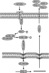
Schematic of signaling through SPARC in IPF fibroblasts. Enhanced expression of SPARC in IPF fibroblasts stimulates PI3K activity, followed by Akt activation. Akt-mediated phosphorylation of GSK-3β releases β-catenin for nuclear translocation and transactivation of downstream gene expression, e.g. PAI-1. The accumulation of secreted PAI-1 protects IPF fibroblasts from plasminogen-induced apoptosis.

Similar articles

Cited by

References

    1. Zisman D. A., Keane M. P., Belperio J. A., Strieter R. M., Lynch J. P., 3rd (2005) Methods Mol. Med. 117, 3–44 - PMC - PubMed
    1. Phan S. H. (2002) Chest 122, 286S–289S - PubMed
    1. Kapanci Y., Desmouliere A., Pache J. C., Redard M., Gabbiani G. (1995) Am. J. Respir. Crit. Care Med. 152, 2163–2169 - PubMed
    1. Cadigan K. M. (2008) Dev. Cell 14, 322–323 - PubMed
    1. Jian H., Shen X., Liu I., Semenov M., He X., Wang X. F. (2006) Genes Dev. 20, 666–674 - PMC - PubMed

Publication types

MeSH terms