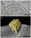
Mycobacterial outer membranes: in search of proteins
- PMID: 20060722
- PMCID: PMC2931330
- DOI: 10.1016/j.tim.2009.12.005
Mycobacterial outer membranes: in search of proteins
Abstract
The cell wall is a major virulence factor of Mycobacterium tuberculosis and contributes to its intrinsic drug resistance. Recently, cryo-electron microscopy showed that mycobacterial cell wall lipids form an unusual outer membrane. Identification of the components of the uptake and secretion machinery across this membrane will be crucial for understanding the physiology and pathogenicity of M. tuberculosis and for the development of better anti-tuberculosis drugs. Although the genome of M. tuberculosis appears to encode over 100 putative outer membrane proteins, only a few have been identified and characterized. Here, we summarize the current knowledge on the structure of the mycobacterial outer membrane and its known proteins. Through comparison to transport processes in Gram-negative bacteria, we highlight several hypothetical outer membrane proteins of M. tuberculosis that await discovery.
Figures


Similar articles
-
Identification of outer membrane proteins of Mycobacterium tuberculosis.Tuberculosis (Edinb). 2008 Nov;88(6):526-44. doi: 10.1016/j.tube.2008.02.004. Epub 2008 Apr 25. Tuberculosis (Edinb). 2008. PMID: 18439872 Free PMC article.
-
[Envelope structure and components of Mycobacterium tuberculosis].Nihon Saikingaku Zasshi. 2010 Dec;65(2-4):355-68. doi: 10.3412/jsb.65.355. Nihon Saikingaku Zasshi. 2010. PMID: 20808057 Review. Japanese. No abstract available.
-
The Mycobacterium tuberculosis MmpL11 Cell Wall Lipid Transporter Is Important for Biofilm Formation, Intracellular Growth, and Nonreplicating Persistence.Infect Immun. 2017 Jul 19;85(8):e00131-17. doi: 10.1128/IAI.00131-17. Print 2017 Aug. Infect Immun. 2017. PMID: 28507063 Free PMC article.
-
Lipid transport in Mycobacterium tuberculosis and its implications in virulence and drug development.Biochem Pharmacol. 2015 Aug 1;96(3):159-67. doi: 10.1016/j.bcp.2015.05.001. Epub 2015 May 16. Biochem Pharmacol. 2015. PMID: 25986884 Review.
-
Crystal structure of the Mycobacterium tuberculosis transcriptional regulator Rv0302.Protein Sci. 2015 Dec;24(12):1942-55. doi: 10.1002/pro.2802. Epub 2015 Sep 29. Protein Sci. 2015. PMID: 26362239 Free PMC article.
Cited by
-
Reduced drug uptake in phenotypically resistant nutrient-starved nonreplicating Mycobacterium tuberculosis.Antimicrob Agents Chemother. 2013 Apr;57(4):1648-53. doi: 10.1128/AAC.02202-12. Epub 2013 Jan 18. Antimicrob Agents Chemother. 2013. PMID: 23335744 Free PMC article.
-
Differential detergent extraction of mycobacterium marinum cell envelope proteins identifies an extensively modified threonine-rich outer membrane protein with channel activity.J Bacteriol. 2013 May;195(9):2050-9. doi: 10.1128/JB.02236-12. Epub 2013 Mar 1. J Bacteriol. 2013. PMID: 23457249 Free PMC article.
-
The effect of anti-tuberculosis drugs therapy on mRNA efflux pump gene expression of Rv1250 in Mycobacterium tuberculosis collected from tuberculosis patients.New Microbes New Infect. 2019 Oct 8;32:100609. doi: 10.1016/j.nmni.2019.100609. eCollection 2019 Nov. New Microbes New Infect. 2019. PMID: 33014381 Free PMC article.
-
Role of porins in iron uptake by Mycobacterium smegmatis.J Bacteriol. 2010 Dec;192(24):6411-7. doi: 10.1128/JB.00986-10. Epub 2010 Oct 15. J Bacteriol. 2010. PMID: 20952578 Free PMC article.
-
A half-century of research on tuberculosis: Successes and challenges.J Exp Med. 2023 Sep 4;220(9):e20230859. doi: 10.1084/jem.20230859. Epub 2023 Aug 8. J Exp Med. 2023. PMID: 37552470 Free PMC article.
References
-
- Brennan PJ, Nikaido H. The envelope of mycobacteria. Annu Rev Biochem. 1995;64:29–63. - PubMed
-
- Minnikin DE. Lipids: Complex lipids, their chemistry, biosynthesis and roles. In: Ratledge C, Stanford J, editors. The biology of the mycobacteria: Physiology, identification and classification. Academic Press; 1982. pp. 95–184.
-
- Rastogi N, et al. The mycobacteria: an introduction to nomenclature and pathogenesis. Rev Sci Tech. 2001;20:21–54. - PubMed
-
- Barry CE. Interpreting cell wall ‘virulence factors’ of Mycobacterium tuberculosis. Trends Microbiol. 2001;9:237–241. - PubMed
-
- Nikaido H, et al. Physical organization of lipids in the cell wall of Mycobacterium chelonae. Mol Microbiol. 1993;8:1025–1030. - PubMed
Publication types
MeSH terms
Substances
Grants and funding
LinkOut - more resources
Full Text Sources
Other Literature Sources

