. 2010 Mar;30(5):1217-30.
doi: 10.1128/MCB.00993-09. Epub 2009 Dec 22.

Differential roles for DNA polymerases eta, zeta, and REV1 in lesion bypass of intrastrand versus interstrand DNA cross-links

Affiliations

Differential roles for DNA polymerases eta, zeta, and REV1 in lesion bypass of intrastrand versus interstrand DNA cross-links

J Kevin Hicks et al. Mol Cell Biol. 2010 Mar.

Abstract

Translesion DNA synthesis (TLS) is a process whereby specialized DNA polymerases are recruited to bypass DNA lesions that would otherwise stall high-fidelity polymerases. We provide evidence that TLS across cisplatin intrastrand cross-links is performed by multiple translesion DNA polymerases. First, we determined that PCNA monoubiquitination by RAD18 is necessary for efficient bypass of cisplatin adducts by the TLS polymerases eta (Poleta), REV1, and zeta (Polzeta) based on the observations that depletion of these proteins individually leads to decreased cell survival, cell cycle arrest in S phase, and activation of the DNA damage response. Second, we showed that in addition to PCNA monoubiquitination by RAD18, the Fanconi anemia core complex is also important for recruitment of REV1 to stalled replication forks in cisplatin treated cells. Third, we present evidence that REV1 and Polzeta are uniquely associated with protection against cisplatin and mitomycin C-induced chromosomal aberrations, and both are necessary for the timely resolution of DNA double-strand breaks associated with repair of DNA interstrand cross-links. Together, our findings indicate that REV1 and Polzeta facilitate repair of interstrand cross-links independently of PCNA monoubiquitination and Poleta, whereas RAD18 plus Poleta, REV1, and Polzeta are all necessary for replicative bypass of cisplatin intrastrand DNA cross-links.


Figures

FIG. 1.
FIG. 1.
Model for lesion bypass of cisplatin intrastrand cross-links and validation of siRNAs used to deplete individual components of the TLS pathway. (A) RAD18 responds to stalled replication forks by monoubiquitinating PCNA at K164. This event serves as a molecular switch by increasing the affinity of Y family TLS polymerases for PCNA via their ubiquitin-binding domains. Evidence suggests that Polη performs the initial nucleotide insertion step opposite the cisplatin intrastrand cross-link while Polζ (composed of the catalytic REV3 domain and the REV7 accessory subunit) performs the extension step beyond the initial insertion. Polζ activity requires REV1, a Y family polymerase thought to promote TLS polymerase switching events. See the introduction for details. (B through D) HeLa cells were transfected with the indicated siRNAs as described in Materials and Methods. After 48 h, total cellular RNA was subjected to RT-PCR using primers specific for either REV3 or GAPDH (B) or whole-cell lysates were separated by SDS-PAGE and subjected to immunoblot analysis using the indicated primary antibodies (C and D). (D) Depletion of RAD18 prevents monoubiquitination of PCNA. Control or RAD18-depleted HeLa cells were exposed to 40 J of UV-C/m2 or 150 μM cisplatin for 2 h and then harvested 8 h later. Whole-cell lysates were subjected to immunoblot analysis with anti-PCNA antibody. (E) Multiple siRNAs were individually tested as to their ability to downregulate ectopically expressed flag-tagged REV1- or V5-tagged REV3 in 293T/17 cells. Non-si indicates a nonspecific control siRNA.
FIG. 2.
FIG. 2.
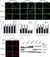
Lesion bypass of cisplatin adducts in HeLa cells requires PCNA monoubiquitination by RAD18 and the polymerase activities of Polη and the REV1/Polζ functional complex. HeLa cells transfected with the indicated siRNAs were treated with 10 μM cisplatin for 1 h. After 24 h, the cells were fixed, stained for γ-H2AX (green), and then imaged by fluorescence microscopy (A) or analyzed by flow cytometry (B and C). Dot plots showing the level of γ-H2AX staining versus DNA content are shown in panel B and corresponding histograms from the same experiment showing DNA content per event are shown in panel C. (D) RAD18-, Polη-, REV1-, REV3-, or REV7-depleted HeLa cells were treated with 10 μM cisplatin as described above. Whole-cell lysates were subjected to immunoblot analysis for CHK1 specifically phosphorylated at Ser 345 and GAPDH as a loading control. Depletion of RAD18, Polη, REV1, REV3, or REV7 leads to an enhanced DNA damage response indicative of incomplete DNA replication in cisplatin-treated cells.
FIG. 3.
FIG. 3.
RAD18 regulates Polη and REV1 localization to sites of cisplatin-induced replication stress marked by PCNA and γ-H2AX. (A and B) Cisplatin induces RAD18, Polη, and REV1 foci that extensively colocalize with PCNA and γ-H2AX. U2OS cells grown on coverslips were infected with lentivirus designed to express EGFP-tagged RAD18, Polη, or REV1. Cells were treated with 33 μM cisplatin for 2 h, fixed 6 h later, and then stained with for PCNA or γ-H2AX as described in Materials and Methods. Cells stained with anti-PCNA antibody were imaged using confocal microscopy (A), and cells stained with anti-γ-H2AX antibody were imaged using an Olympus BX-51 fluorescence microscope (B). (C) REV1 and Polη focus formation is dependent upon RAD18. U2OS cells were infected with lentivirus encoding one of two different shRNAs specific for RAD18. Cells were then infected with EGFP-Polη or EGFP-REV1 lentivirus as described above. The percentage of EGFP-positive cells exhibiting 10 or more foci was determined. The results are expressed as means ± the standard errors of the mean (SEM) from three independent experiments. (D) Expression of shRNA effectively depletes RAD18 protein in U2OS cells as determined by immunoblotting of whole-cell extracts. (E) Cisplatin induces the formation of Polη and REV1 foci that colocalize. U2OS cells were first infected with lentivirus encoding EGFP-tagged Polη, followed by lentivirus encoding Flag-tagged REV1. Cells were treated with cisplatin, fixed, and then stained with anti-Flag antibody. Cells were imaged by using confocal microscopy.
FIG. 4.
FIG. 4.
The FA core complex is necessary for cisplatin-induced REV1 focus formation. U2OS cells were transfected twice with Non-si siRNA or siRNA specific for FANCA or FANCD2 mRNA. After the second transfection, cells were infected with lentivirus encoding EGFP-RAD18, Polη, or REV1 as described in Materials and Methods. At 40 h after infection, cells were treated with 33 μM cisplatin for 2 and 6 h later the samples were fixed in paraformaldehyde plus 0.5% Triton X-100 and then stained for FANCD2 protein. Cells were imaged by using an Olympus BX-51 fluorescence microscope. (A) FANCA-deficient cells are defective in REV1 focus formation in response to cisplatin treatment. (B) The percentage of EGFP-positive cells exhibiting 10 or more foci was determined. The results represent the means ± the SEM of two independent experiments. Effective knockdown of FANCA or FANCD2 protein was assessed by analyzing FANCD2 focus formation by immunofluorescence (C) and FANCD2 monoubiquitination by immunoblotting (D). Depletion of FANCA protein abrogates both cisplatin-induced FANCD2 focus formation and MMC-induced FANCD2 monoubiquitination.
FIG. 5.
FIG. 5.
Depletion of REV1, REV3, or REV7 renders HeLa or BL2 cells hypersensitive to cisplatin-induced cytotoxicity and genomic instability. (A) HeLa cells were transfected with control (Non-si) siRNA or siRNA targeting RAD18, Polη, REV1, REV3, or REV7. Cells were seeded at known densities, treated with cisplatin for 2 h the following day, and then allowed to form colonies for approximately 12 days. The data are expressed as the average surviving fraction ± the SEM for each dose of cisplatin. (B) Wild-type BL2 cells or BL2 cells lacking expression of polymerase Iota (Polι), Polη, or REV3 (clones 332 and 504) were treated with various doses of cisplatin for 2 h. Two days later, the cells were harvested and subjected to the trypan blue exclusion assay to assess viability. The data are expressed as the average percentage of cells stained negative for trypan blue, normalized to the untreated control. At least three independent experiments were performed. Error bars represent the SEM. (C) siRNA-transfected HeLa cells were treated with 10 μM cisplatin for 1 h, and metaphase cells were assessed for chromosomal gaps and breaks 24 h later as described in Materials and Methods. The data are expressed as the average gaps and breaks per metaphase ± the SEM (n = 50). (D) Wild-type BL2 or BL2 knockout cells were treated with 30 μM cisplatin for 1 h and then analyzed for chromosomal aberrations 24 h later. The data are expressed as the average gaps and breaks per metaphase ± SEM (n = 50). P values were calculated by using the Student t test, where “**” represents P values of <0.001. (E) Representative images of chromosomal aberrations observed in Non-si or REV3-2 siRNA-transfected HeLa cells treated with 10 μM cisplatin. Red arrows indicate chromatid gaps and breaks.
FIG. 6.
FIG. 6.
REV1 and Polζ (REV3 and REV7) are necessary for repair of cisplatin-induced interstrand cross-links. (A) RAD18-, Polη, REV1-, REV3-, or REV7-depleted HeLa cells were treated with 3 μM cisplatin for 1 h and fixed 48 h later. Cells were then stained for S1981P-ATM and 53BP1 as surrogate markers of DNA DSBs. Nuclear DNA was stained with DAPI. (B) The percentage of cells exhibiting 10 or more colocalized foci containing both phospho-ATM and 53BP1 was determined. The data represent the means ± the SEM of three independent experiments where >300 cells were counted in each experiment. REV1-, REV3-, or REV7-depleted cells fail to resolve cisplatin-induced DSBs in a timely manner. (C) Inhibition of homologous recombination repair leads to phospho-ATM and 53BP1 focus retention in cisplatin-treated cells. Control or RAD51-depleted HeLa cells were treated with 3 μM cisplatin for 1 h and then fixed 24 or 48 h later. The percentage of cells exhibiting 10 or more phospho-ATM and 53BP1 colocalized foci are shown. The data represent the means ± the SEM of two independent experiments. Representative images of Non-si or Rad51 siRNA-transfected cells treated with cisplatin are shown in panel D.
FIG. 7.
FIG. 7.
The REV1/Polζ functional complex protects cells from MMC-induced cytotoxicity and genomic instability. (A) HeLa cells depleted of RAD18, Polη, REV1, REV3, or REV7 were treated with MMC for 24 h at the indicated doses and then subjected to the clonogenic survival assay. The average surviving fraction of at least three independent experiments are shown. Error bars represent the SEM. (B) Wild-type BL2 or BL2 cells deficient in polymerase Iota (Polι), Polη, or REV3 (clones 332 and 504) were treated with MMC for 48 h and then subjected to the trypan blue exclusion assay. The average percentage of cells excluding trypan blue is shown (n ≥ 3). Error bars represent the SEM. (C) Control (Non-si) or Polη-, REV1-, REV3-, REV7-, or RAD51-depleted HeLa cells were treated with 10 or 20 ng of MMC/ml for 24 h, fixed, and then analyzed for chromosomal aberrations. (D) BL2 or BL2 knockout cells were treated with 40 ng of MMC/ml for 24 h and then analyzed for chromosomal aberrations. The data are expressed as the average gaps and breaks per metaphase ± the SEM (n = 50). P values were calculated by the Student t test, where “**” represents P values of <0.001. (E) Representative images of chromosomal aberrations observed in Non-si and REV3-2 siRNA transfected HeLa cells treated with 10 ng of MMC/ml. Red arrows point to chromatid gaps and breaks.
FIG. 8.
FIG. 8.
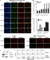
The REV1/Polζ functional complex is necessary for efficient repair of interstrand DNA cross-links induced by MMC. (A) RAD18-, Polη-, REV1-, REV3-, or REV7-depleted HeLa cells grown on coverslips were treated with 30 ng of MMC/ml for 1 h and then allowed to recover for 48 h. Cells were fixed in 100% methanol and then stained for S1981P-ATM and 53BP1 as surrogate markers of DSBs. Nuclear DNA was stained with DAPI. (B) The average percentage of cells exhibiting 10 or more foci containing both phospho-ATM and 53BP1 was determined. Error bars represent SEM (n = 3). (C) Inhibition of homologous recombination repair lead to phospho-ATM and 53BP1 focus retention in MMC-treated cells. Control or RAD51-depleted HeLa cells were treated with 30 ng of MMC/ml for 1 h and then fixed 24 or 48 h later. The average percentage of cells exhibiting 10 or more phospho-ATM and 53BP1 colocalized foci was determined. The data represent the means ± the SEM of three independent experiments. Representative images of Non-si or Rad51 siRNA transfected cells treated with MMC are shown in panel D. Effective knockdown of RAD51 protein in HeLa cells is shown in panel E. (F) siRNA-transfected HeLa cells were treated with 3 ng of MMC/ml for 48 h. Cells were fixed, stained for DNA content with propidium iodide, and then analyzed by flow cytometry. REV1-, REV3-, or REV7-depleted cells exhibited a prolonged G2 cell cycle checkpoint suggestive of a defect in interstrand DNA cross-link repair.

Similar articles

Cited by

References

    1. Acharya, N., R. E. Johnson, S. Prakash, and L. Prakash. 2006. Complex formation with Rev1 enhances the proficiency of Saccharomyces cerevisiae DNA polymerase ζ for mismatch extension and for extension opposite from DNA lesions. Mol. Cell. Biol. 26:9555-9563. - PMC - PubMed
    1. Albertella, M. R., C. M. Green, A. R. Lehmann, and M. J. O'Connor. 2005. A role for polymerase η in the cellular tolerance to cisplatin-induced damage. Cancer Res. 65:9799-9806. - PubMed
    1. Alt, A., K. Lammens, C. Chiocchini, A. Lammens, J. C. Pieck, D. Kuch, K. P. Hopfner, and T. Carell. 2007. Bypass of DNA lesions generated during anticancer treatment with cisplatin by DNA polymerase η. Science 318:967-970. - PubMed
    1. Bakkenist, C. J., and M. B. Kastan. 2003. DNA damage activates ATM through intermolecular autophosphorylation and dimer dissociation. Nature 421:499-506. - PubMed
    1. Bassett, E., N. M. King, M. F. Bryant, S. Hector, L. Pendyala, S. G. Chaney, and M. Cordeiro-Stone. 2004. The role of DNA polymerase η in translesion synthesis past Platinum-DNA adducts in human fibroblasts. Cancer Res. 64:6469-6475. - PubMed

Publication types

MeSH terms