. 2010 Jan 18;207(1):129-39.
doi: 10.1084/jem.20091837. Epub 2009 Dec 21.

VSOP/Hv1 proton channels sustain calcium entry, neutrophil migration, and superoxide production by limiting cell depolarization and acidification

Affiliations

VSOP/Hv1 proton channels sustain calcium entry, neutrophil migration, and superoxide production by limiting cell depolarization and acidification

Antoun El Chemaly et al. J Exp Med. .

Abstract

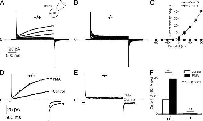
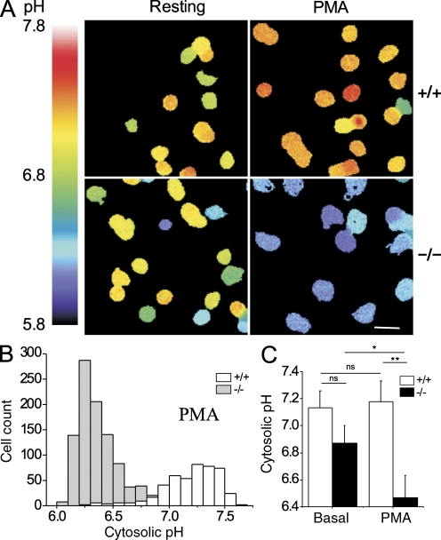
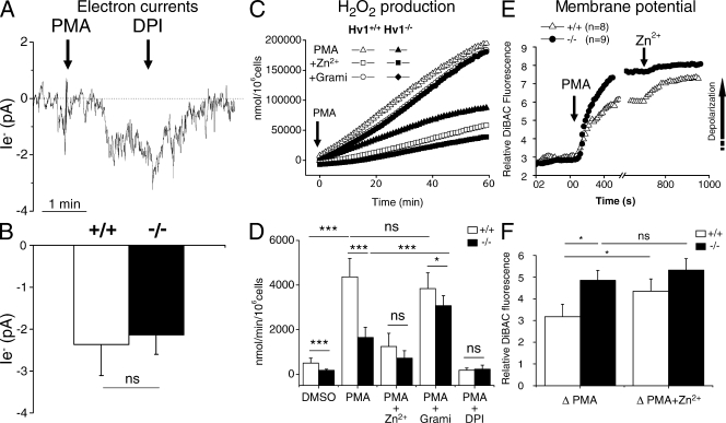
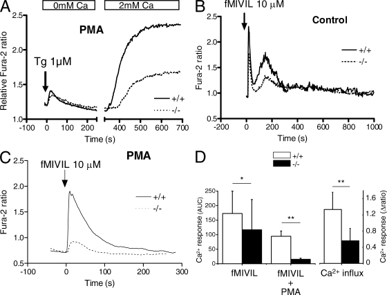
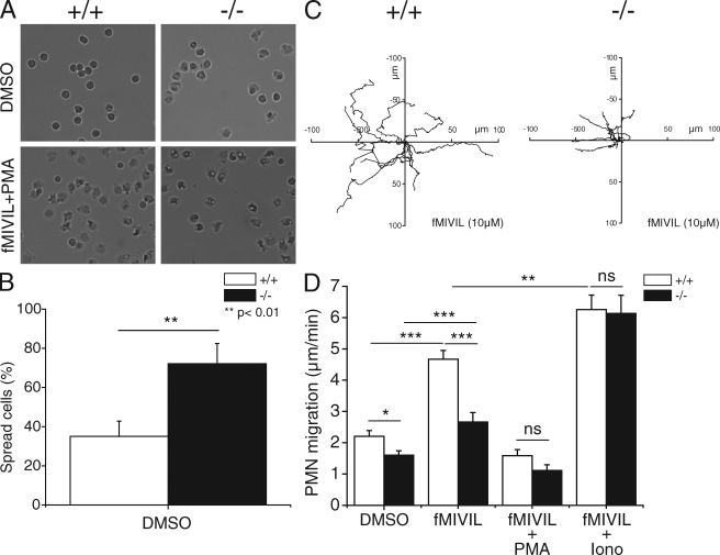
Neutrophils kill microbes with reactive oxygen species generated by the NADPH oxidase, an enzyme which moves electrons across membranes. Voltage-gated proton channels (voltage-sensing domain only protein [VSOP]/Hv1) are required for high-level superoxide production by phagocytes, but the mechanism of this effect is not established. We show that neutrophils from VSOP/Hv1-/- mice lack proton currents but have normal electron currents, indicating that these cells have a fully functional oxidase that cannot conduct protons. VSOP/Hv1-/- neutrophils had a more acidic cytosol, were more depolarized, and produced less superoxide and hydrogen peroxide than neutrophils from wild-type mice. Hydrogen peroxide production was rescued by providing an artificial conductance with gramicidin. Loss of VSOP/Hv1 also aborted calcium responses to chemoattractants, increased neutrophil spreading, and decreased neutrophil migration. The migration defect was restored by the addition of a calcium ionophore. Our findings indicate that proton channels extrude the acid and compensate the charge generated by the oxidase, thereby sustaining calcium entry signals that control the adhesion and motility of neutrophils. Loss of proton channels thus aborts superoxide production and causes a severe signaling defect in neutrophils.


Figures

Figure 1.
Figure 1.
Proton currents in resting and activated VSOP/Hv1−/− neutrophils. (A and B) Typical currents recorded in blood neutrophils from WT and VSOP/Hv1−/− mice, using the whole-cell configuration and acidic pipette solutions to activate proton channels (pHi/o = 6.1/7.2). Proton currents were elicited by 3.5-s depolarizing steps ranging from −80 to +80 mV, applied every 20 s from a holding potential of −60 mV. (C) Current–voltage relationship of time-dependent outward currents recorded in 5 WT and 18 VSOP/Hv1−/− blood neutrophils. (D and E) Effect of 100 nM PMA on the currents recorded at +60 mV in the perforated patch configuration (pHi/o = 7.0/7.0). The lack of proton currents in VSOP/Hv1−/− cells persisted after application of PMA to activate the phagocyte NADPH oxidase. Arrowheads in D show the PMA-activated current. The dashed line indicates zero current level. (F) Mean current amplitude at +60mV before and after PMA addition. Data are mean values ± SEM of 9 WT and 18 VSOP/Hv1−/− neutrophils that were tested in 27 independent experiments. ***, P < 0.0001, unpaired Student's t test. Five WT and nine VSOP/Hv1−/− mice were used for these experiments.
Figure 2.
Figure 2.
Cytosolic pH changes in VSOP/Hv1−/− neutrophils. Bone marrow neutrophils were loaded with the pH-sensitive dye BCECF and changes in fluorescence ratio (F490/F440) measured in sodium-free solutions to minimize the contribution of Na+/H+ exchange. (A) Representative ratio images of WT and VSOP/Hv1−/− neutrophils before (left) and after (right) addition of PMA. Bar, 10 µm. (B) Cytosolic pH distribution of all the PMA-treated cells. (C) Mean cytosolic pH of resting and PMA-activated neutrophils. Data are means ± SD of three experiments with >100 cells each from two WT and two VSOP/Hv1−/− mice. **, P < 0.01; *, P < 0.05, unpaired Student's t test.
Figure 3.
Figure 3.
Electron currents, H2O2 production, and membrane potential of VSOP/Hv1−/− neutrophils. (A and B) Electron current recorded at −60 mV in a WT blood neutrophil in the perforated patch configuration. Currents were evoked by 100 nM PMA and blocked by 1 µM DPI. (B) Mean amplitude of the PMA-activated electron currents. Data are means ± SEM of 6 WT and 11 VSOP/Hv1−/− neutrophils from five WT and nine VSOP/Hv1−/− mice that were tested in 17 independent experiments. ns, not significant at P < 0.05 by an unpaired Student's t test. (C) Time-dependent H2O2 production in WT and VSOP/Hv1−/− blood neutrophils activated with PMA in the absence or presence of 1 mM of the proton channel blocker Zn2+ or 40 µg/ml of the proton-permeable channel gramicidin. Data are from two representative experiments that were independently performed more than three times. (D) Mean H2O2 production from WT and VSOP/Hv1−/− neutrophils. Data are means ± SD of three to five separate experiments done in triplicate from six WT and five VSOP/Hv1−/− mice. ***, P < 0.0001; *, P < 0.05, unpaired Student's t test. (E and F) Membrane potential changes measured with DiBAC4(3) during sequential addition of 1 µM PMA and 100 µM Zn2+ to blood neutrophils. (E) Mean responses of eight WT and nine VSOP/Hv1−/− neutrophils from four independent experiments. PMA evoked a larger depolarization in VSOP/Hv1−/− cells. Arrow indicates the direction of depolarization. (F) Mean change in DiBAC4(3) fluorescence evoked by PMA and PMA+Zn2+. Data are means ± SEM of eight WT and nine VSOP/Hv1−/− blood neutrophils from four independent experiments using five WT and four VSOP/Hv1−/− mice. *, P < 0.05, unpaired Student's t test.
Figure 4.
Figure 4.
Calcium handling in VSOP/Hv1−/− neutrophils. Changes in cytosolic Ca2+ were measured with fura-2. (A) SOCE in blood neutrophils exposed to PMA for 20 min to activate the oxidase. Cells were deprived of Ca2+, treated with 1 µM thapsigargin to deplete Ca2+ stores, and exposed to 2 mM Ca2+ to reveal SOCE. Traces are means of 9 WT and 14 VSOP/Hv1−/− recordings (>10 cells each) from five WT and three VSOP/Hv1−/− mice. (B and C) Calcium elevations evoked by 10 µM fMIVIL in bone marrow neutrophils pretreated or not with PMA. Traces in B are means of 19 and 64 cells measured in five and seven independent experiments from four WT and four VSOP/Hv1−/− mice. Traces in C are means of four separate recordings (>10 cells each) from two WT and two VSOP/Hv1−/− mice. The chemotactic peptide was added at t = 0 (arrows). (D) Mean changes in cytosolic Ca2+ evoked by fMIVIL (area under the curve, AUC) and by Ca2+ readmission (Δ ratio amplitude). Data are means ± SD of the experiments in A–C. **, P < 0.001; *, P < 0.05, unpaired Student's t test.
Figure 5.
Figure 5.
Spreading and migration of VSOP/Hv1−/− neutrophils. Bone marrow neutrophils were seeded on BSA-coated Greiner 96-well plates and fMIVIL was added to promote chemokinesis. (A) Representative phase-contrast images of neutrophils before (top) and after (bottom) stimulation with fMIVIL+PMA. The combination of fMIVIL and PMA induced 100% of the cells to spread. (B) Percentage of spread cells among naive WT and VSOP/Hv1−/− neutrophils. Data are means ± SD of 107 WT and 94 VSOP/Hv1−/− neutrophils from three independent experiments. **, P < 0.01, unpaired Student's t test. (C) Migration tracings of nine WT and nine VSOP/Hv1−/− neutrophils exposed to 10 µM fMIVIL for 45 min. (D) Mean migration speed (in micrometers per minute) of neutrophils exposed to DMSO, to fMIVIL alone, to fMIVIL together with PMA, or to fMIVIL together with 100 nM of the Ca2+ ionophore ionomycin in a buffer containing 5 µM Ca2+. Note that PMA prevented both WT and VSOP/Hv1−/− neutrophils from migrating effectively upon fMIVIL stimulation, whereas ionomycin restored normal migration in VSOP/Hv1−/− neutrophils. Data are mean ± SEM of 13–33 individual tracings for each condition from three to five independent experiments. ***, P < 0.0001; **, P < 0.001; *, P < 0.05, unpaired Student's t test. Five WT and five VSOP/Hv1−/− mice were sacrificed for this experiment.

Similar articles

Cited by

References

    1. Ahluwalia J. 2008. Chloride channels activated by swell can regulate the NADPH oxidase generated membrane depolarisation in activated human neutrophils. Biochem. Biophys. Res. Commun. 365:328–333 10.1016/j.bbrc.2007.10.176 - DOI - PubMed
    1. Ahluwalia J., Tinker A., Clapp L.H., Duchen M.R., Abramov A.Y., Pope S., Nobles M., Segal A.W. 2004. The large-conductance Ca2+-activated K+ channel is essential for innate immunity. Nature. 427:853–858 10.1038/nature02356 - DOI - PMC - PubMed
    1. Babior B.M., Lambeth J.D., Nauseef W. 2002. The neutrophil NADPH oxidase. Arch. Biochem. Biophys. 397:342–344 10.1006/abbi.2001.2642 - DOI - PubMed
    1. Bánfi B., Schrenzel J., Nüsse O., Lew D.P., Ligeti E., Krause K.H., Demaurex N. 1999. A novel H+ conductance in eosinophils: unique characteristics and absence in chronic granulomatous disease. J. Exp. Med. 190:183–194 10.1084/jem.190.2.183 - DOI - PMC - PubMed
    1. Bánfi B., Maturana A., Jaconi S., Arnaudeau S., Laforge T., Sinha B., Ligeti E., Demaurex N., Krause K.H. 2000. A mammalian H+ channel generated through alternative splicing of the NADPH oxidase homolog NOH-1. Science. 287:138–142 10.1126/science.287.5450.138 - DOI - PubMed

Publication types

MeSH terms