

Review of hair follicle dermal cells
- PMID: 20022473
- PMCID: PMC2818774
- DOI: 10.1016/j.jdermsci.2009.11.005
Review of hair follicle dermal cells
Abstract
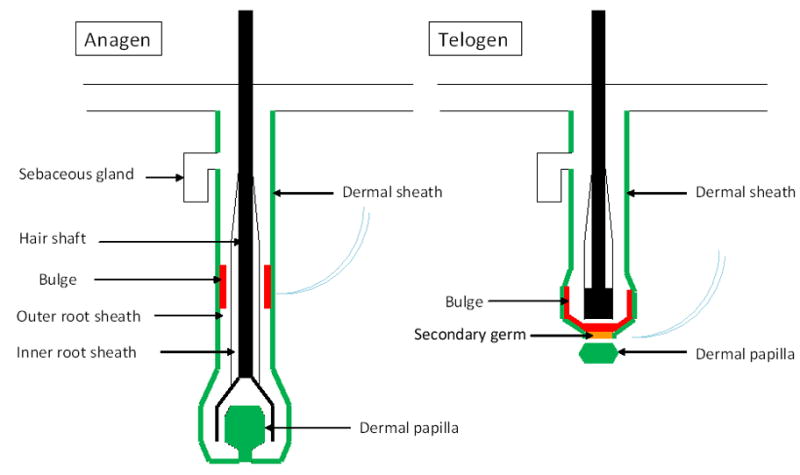
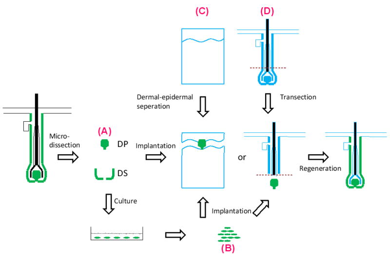
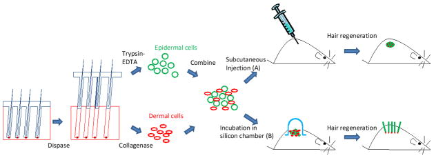
Hair follicle stem cells in the epithelial bulge are responsible for the continual regeneration of the hair follicle during cycling. The bulge cells reside in a niche composed of dermal cells. The dermal compartment of the hair follicle consists of the dermal papilla and dermal sheath. Interactions between hair follicle epithelial and dermal cells are necessary for hair follicle morphogenesis during development and in hair reconstitution assays. Dermal papilla and dermal sheath cells express specific markers and possess distinctive morphology and behavior in culture. These cells can induce hair follicle differentiation in epithelial cells and are required in hair reconstitution assays either in the form of intact tissue, dissociated freshly prepared cells or cultured cells. This review will focus on hair follicle dermal cells since most therapeutic efforts to date have concentrated on this aspect of the hair follicle, with the idea that enriching hair-inductive dermal cell populations and expanding their number by culture while maintaining their properties, will establish an efficient hair reconstitution assay that could eventually have therapeutic implications.
Copyright 2009 Japanese Society for Investigative Dermatology. Published by Elsevier Ireland Ltd. All rights reserved.
Figures
Similar articles
-
Hair follicle dermal cells differentiate into adipogenic and osteogenic lineages.Exp Dermatol. 2003 Dec;12(6):849-59. doi: 10.1111/j.0906-6705.2003.00161.x. Exp Dermatol. 2003. PMID: 14714566
-
Culture and Differentiation of Human Hair Follicle Dermal Papilla Cells in a Soft 3D Self-Assembling Peptide Scaffold.Biomolecules. 2020 Apr 28;10(5):684. doi: 10.3390/biom10050684. Biomolecules. 2020. PMID: 32354097 Free PMC article.
-
Roles for PDGF-A and sonic hedgehog in development of mesenchymal components of the hair follicle.Development. 1999 Jun;126(12):2611-21. doi: 10.1242/dev.126.12.2611. Development. 1999. PMID: 10331973
-
Strategies to enhance epithelial-mesenchymal interactions for human hair follicle bioengineering.J Dermatol Sci. 2013 May;70(2):78-87. doi: 10.1016/j.jdermsci.2013.02.004. Epub 2013 Feb 28. J Dermatol Sci. 2013. PMID: 23557720 Review.
-
Maintaining Hair Inductivity in Human Dermal Papilla Cells: A Review of Effective Methods.Skin Pharmacol Physiol. 2020;33(5):280-292. doi: 10.1159/000510152. Epub 2020 Oct 14. Skin Pharmacol Physiol. 2020. PMID: 33053562 Review.
Cited by
-
The role of hsa-miR-193a-5p as an important factor for control of inositol in alopecia areata.Skin Res Technol. 2024 Jul;30(7):e13800. doi: 10.1111/srt.13800. Skin Res Technol. 2024. PMID: 38925555 Free PMC article.
-
HaCaT‑conditioned medium supplemented with the small molecule inhibitors SB431542 and CHIR99021 and the growth factor PDGF‑AA prevents the dedifferentiation of dermal papilla cells in vitro.Mol Med Rep. 2021 May;23(5):326. doi: 10.3892/mmr.2021.11965. Epub 2021 Mar 24. Mol Med Rep. 2021. PMID: 33760132 Free PMC article.
-
Hair follicle associated pluripotent (HAP) stem cells jump from transplanted whiskers to pelage follicles and stimulate hair growth.Sci Rep. 2022 Dec 7;12(1):21174. doi: 10.1038/s41598-022-25383-z. Sci Rep. 2022. PMID: 36476963 Free PMC article.
-
Establishment and characterization of matched immortalized human frontal and occipital scalp dermal papilla cell lines from androgenetic alopecia.Sci Rep. 2023 Dec 5;13(1):21421. doi: 10.1038/s41598-023-48942-4. Sci Rep. 2023. PMID: 38049592 Free PMC article.
-
Polygonum multiflorum extract support hair growth by elongating anagen phase and abrogating the effect of androgen in cultured human dermal papilla cells.BMC Complement Med Ther. 2020 May 12;20(1):144. doi: 10.1186/s12906-020-02940-5. BMC Complement Med Ther. 2020. PMID: 32398000 Free PMC article.
References
-
- Cotsarelis G. Epithelial stem cells: a folliculocentric view. J Invest Dermatol. 2006;126:1459–68. - PubMed
-
- Millar SE. Molecular mechanisms regulating hair follicle development. J Invest Dermatol. 2002;118:216–25. - PubMed
-
- Paus R, Cotsarelis G. The biology of hair follicles. N Engl J Med. 1999;341:491–7. - PubMed
-
- Ito M, Sato Y. Dynamic ultrastructural changes of the connective tissue sheath of human hair follicles during hair cycle. Arch Dermatol Res. 1990;282:434–41. - PubMed
Publication types
MeSH terms
Substances
Grants and funding
LinkOut - more resources
Full Text Sources
Other Literature Sources
Miscellaneous
