

Global analysis of TDP-43 interacting proteins reveals strong association with RNA splicing and translation machinery
- PMID: 20020773
- PMCID: PMC2897173
- DOI: 10.1021/pr901076y
Global analysis of TDP-43 interacting proteins reveals strong association with RNA splicing and translation machinery
Abstract
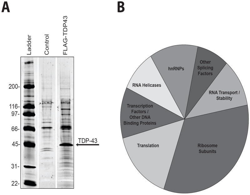
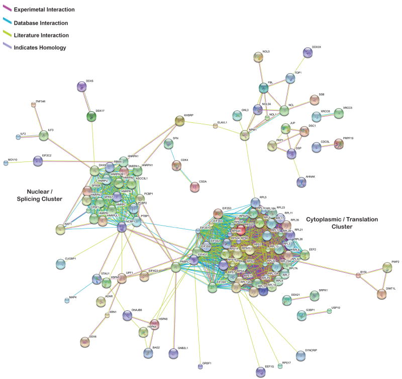
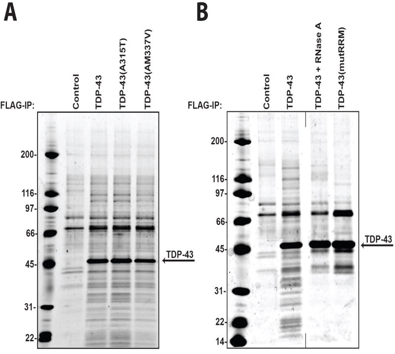
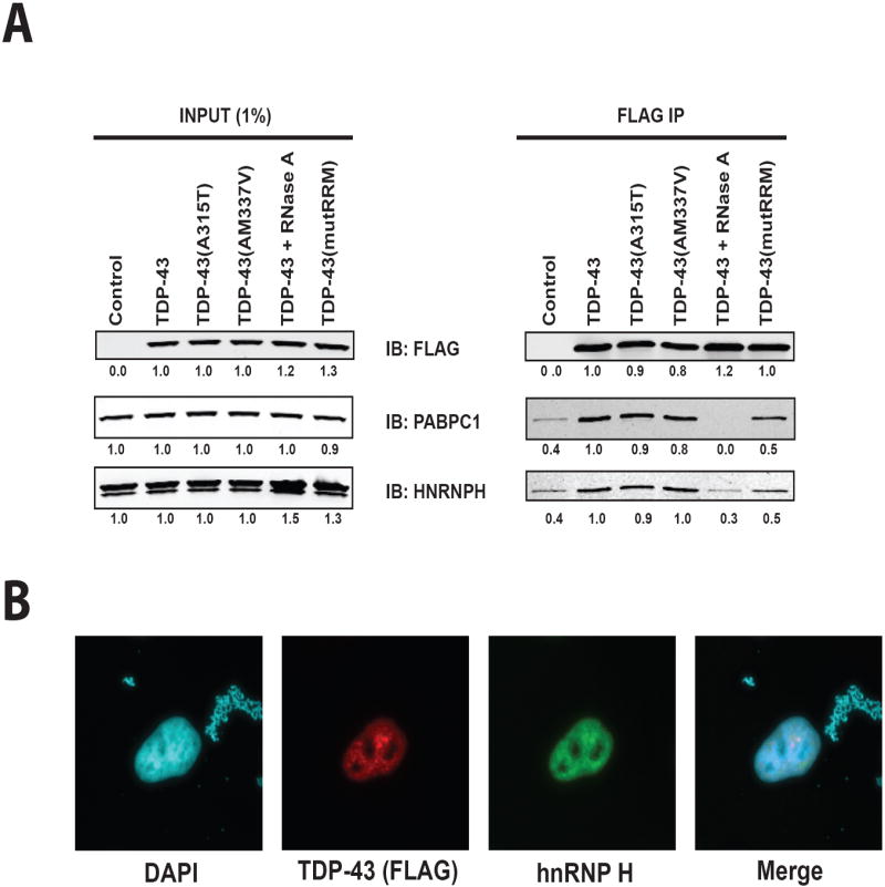
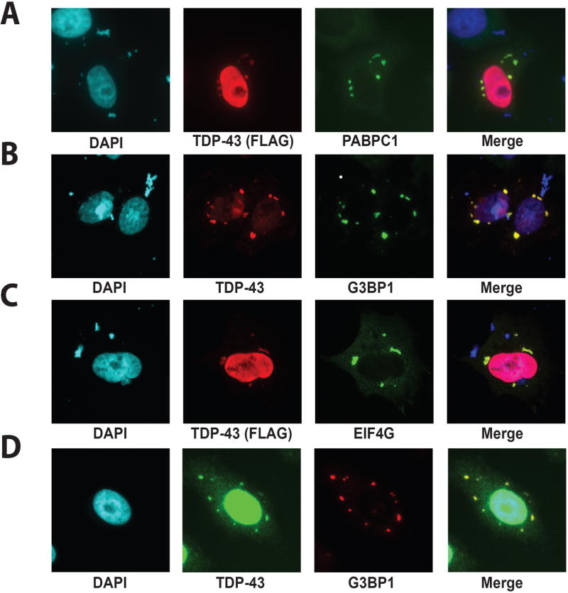
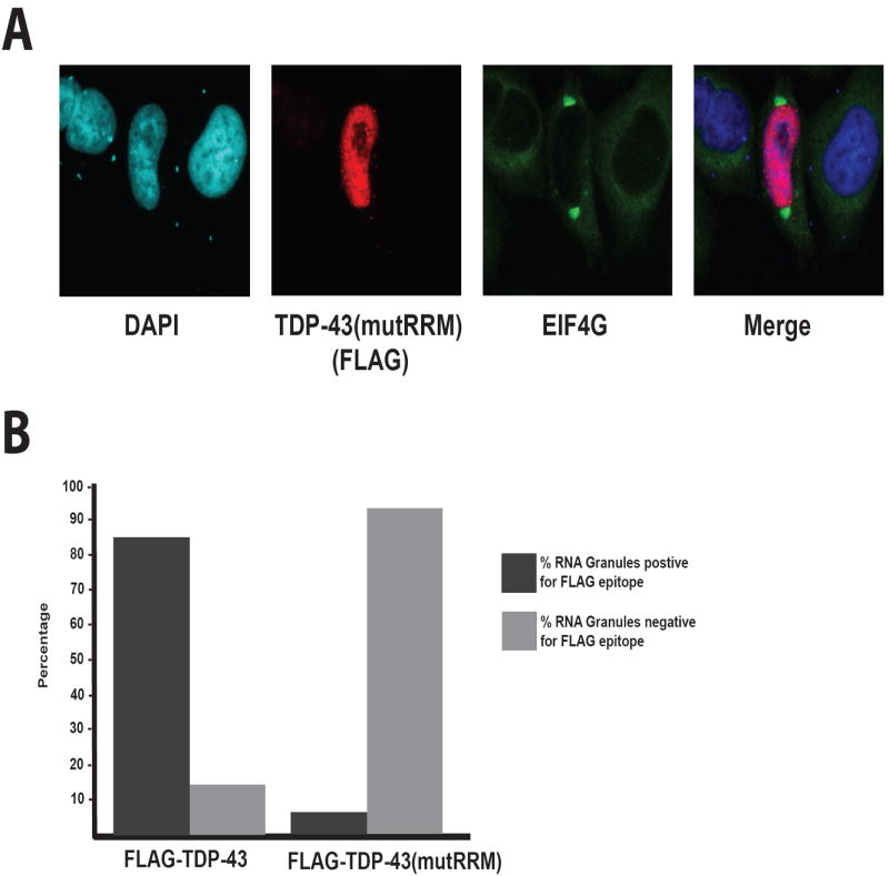
TDP-43 is a highly conserved and ubiquitously expressed member of the heterogeneous nuclear ribonucleoprotein (hnRNP) family of proteins. Recently, TDP-43 was shown to be a major disease protein in the ubiquitinated inclusions characteristic of most cases of amyotrophic lateral sclerosis (ALS), tau-negative frontotemporal lobar degeneration (FTLD), and inclusion body myopathy. In these diseases, TDP-43 is redistributed from its predominantly nuclear location to ubiquitin-positive, cytoplasmic foci. The extent to which TDP-43 drives pathophysiology is unknown, but the identification of mutations in TDP-43 in familial forms of ALS and FTLD-U suggests an important role for this protein in pathogenesis. Little is known about TDP-43 function and only a few TDP-43 interacting proteins have been previously identified, which makes further insight into both the normal and pathological functions of TDP-43 difficult. Here we show, via a global proteomic approach, that TDP-43 has extensive interaction with proteins that regulate RNA metabolism. Some interactions with TDP-43 were found to be dependent on RNA-binding, whereas other interactions are RNA-independent. Disease-causing mutations in TDP-43 (A315T and M337V) do not alter its interaction profile. TDP-43 interacting proteins largely cluster into two distinct interaction networks, a nuclear/splicing cluster and a cytoplasmic/translation cluster, strongly suggesting that TDP-43 has multiple roles in RNA metabolism and functions in both the nucleus and the cytoplasm. Finally, we found numerous TDP-43 interactors that are known components of stress granules, and indeed, we find that TDP-43 is also recruited to stress granules.
Figures
Similar articles
-
Tar DNA binding protein-43 (TDP-43) associates with stress granules: analysis of cultured cells and pathological brain tissue.PLoS One. 2010 Oct 11;5(10):e13250. doi: 10.1371/journal.pone.0013250. PLoS One. 2010. PMID: 20948999 Free PMC article.
-
Increased cytoplasmic TDP-43 reduces global protein synthesis by interacting with RACK1 on polyribosomes.Hum Mol Genet. 2017 Apr 15;26(8):1407-1418. doi: 10.1093/hmg/ddx035. Hum Mol Genet. 2017. PMID: 28158562 Free PMC article.
-
Heterogeneous Nuclear Ribonucleoprotein E2 (hnRNP E2) Is a Component of TDP-43 Aggregates Specifically in the A and C Pathological Subtypes of Frontotemporal Lobar Degeneration.Front Neurosci. 2019 Jun 4;13:551. doi: 10.3389/fnins.2019.00551. eCollection 2019. Front Neurosci. 2019. PMID: 31213972 Free PMC article.
-
Mechanisms Associated with TDP-43 Neurotoxicity in ALS/FTLD.Adv Neurobiol. 2018;20:239-263. doi: 10.1007/978-3-319-89689-2_9. Adv Neurobiol. 2018. PMID: 29916022 Review.
-
TDP-43: a DNA and RNA binding protein with roles in neurodegenerative diseases.Int J Biochem Cell Biol. 2010 Oct;42(10):1606-9. doi: 10.1016/j.biocel.2010.06.016. Epub 2010 Jun 25. Int J Biochem Cell Biol. 2010. PMID: 20601083 Review.
Cited by
-
Stress granules inhibit fatty acid oxidation by modulating mitochondrial permeability.Cell Rep. 2021 Jun 15;35(11):109237. doi: 10.1016/j.celrep.2021.109237. Cell Rep. 2021. PMID: 34133922 Free PMC article.
-
TDP-43 regulates β-adducin (Add2) transcript stability.RNA Biol. 2014;11(10):1280-90. doi: 10.1080/15476286.2014.996081. RNA Biol. 2014. PMID: 25602706 Free PMC article.
-
TDP-43 identified from a genome wide RNAi screen for SOD1 regulators.PLoS One. 2012;7(4):e35818. doi: 10.1371/journal.pone.0035818. Epub 2012 Apr 26. PLoS One. 2012. PMID: 22563406 Free PMC article.
-
TDP-43 and other hnRNPs regulate cryptic exon inclusion of a key ALS/FTD risk gene, UNC13A.PLoS Biol. 2023 Mar 17;21(3):e3002028. doi: 10.1371/journal.pbio.3002028. eCollection 2023 Mar. PLoS Biol. 2023. PMID: 36930682 Free PMC article.
-
Ataxin-2 intermediate-length polyglutamine expansions are associated with increased risk for ALS.Nature. 2010 Aug 26;466(7310):1069-75. doi: 10.1038/nature09320. Nature. 2010. PMID: 20740007 Free PMC article.
References
-
- Neumann M, Sampathu DM, Kwong LK, Truax AC, Micsenyi MC, Chou TT, Bruce J, Schuck T, Grossman M, Clark CM, McCluskey LF, Miller BL, Masliah E, Mackenzie IR, Feldman H, Feiden W, Kretzschmar HA, Trojanowski JQ, Lee VM. Ubiquitinated TDP-43 in frontotemporal lobar degeneration and amyotrophic lateral sclerosis. Science. 2006;314(5796):130–3. - PubMed
-
- Neumann M, Mackenzie IR, Cairns NJ, Boyer PJ, Markesbery WR, Smith CD, Taylor JP, Kretzschmar HA, Kimonis VE, Forman MS. TDP-43 in the ubiquitin pathology of frontotemporal dementia with VCP gene mutations. J Neuropathol Exp Neurol. 2007;66(2):152–7. - PubMed
-
- Sreedharan J, Blair IP, Tripathi VB, Hu X, Vance C, Rogelj B, Ackerley S, Durnall JC, Williams KL, Buratti E, Baralle F, de Belleroche J, Mitchell JD, Leigh PN, Al-Chalabi A, Miller CC, Nicholson G, Shaw CE. TDP-43 mutations in familial and sporadic amyotrophic lateral sclerosis. Science. 2008;319(5870):1668–72. - PMC - PubMed
Publication types
MeSH terms
Substances
Grants and funding
LinkOut - more resources
Full Text Sources
Other Literature Sources
Miscellaneous
