. 2010 Feb 19;285(8):5417-27.
doi: 10.1074/jbc.M109.036814. Epub 2009 Dec 14.

A novel type of E3 ligase for the Ufm1 conjugation system

Affiliations

A novel type of E3 ligase for the Ufm1 conjugation system

Kanako Tatsumi et al. J Biol Chem. .

Abstract

The ubiquitin fold modifier 1 (Ufm1) is the most recently discovered ubiquitin-like modifier whose conjugation (ufmylation) system is conserved in multicellular organisms. Ufm1 is known to covalently attach with cellular protein(s) via a specific E1-activating enzyme (Uba5) and an E2-conjugating enzyme (Ufc1), but its E3-ligating enzyme(s) as well as the target protein(s) remain unknown. Herein, we report both a novel E3 ligase for Ufm1, designated Ufl1, and an Ufm1-specific substrate ligated by Ufl1, C20orf116. Ufm1 was covalently conjugated with C20orf116. Although Ufl1 has no obvious sequence homology to any other known E3s for ubiquitin and ubiquitin-like modifiers, the C20orf116 x Ufm1 formation was greatly accelerated by Ufl1. The C20orf116 x Ufm1 conjugate was cleaved by Ufm1-specific proteases, implying the reversibility of ufmylation. The conjugation was abundant in the liver and lungs of Ufm1-transgenic mice, fractionated into membrane fraction, and impaired in Uba5 knock-out cells. Intriguingly, immunological analysis revealed localizations of Ufl1 and C20orf116 mainly to the endoplasmic reticulum. Our results provide novel insights into the Ufm1 system involved in cellular regulation of multicellular organisms.


Figures

FIGURE 1.
FIGURE 1.
C20orf116, a substrate for the Ufm1 modification system. A, sequence alignment of human C20orf116 (HsC20orf116) and its homologues of other species. The amino acid sequence of human C20orf116 was compared with those of other species by using the ClustalW program. *, indicate identical amino acids. Single and double dots indicate weakly and strongly similar amino acids, respectively. The underline indicates the PCI domain. B, tissue distribution of C20orf116. Homogenates prepared from various mouse tissues were analyzed by immunoblot with anti-C20orf116 and anti-actin antibodies. 20 μg of proteins were applied on each lane. C, C20orf116 is conjugated with Ufm1 in vivo. HEK293 cells were transfected with vectors at the indicated combinations. The cell lysates were immunoprecipitated (IP) with anti-GFP or anti-FLAG antibody, followed by immunoblot analysis with anti-GFP and anti-C20orf116 antibodies. The bands corresponding to GFP, GFP-Ufm1, endogenous C20orf116, C20orf116-FLAG, and C20orf116-FLAG·GFP-Ufm1 conjugate are indicated on the left. The arrowheads and asterisks indicate C20orf116-FLAG·GFP-Ufm1 conjugate and unknown proteins conjugated with GFP-Ufm1, respectively. D, determination of a lysine residue of C20orf116 for Ufm1 conjugation. GFP-Ufm1 was co-expressed with wild-type C20orf116 or the indicated mutants. The cell lysates were immunoprecipitated by anti-FLAG antibody and analyzed as described in C. E, in vitro reconstitution of C20orf116·Ufm1 conjugate. Purified recombinant C20orf116ΔN50 was incubated at 30 °C for 90 min with components of various combinations as indicated. The mixtures were boiled for 5 min with SDS-sample buffer containing 5% β-mercaptoethanol to stop the reaction. The samples were analyzed by immunoblot with anti-C20orf116 antibody. The bands corresponding to C20orf116ΔN50, LC3, and C20orf116ΔN50·Ufm1ΔC2 conjugate are indicated. *, degradative product of C20orf116ΔN50. The data shown in B–E are representative of three experiments with similar results.
FIGURE 2.
FIGURE 2.
Identification of Ufc1- and C20orf116-interacting protein, Ufl1. A, sequence alignment of human Ufl1 (HsUfl1) and its homologues. Alignment was performed as described in the legend to Fig. 1A. B, tissue distribution of Ufl1. Homogenates prepared as described in the legend to Fig. 1B were analyzed by immunoblot with anti-Ufl1 and anti-actin antibodies. C, schematic representation of constructs for Ufl1 (wt) and its deletion mutants (M1–M5). D, Ufl1 interacts with C20orf116. HEK293 cells were transfected with 3×FLAG-tagged Ufl1 or its mutants (M1–M5). The cell lysates were immunoprecipitated (IP) with anti-FLAG antibody, followed by immunoblot analysis with anti-FLAG and anti-C20orf116 antibodies. E, Ufl1 interacts with Ufc1 in vitro. Each purified recombinant MBP, MBP-Ufl1, and MBP-Ufl1 mutant (M1–M5) was incubated with recombinant Ufc1 at 4 °C for 3 h in TN buffer. The mixtures were pulled down with amyrose resin and then subjected to SDS-PAGE followed by Coomassie Brilliant Blue (CBB) staining or immunoblot with anti-Ufc1 antibody. The data shown in B, D, and E) are representative of three experiments with similar results.
FIGURE 3.
FIGURE 3.
Ufl1, a novel E3-like enzyme for the Ufm1 conjugation system. A, formation of C20orf116·Ufm1 conjugate is significantly enhanced by Ufl1 in vivo. HEK293 cells were transfected with vectors at the indicated combinations, and then the cell lysates were immunoprecipitated (IP) with anti-GFP or anti-FLAG antibody. The cell lysates (Crude) and immunoprecipitants (IP) were subjected to SDS-PAGE, followed by immunoblot analysis with anti-GFP and anti-C20orf116 antibodies. The bands corresponding to GFP, GFP-Ufm1, endogenous C20orf116, C20orf116-FLAG, and C20orf116-FLAG·GFP-Ufm1 conjugate are indicated. The arrowheads and asterisks indicate C20orf116-FLAG·GFP-Ufm1 conjugate and unknown proteins conjugated with GFP-Ufm1, respectively. Quantitative densitometry of immunoblotting data from three individual experiments was performed, and the ratios of C20orf116-FLAG·GFP-Ufm1 relative to GFP-Ufm1 (middle bottom graph) and C20orf116-FLAG·GFP-Ufm1 relative to FLAG-C20orf116 (right top graph) were plotted. Data are means ± S.D. of three experiments. *, p < 0.05. B, formation of C20orf116·Ufm1 conjugate is markedly reduced by Ufl1 knockdown in vivo. At 24 h after introduction of control or Ufl1 short hairpin RNA vector, HEK293 cells were transfected with vectors at the indicated combinations, and then the cell lysates were analyzed as described in A. Quantitative densitometry of immunoblotting data from three individual experiments was performed, and the ratios of C20orf116-FLAG·GFP-Ufm1 relative to GFP-Ufm1 (middle bottom graph) and C20orf116-FLAG·GFP-Ufm1 relative to FLAG-C20orf116 (right upper graph) were plotted. Data are mean ± S.D. of three experiments. *, p < 0.05. C, Ufl1 enhances the formation of C20orf116·Ufm1 conjugate in vitro. The conjugation reactions described in Fig. 1E were performed in the presence or absence of MBP-Ufl1. The bands corresponding to C20orf116ΔN50 and C20orf116ΔN50·Ufm1ΔC2 conjugates are indicated. *, degradative form of C20orf116ΔN50. D, N-terminal region of Ufl1 is sufficient for enhancing C20orf116·Ufm1 in vitro. The conjugation reactions described in C were performed in the presence of MBP-Ufl1 and its mutants. The bands corresponding to C20orf116ΔN50 and C20orf116ΔN50·Ufm1ΔC2 conjugate are indicated. *, degradative form of C20orf116ΔN50. The data shown in A–D are representative of three experiments with similar results.
FIGURE 4.
FIGURE 4.
Ufm1·C20orf116 conjugation is reversible. A, deconjugation of C20orf116·Ufm1 by UfSP1 and UfSP2. The in vitro conjugation reaction described in Fig. 3C was stopped by the addition of 5 units of apyrase (lane 2) and then incubated with purified UfSP1 (10 nm) (lanes 3 and 5), UfSP1C53S (10 nm) (lane 7), UfSP2 (10 nm) (lanes 4 and 6), and UfSP2C249S (10 nm) (lane 8) at 30 °C for 15 min. UfSP1 and UfSP2 were pretreated with 1 mm N-ethylmaleimide (NEM) (lanes 5 and 6). The in vitro conjugation reaction without ATP was used as a negative control (lane 1). The mixtures were boiled for 5 min in SDS-sample buffer containing 5% β-mercaptoethanol and then subjected to SDS-PAGE, followed by immunoblot with anti-C20orf116 antibody. B, comparison of isopeptidase activity among UfSPs. The in vitro conjugation and its stop reactions were performed as described in A. The reaction mixtures were incubated with UfSP1 (lanes 3–6) or UfSP2 (lanes 8–11) at various concentrations and then analyzed as described in A. The in vitro conjugation reactions in the presence and absence of ATP were used as a positive and negative control, respectively (lanes 1 and 2). The bands corresponding to C20orf116ΔN50 and C20orf116ΔN50·Ufm1ΔC2 are indicated. *, degradative form of C20orf116ΔN50. The data shown in A and B are representative of three experiments with similar results.
FIGURE 5.
FIGURE 5.
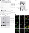
The Ufm1 system in mice. A, differences in Ufm1 conjugates among tissues. Lysates from the indicated tissues of non-tg or FLAGHis-Ufm1 tg were immunoprecipitated (IP) with anti-FLAG antibody, and the immunoprecipitants were analyzed by immunoblot with anti-Ufm1 and anti-C20orf116 antibodies. 20 μg of proteins were applied on each lane. B, subcellular fractionation of liver of FLAGHis-Ufm1 tg. Subcellular fractionation was performed as described under “Experimental Procedures.” Each fraction was subjected to SDS-PAGE and then analyzed by immunoblot with the indicated antibodies. T, total homogenate; N, nuclear fraction; C, cytosol fraction; ML, mitochondria and lysosome fraction; Mi, microsome fraction. The total amounts of proteins in each fraction prepared from 20 μg of total homogenate were applied on each lane. C, subcellular distribution of Ufm1-conjugated proteins. Each fraction prepared as described in B was immunoprecipitated with anti-FLAG antibody, and the immunoprecipitants were analyzed by immunoblot with anti-Ufm1 and anti-C20orf116 antibodies. The data shown in A–C are representative of three experiments with similar results. D, subcellular localization of Ufl1 and C20orf116. HeLa cells were transfected with C20orf116-GFP or GFP-Ufl1 and then immunostained with anti-calreticulin or anti-β-COP antibody. The right-hand panels show merged images. Bar, 10 μm.
FIGURE 6.
FIGURE 6.
Impairment of Ufm1 conjugation in Uba5 knock-out mice. A, Ufm1 conjugation in Uba5 knock-out MEFs. MEFs prepared from embryonic day 10.5 Uba5+/+ and Uba5−/− were lysed, and the lysates were analyzed by immunoblot with anti-Ufm1, anti-Uba5, and anti-actin antibodies. A knockdown of Ufm1 was not accompanied by any change of the ∼35-, ∼40-, ∼62-, and ∼90-kDa Ufm1-reactive proteins, whereas levels of free Ufm1 and the 39- and 51-kDa proteins (the latter two proteins were not recognized in Uba5 knock-out MEFs) were decreased by the knockdown (right-hand panels). Thus, we concluded that the ∼35, ∼40, ∼62, and ∼90 kDa bands indicated by asterisks are nonspecific. B, Uba5 is indispensable for C20orf116 conjugation with Ufm1. Uba5+/+ (lanes 1–3) and Uba5−/− (lanes 4–6) MEFs were stably transfected with C20orf116-FLAG (lanes 1–6) together with GFP-Ufm1 (lanes 2 and 5) or GFP-Ufm1ΔC3 (lanes 3 and 6), and then the cell lysates were immunoprecipitated with anti-GFP antibody (lanes 7–12). The lysates (Crude) and immunoprecipitants (IP) were subjected to SDS-PAGE, followed by immunoblot analysis with the indicated antibodies. The bands corresponding to GFP-Ufm1, C20orf116-FLAG, C20orf116-FLAG·GFP-Ufm1, and IgG HC are indicated. *, unknown proteins conjugated with GFP-Ufm1. The data shown in A and B are representative of three experiments with similar results. C, overall features of the Ufm1 conjugation pathway. GC, thioester linkage; G-K, isopeptide bond.

Similar articles

Cited by

References

    1. Hershko A., Ciechanover A. (1998) Annu. Rev. Biochem. 67, 425–479 - PubMed
    1. Mukhopadhyay D., Riezman H. (2007) Science 315, 201–205 - PubMed
    1. Pickart C. M., Eddins M. J. (2004) Biochim. Biophys. Acta 1695, 55–72 - PubMed
    1. Groettrup M., Pelzer C., Schmidtke G., Hofmann K. (2008) Trends Biochem. Sci. 33, 230–237 - PubMed
    1. Jin J., Li X., Gygi S. P., Harper J. W. (2007) Nature 447, 1135–1138 - PubMed

Publication types