A small-cell lung cancer genome with complex signatures of tobacco exposure
- PMID: 20016488
- PMCID: PMC2880489
- DOI: 10.1038/nature08629
A small-cell lung cancer genome with complex signatures of tobacco exposure
Abstract
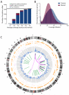
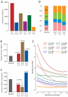
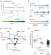
Cancer is driven by mutation. Worldwide, tobacco smoking is the principal lifestyle exposure that causes cancer, exerting carcinogenicity through >60 chemicals that bind and mutate DNA. Using massively parallel sequencing technology, we sequenced a small-cell lung cancer cell line, NCI-H209, to explore the mutational burden associated with tobacco smoking. A total of 22,910 somatic substitutions were identified, including 134 in coding exons. Multiple mutation signatures testify to the cocktail of carcinogens in tobacco smoke and their proclivities for particular bases and surrounding sequence context. Effects of transcription-coupled repair and a second, more general, expression-linked repair pathway were evident. We identified a tandem duplication that duplicates exons 3-8 of CHD7 in frame, and another two lines carrying PVT1-CHD7 fusion genes, indicating that CHD7 may be recurrently rearranged in this disease. These findings illustrate the potential for next-generation sequencing to provide unprecedented insights into mutational processes, cellular repair pathways and gene networks associated with cancer.
Figures


Similar articles
-
Depressing time: Waiting, melancholia, and the psychoanalytic practice of care.In: Kirtsoglou E, Simpson B, editors. The Time of Anthropology: Studies of Contemporary Chronopolitics. Abingdon: Routledge; 2020. Chapter 5. In: Kirtsoglou E, Simpson B, editors. The Time of Anthropology: Studies of Contemporary Chronopolitics. Abingdon: Routledge; 2020. Chapter 5. PMID: 36137063 Free Books & Documents. Review.
-
Correlation between cervical carcinogenesis and tobacco use by sexual partners.Hell J Nucl Med. 2019 Sep-Dec;22 Suppl 2:184-190. Hell J Nucl Med. 2019. PMID: 31802062
-
Cancers adapt to their mutational load by buffering protein misfolding stress.Elife. 2024 Nov 25;12:RP87301. doi: 10.7554/eLife.87301. Elife. 2024. PMID: 39585785 Free PMC article.
-
Using Experience Sampling Methodology to Capture Disclosure Opportunities for Autistic Adults.Autism Adulthood. 2023 Dec 1;5(4):389-400. doi: 10.1089/aut.2022.0090. Epub 2023 Dec 12. Autism Adulthood. 2023. PMID: 38116059 Free PMC article.
-
Slowing the Titanic: China's Epic Struggle with Tobacco.J Thorac Oncol. 2016 Dec;11(12):2053-2065. doi: 10.1016/j.jtho.2016.07.020. Epub 2016 Aug 4. J Thorac Oncol. 2016. PMID: 27498288 Review.
Cited by
-
Genetic changes in squamous cell lung cancer: a review.J Thorac Oncol. 2012 May;7(5):924-33. doi: 10.1097/JTO.0b013e31824cc334. J Thorac Oncol. 2012. PMID: 22722794 Free PMC article. Review.
-
Genomic analyses reveal mutational signatures and frequently altered genes in esophageal squamous cell carcinoma.Am J Hum Genet. 2015 Apr 2;96(4):597-611. doi: 10.1016/j.ajhg.2015.02.017. Am J Hum Genet. 2015. PMID: 25839328 Free PMC article.
-
Epigenetic regulation in the inner ear and its potential roles in development, protection, and regeneration.Front Cell Neurosci. 2015 Jan 7;8:446. doi: 10.3389/fncel.2014.00446. eCollection 2014. Front Cell Neurosci. 2015. PMID: 25750614 Free PMC article. Review.
-
The life history of 21 breast cancers.Cell. 2012 May 25;149(5):994-1007. doi: 10.1016/j.cell.2012.04.023. Epub 2012 May 17. Cell. 2012. PMID: 22608083 Free PMC article.
-
Molecular Signature of Small Cell Lung Cancer after Treatment Failure: The MCM Complex as Therapeutic Target.Cancers (Basel). 2021 Mar 10;13(6):1187. doi: 10.3390/cancers13061187. Cancers (Basel). 2021. PMID: 33801812 Free PMC article.
References
-
- Jha P. Avoidable global cancer deaths and total deaths from smoking. Nat Rev Cancer. 2009;9:655–64. - PubMed
-
- Pfeifer GP, et al. Tobacco smoke carcinogens, DNA damage and p53 mutations in smoking-associated cancers. Oncogene. 2002;21:7435–51. - PubMed
-
- DeMarini DM. Genotoxicity of tobacco smoke and tobacco smoke condensate: a review. Mutat Res. 2004;567:447–74. - PubMed
-
- Rodin SN, Rodin AS. Origins and selection of p53 mutations in lung carcinogenesis. Semin Cancer Biol. 2005;15:103–12. - PubMed
Publication types
MeSH terms
Substances
Grants and funding
LinkOut - more resources
Full Text Sources
Other Literature Sources
Medical
Molecular Biology Databases
Research Materials

