

Evidence for the prevention of enthesitis in HLA-B27/hβ(2)m transgenic rats treated with a monoclonal antibody against TNF-α
- PMID: 20015205
- PMCID: PMC3822794
- DOI: 10.1111/j.1582-4934.2009.00984.x
Evidence for the prevention of enthesitis in HLA-B27/hβ(2)m transgenic rats treated with a monoclonal antibody against TNF-α
Abstract
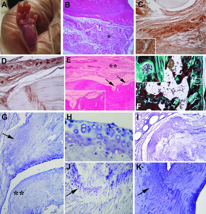
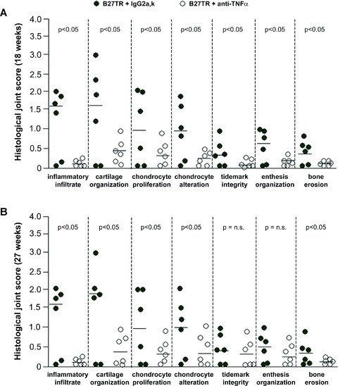
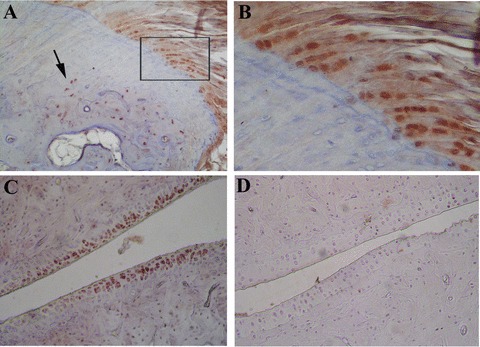
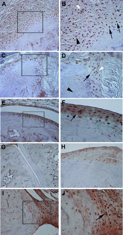
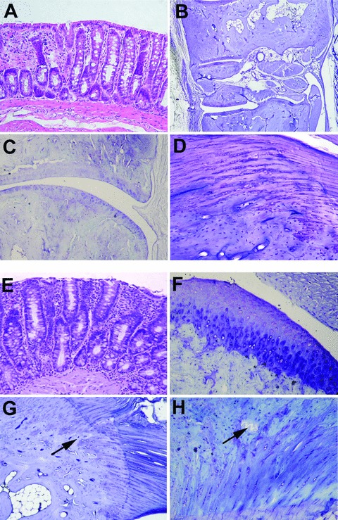
Transgenic rats with high expression of HLA-B27 and human β(2) -microglobulin (B27TR) develop a multisystem inflammatory disease resembling human inflammatory bowel disease (IBD) and spondyloarthropaties (SpA). Tumour necrosis factor α (TNF-α) has a crucial role in sustaining chronic inflammation in the gut and joints. The aim of this work was to evaluate whether TNF-α blockade could prevent or reduce the inflammation of peripheral joints in B27TR. A first group of 9-week-old B27TR received an anti-TNF-α monoclonal antibody (mAb) or an isotypic IgG2a,k up to the age of 18 weeks. An untreated group was monitored up to the age of 18 weeks and then randomly assigned to a 9-week treatment with anti-TNF-α mAb or IgG2a,k. Each rat was monitored for clinical IBD and peripheral joint manifestations. After sacrifice the colon and hind paws were examined for macroscopical and microscopical pathological changes. Early TNF-α blockade prevented, and late treatment improved IBD signs in B27TR. Erythema, oedema, inflammatory infiltrate close to the tendons and enthesis, proliferating chondrocyte-like cells, signs of new endochondral bone ossification and bone erosion were observed in peripheral joints of four out of six IgG2a,k-treated B27TR, both at 18 and 27 weeks. Immunopositivity for phosphorylated Smad1/5/8 indicated that the process of joint remodelling was activated in B27TR. Some entheses showed chondroid nodules. Anti-TNF-α treatment reduced inflammation and preserved the enthesis organization in most animals. Occasional and transient erythema and oedema were still present in three of six of the late anti-TNF-α-treated animals. Smad1/5/8 signalling was not inhibited by late anti-TNF-α treatment. In B27TR, articular involvement follows IBD onset and develops at entheses. Early TNF-α blockade prevents the onset of IBD and consequently the development of enthesitis in peripheral joints in the B27TR model of human SpA.
© 2011 The Authors Journal of Cellular and Molecular Medicine © 2011 Foundation for Cellular and Molecular Medicine/Blackwell Publishing Ltd.
Figures
Similar articles
-
TNFalpha blockade prevents the development of inflammatory bowel disease in HLA-B27 transgenic rats.J Cell Mol Med. 2009 Jan;13(1):164-76. doi: 10.1111/j.1582-4934.2008.00310.x. Epub 2008 Mar 19. J Cell Mol Med. 2009. PMID: 18363845 Free PMC article.
-
Relationship between inflammation, bone destruction, and osteoproliferation in the HLA-B27/human β2 -microglobulin-transgenic rat model of spondylarthritis.Arthritis Rheum. 2012 Oct;64(10):3210-9. doi: 10.1002/art.34600. Arthritis Rheum. 2012. PMID: 22736144 Free PMC article.
-
HLA-B27 heavy chain homodimers are expressed in HLA-B27 transgenic rodent models of spondyloarthritis and are ligands for paired Ig-like receptors.J Immunol. 2004 Aug 1;173(3):1699-710. doi: 10.4049/jimmunol.173.3.1699. J Immunol. 2004. PMID: 15265899
-
HLA-B27 transgenic rat: an animal model mimicking gut and joint involvement in human spondyloarthritides.Ann N Y Acad Sci. 2009 Sep;1173:570-4. doi: 10.1111/j.1749-6632.2009.04757.x. Ann N Y Acad Sci. 2009. PMID: 19758201 Review.
-
Advances in the treatment of uveitis in patients with spondyloarthritis - is it the time for biologic therapy?Rom J Ophthalmol. 2018 Apr-Jun;62(2):114-122. Rom J Ophthalmol. 2018. PMID: 30206554 Free PMC article. Review.
Cited by
-
Chronic Calcium Channel Inhibitor Verapamil Antagonizes TNF-α-Mediated Inflammatory Reaction and Protects Against Inflammatory Arthritis in Mice.Inflammation. 2016 Oct;39(5):1624-34. doi: 10.1007/s10753-016-0396-1. Inflammation. 2016. PMID: 27438468
-
Lessons on SpA pathogenesis from animal models.Semin Immunopathol. 2021 Apr;43(2):207-219. doi: 10.1007/s00281-020-00832-x. Epub 2021 Jan 15. Semin Immunopathol. 2021. PMID: 33449154 Review.
-
Predictors of response to TNF antagonists in patients with ankylosing spondylitis and psoriatic arthritis: systematic review and meta-analysis.RMD Open. 2015 Feb 18;1(1):e000017. doi: 10.1136/rmdopen-2014-000017. eCollection 2015. RMD Open. 2015. PMID: 26509050 Free PMC article.
-
Enthesitis: Much More Than Focal Insertion Point Inflammation.Curr Rheumatol Rep. 2018 May 30;20(7):41. doi: 10.1007/s11926-018-0751-3. Curr Rheumatol Rep. 2018. PMID: 29846815 Free PMC article. Review.
-
Inflammation-driven bone formation in a mouse model of ankylosing spondylitis: sequential not parallel processes.Arthritis Res Ther. 2016 Jan 29;18:35. doi: 10.1186/s13075-015-0805-0. Arthritis Res Ther. 2016. PMID: 26831337 Free PMC article.
References
-
- Hammer RE, Maika SD, Richardson JA, et al. Spontaneous inflammatory disease in transgenic rats expressing HLA-B27 and human beta2m: an animal model of HLA-B27-associated human disorders. Cell. 1990;63:1099–112. - PubMed
-
- Taurog JD, Maika SD, Simmons WA, et al. Susceptibility to inflammatory disease in HLA-B27 transgenic rat lines correlates with the level of B27 expression. J Immunol. 1993;150:4168–78. - PubMed
-
- Breban M. Animal models and in vitro models for the study of aetiopathogenesis of spondyloarthropathies. Baillieres Clin Rheumatol. 1998;12:611–26. - PubMed
Publication types
MeSH terms
Substances
LinkOut - more resources
Full Text Sources
Medical
Research Materials
