. 2010 Feb 12;285(7):4983-94.
doi: 10.1074/jbc.M109.037622. Epub 2009 Dec 10.

Niemann-Pick C1 functions independently of Niemann-Pick C2 in the initial stage of retrograde transport of membrane-impermeable lysosomal cargo

Affiliations

Niemann-Pick C1 functions independently of Niemann-Pick C2 in the initial stage of retrograde transport of membrane-impermeable lysosomal cargo

Stephen D B Goldman et al. J Biol Chem. .

Abstract

The rare neurodegenerative disease Niemann-Pick Type C (NPC) results from mutations in either NPC1 or NPC2, which are membrane-bound and soluble lysosomal proteins, respectively. Previous studies have shown that mutations in either protein result in biochemically indistinguishable phenotypes, most notably the hyper-accumulation of cholesterol and other cargo in lysosomes. We comparatively evaluated the kinetics of [(3)H]dextran release from lysosomes of wild type, NPC1, NPC2, and NPC1/NPC2 pseudo-double mutant cells and found significant differences between all cell types examined. Specifically, NPC1 or NPC2 mutant fibroblasts treated with NPC1 or NPC2 siRNA (to create NPC1/NPC2 pseudo-double mutants) secreted dextran less efficiently than did either NPC1 or NPC2 single mutant cell lines, suggesting that the two proteins may work independently of one another in the egress of membrane-impermeable lysosomal cargo. To investigate the basis for these differences, we examined the role of NPC1 and NPC2 in the retrograde fusion of lysosomes with late endosomes to create so-called hybrid organelles, which is believed to be the initial step in the egress of cargo from lysosomes. We show here that cells with mutated NPC1 have significantly reduced rates of late endosome/lysosome fusion relative to wild type cells, whereas cells with mutations in NPC2 have rates that are similar to those observed in wild type cells. Instead of being involved in hybrid organelle formation, we show that NPC2 is required for efficient membrane fission events from nascent hybrid organelles, which is thought to be required for the reformation of lysosomes and the release of lysosomal cargo-containing membrane vesicles. Collectively, these results suggest that NPC1 and NPC2 can function independently of one another in the egress of certain membrane-impermeable lysosomal cargo.


Figures

FIGURE 1.
FIGURE 1.
Release of lysosomal [3H]dextran polymers from cells with or without functional mutations in NPC1 and NPC2. A, wild type cells (▴) released about 80% of their initial dextran over a 24-h time period. Cells with mutations in NPC1 (NPC1−/−/NPC2+/+, GM03123) (●) had significantly compromised dextran release relative to wild type cells. Cells with functional mutations in NPC2 (NPC1+/+/NPC2−/−, GM18455) (▾) secreted dextran less efficiently than cells with mutation in NPC1. Pseudo-double mutants (designated NPC1−/−/NPC2−/−) were created from NPC2−/− (GM18455) cells that had been transfected with NPC1 siRNA (■) or NPC1−/− fibroblasts (GM03123) that had been transfected with NPC2 siRNA (♦). Both pseudo-double mutant fibroblasts exhibited less efficient dextran secretion than either NPC1 or NPC2 single mutant cell lines. B, wild type cells released about 80% of their total dextran at 24 h. Cells incubated in serum-free media (SFM) for 72 h exhibited similar cumulative dextran secretion at the 24-h time point. Similarly, mucolipidosis type IV (MLIV) and Sandhoff-diseased cells exhibited no changes in dextran secretion at 24 h. C and D, dextran secretion profiles observed for NPC1−/− or NPC2−/− fibroblasts with (○) or without (●) 10 μm U18666A in the culture medium. The addition of U18666A significantly decreased dextran secretion in cells with functional mutations in NPC1 but not in fibroblasts with mutations in NPC2. E and F, dextran secretion profiles were observed with fibroblasts harboring mutations in NPC1 or NPC2 that were (○) or were not (●) treated with 10 μg/ml progesterone in the cell culture medium. Progesterone treatment did not significantly influence dextran secretion in either cell line. Data shown are the means ± S.E. from experiments run in triplicate (*, p < 0.05 by unpaired t test).
FIGURE 2.
FIGURE 2.
NPC1, but not NPC2, is required for efficient retrograde fusion of lysosomes with late endosomes. A, the appearance of a FRET signal indicates retrograde fusion of lysosomes with late endosomes to create hybrid organelles. The appearance of FRET signal as a function of time is shown for wild type fibroblasts (black circles), NPC2−/− fibroblasts (red circles), NPC1−/− fibroblasts (green circles), and NPC2−/− fibroblasts treated with siRNA against NPC1 (blue circles). B, measured FRET in all cell types at the zero time point (see “Experimental Procedures”) is shown. The FRET signal observed with wild type cells is not significantly different from that of cells with mutations in NPC2 (both GM17910 and GM18455 cell lines). Alternatively, cells with mutations in NPC1 have significantly reduced hybrid organelle formation. The efficiency of retrograde fusion of cells with mutations in NPC2 can be decreased to levels comparable with NPC1 mutant cells by treating them with NPC1 siRNA. As a control, a scrambled version of the NPC1 siRNA has no significant impact on the rate of hybrid organelle formation in cells with mutations in NPC2. Additionally, NPC1-null Chinese hamster ovary cells (M12) exhibit similarly low fusion profiles. C, wild type MEFs exhibited normal rates of fusion, whereas MEFs heterozygous in NPC1 (NPC1+/− /NPC2+/+ and NPC1+/− /NPC2+/−) showed intermediary fusion kinetics. NPC1−/−/NPC2−/− MEFs showed fusion kinetics similar to NPC1−/−/NPC2+/+ cells. Data points and bars represent the average ± S.E. from three independent experiments (*, p < 0.05 by unpaired t test).
FIGURE 3.
FIGURE 3.
NPC1, but not NPC2, is required for amine (CQ)-induced co-localization of lysosomes with late endosomes. A, under normal cell culture conditions very few lysosomes co-localize with late endosomes, as was observed from immunofluorescence analysis of cells probed for the cation-independent (CI)-MPR and LAMP1 (late endosome- and lysosome-specific proteins, respectively). When wild type cells are exposed to high concentrations of the lysosomotropic amine (100 μm CQ for 3 h), the degree of lysosome co-localization with late endosomes was significantly enhanced. B, cells with mutations in NPC1 did not show enhanced late endosome/lysosome co-localization when exposed to CQ. C and D, cells with mutations in NPC2 (both GM17910 and GM18455 cell lines) behaved similarly to wild type cells in their response to CQ treatment. Images are representative of cells observed from at least three independent experiments. The percent co-localization is shown in each merge panel.
FIGURE 4.
FIGURE 4.
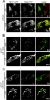
Amine-induced co-localization of NPC1 and Rab9 fluorescent fusion proteins. Fibroblasts were transfected with plasmids to express NPC1-GFP (a late endosome/lysosomal protein) and Rab9-YFP (a late endosomal protein) and, where indicated, incubated with 100 μm CQ for 3 h to observe the influence of the amine on the degree of co-localization of these two fluorescent fusion proteins in living cells. A, Rab9-YFP did not significantly co-localize with NPC1-GFP in wild type cells without amine treatment. When wild type cells were exposed to CQ, the fluorescent fusion proteins became significantly co-localized. B and C, the amine CQ enhanced Rab9-YFP/NPC1-GFP co-localization in cells with mutations in NPC2 (both GM17910 and GM18455 cell lines), as was the case with wild type cells. Images are representative of cells observed from at least three independent experiments. The percent co-localization is shown in each merge panel.
FIGURE 5.
FIGURE 5.
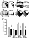
Evaluation of amine-induced vacuole size in fibroblasts with mutations in NPC1 and NPC2. Indicated fibroblasts were incubated with 70 μm NR for 6 h and visualized by phase contrast microscopy. To the right of each phase image is the computer software-generated threshold image of the amine-induced vacuoles that was used to estimate their average diameter. A, wild type cells are shown. B, cells with mutations in NPC1 are shown. C and D, cells with mutations in NPC2 (both GM17910 and GM18455 cell lines) are shown. E, bars represent the average diameter of NR-containing vacuoles in the indicated cell type. Indicated cells (nocodazole (+NOC), black bars) were treated with the microtubule depolymerizing agent nocodazole (1 μm, 1 h before and concurrent with the administration of NR). Bars represent the average ± S.E. of six different cells obtained in three independent experiments (*, p < 0.05 by unpaired t test).
FIGURE 6.
FIGURE 6.
Kinetics of amine-induced vacuole formation and size reduction. A, phase contrast images are shown of wild type and NPC2 mutant fibroblasts incubated for 3 h with 100 μm CQ to form amine-induced vacuoles. The size of the amine-induced vacuoles is greater in cells with mutations in NPC2 relative to wild type cells (images are representative of three independent experiments; the scale bar is 20 μm). B, kinetics are shown of retrograde fusion of lysosomes with late endosomes using the FRET assay in wild type and NPC2 mutant fibroblasts treated with 100 μm CQ for 30 min before data analysis. Data points represent the average ± S.D. for three independent experiments. C, wild type and NPC2 mutant fibroblasts incubated with 70 μm NR and visualized by phase contrast microscopy are shown. The indicated times represent how long the cells have been in cell culture medium devoid of NR (sink conditions). Below each phase image is the computer software-generated threshold image of the amine-induced vacuoles that were used to estimate their average diameter. Images are representative of at least 10 independent observations. The scale bar represents 10 μm. D, kinetic analysis is shown of the NR-containing vacuole diameter reduction as a function of time in NR-free medium in normal (●) and NPC2 mutant (○) fibroblasts. Data points represent the average ± S.E. for 10 independent experiments (*, p < 0.05 by unpaired t test).
FIGURE 7.
FIGURE 7.
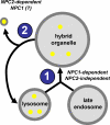
Proposed model illustrating the initial steps involved in the retrograde transport of membrane-impermeable lysosomal cargo. Lysosomes containing membrane-impermeable cargo (yellow circles) fuse with late endosomes to create hybrid organelles (labeled as step 1). Fission of membrane vesicles from hybrid organelles leads to the reformation of lysosomes and the release of impermeable cargo-containing transport vesicles (labeled as step 2). The suggested role of NPC proteins in each step is indicated.

Similar articles

Cited by

References

    1. Liscum L., Sturley S. L. (2004) Biochim. Biophys. Acta 1685, 22–27 - PubMed
    1. Carstea E. D., Morris J. A., Coleman K. G., Loftus S. K., Zhang D., Cummings C., Gu J., Rosenfeld M. A., Pavan W. J., Krizman D. B., Nagle J., Polymeropoulos M. H., Sturley S. L., Ioannou Y. A., Higgins M. E., Comly M., Cooney A., Brown A., Kaneski C. R., Blanchette-Mackie E. J., Dwyer N. K., Neufeld E. B., Chang T. Y., Liscum L., Strauss J. F., 3rd, Ohno K., Zeigler M., Carmi R., Sokol J., Markie D., O'Neill R. R., van Diggelen O. P., Elleder M., Patterson M. C., Brady R. O., Vanier M. T., Pentchev P. G., Tagle D. A. (1997) Science 277, 228–231 - PubMed
    1. Naureckiene S., Sleat D. E., Lackland H., Fensom A., Vanier M. T., Wattiaux R., Jadot M., Lobel P. (2000) Science 290, 2298–2301 - PubMed
    1. Pentchev P. G., Comly M. E., Kruth H. S., Tokoro T., Butler J., Sokol J., Filling-Katz M., Quirk J. M., Marshall D. C., Patel S. (1987) FASEB J. 1, 40–45 - PubMed
    1. Liscum L., Ruggiero R. M., Faust J. R. (1989) J. Cell Biol. 108, 1625–1636 - PMC - PubMed

Publication types

MeSH terms

LinkOut - more resources