. 2010 Feb;84(4):1771-84.
doi: 10.1128/JVI.01510-09. Epub 2009 Dec 9.

Nucleolin associates with the human cytomegalovirus DNA polymerase accessory subunit UL44 and is necessary for efficient viral replication

Affiliations

Nucleolin associates with the human cytomegalovirus DNA polymerase accessory subunit UL44 and is necessary for efficient viral replication

Blair L Strang et al. J Virol. 2010 Feb.

Abstract

In the eukaryotic cell, DNA replication entails the interaction of multiple proteins with the DNA polymerase processivity factor PCNA. As the structure of the presumptive human cytomegalovirus (HCMV) DNA polymerase processivity factor UL44 is highly homologous to that of PCNA, we hypothesized that UL44 also interacts with numerous proteins. To investigate this possibility, recombinant HCMV expressing FLAG-tagged UL44 was generated and used to immunoprecipitate UL44 and associated proteins from infected cell lysates. Unexpectedly, nucleolin, a major protein component of the nucleolus, was identified among these proteins by mass spectrometry and Western blotting. The association of nucleolin and UL44 in infected cell lysate was confirmed by reciprocal coimmunoprecipitation in the presence and absence of nuclease. Western blotting and immunofluorescence assays demonstrated that the level of nucleolin increases during infection and that nucleolin becomes distributed throughout the nucleus. Furthermore, the colocalization of nucleolin and UL44 in infected cell nuclei was observed by immunofluorescence assays. Assays of HCMV-infected cells treated with small interfering RNA (siRNA) targeting nucleolin mRNA indicated that nucleolin was required for efficient virus production, viral DNA synthesis, and the expression of a late viral protein, with a correlation between the efficacy of knockdown and the effect on virus replication. In contrast, the level of neither global protein synthesis nor the replication of an unrelated virus (reovirus) was reduced in siRNA-treated cells. Taken together, our results indicate an association of nucleolin and UL44 in HCMV-infected cells and a role for nucleolin in viral DNA synthesis.


Figures
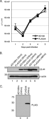
FIG. 1.
FIG. 1.
Characterization of FLAG44 virus. (A) Replication of AD169rv and FLAG44 viruses. HFF cells were infected at an MOI of 1, and virus supernatant was harvested at the indicated time points. The virus titer is represented as PFU/ml on HFF cells. Data points (open squares, AD169rv; closed squares, FLAG44) represent the mean titers for three experiments. Error bars represent the standard deviations for the titers from those experiments. (B) Western blotting of AD169rv- and FLAG44-infected cells. HFF cells were infected at an MOI of 1, and cell lysates were prepared at the indicated time points. UL44 (top) and β-actin (bottom) protein levels were assayed by Western blotting using antibodies recognizing these proteins. The positions of molecular mass markers (kDa) are indicated on the left. (C) Detection of FLAG-tagged UL44 by Western blotting. The cell lysate analyzed in lanes 3 and 4 of B was probed with an anti-FLAG antibody. The positions of molecular mass markers (kDa) are indicated on the left.
FIG. 2.
FIG. 2.
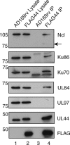
Detection of protein immunoprecipitated from FLAG44-infected cell lysates by Western blotting. Lysates from AD169rv (lane 1)-and FLAG44 (lane 2)-infected cells and protein immunoprecipitated using an anti-FLAG antibody from those lysates (lanes 3 and 4, respectively) were separated on a 10% polyacrylamide gel. Proteins in each lane were examined by Western blotting for the presence of UL44, FLAG, Ku86, Ku70, and nucleolin (Ncl) by using antibodies recognizing these proteins as indicated to the right. The positions of molecular mass markers (kDa) are indicated to the left. Arrow indicates the detection of a 77-kDa band.
FIG. 3.
FIG. 3.
Reciprocal co-IP of UL44 and nucleolin. HFF cells were mock infected or infected with AD169 (MOI of 3). Cell lysates were prepared 72 h postinfection and precleared with immunoglobulin. IP of the lysate was then carried out with monoclonal antibodies recognizing either UL44 (A) or nucleolin (Ncl) (B) or immunoglobulin of the same isotype as the monoclonal antibody used. Proteins were analyzed by Western blotting using the monoclonal antibodies indicated to the right. The positions of molecular mass markers (kDa) are indicated to the left of A to C. (A) Lanes 1 and 2, uninfected cell lysate immunoprecipitated with immunoglobulin G (IgG) and anti-UL44 antibody (Ab), respectively; lanes 3 and 4, infected cell lysate immunoprecipitated with IgG and antibody (Ab), respectively; lane 5, infected cell lysate immunoprecipitated with MAb in the presence of benzonase (Ab+B); lane 6, infected cell lysate (Lys). (B) Lanes 1 and 2, uninfected cells immunoprecipitated with IgG and anti-nucleolin antibody (Ab), respectively; lanes 4 and 5, infected cells immunoprecipitated with IgG and antibody (Ab), respectively; lane 6, infected cell lysate immunoprecipitated with MAb in the presence of benzonase (Ab+B); lanes 3 and 7, uninfected and infected cell lysate (Lys), respectively. (C) Samples from lanes 5, 6, and 7 in B probed as described above with UL44 MAb. (D) The cell lysate used in the IP in A (lanes 4 and 5) was run on an ethidium bromide-stained 0.8% agarose gel. Lane 1, no sample; lane 2, IP in the absence of benzonase (A, lane 4); lane 3, IP in the presence of benzonase (A, lane 5). The position of the dye front, which corresponds to where a 0.5-kbp DNA would migrate, is indicated with an arrow.
FIG. 4.
FIG. 4.
Analysis of nucleolin levels in infected and uninfected cells. Shown are nucleolin levels at different time points postinfection. Lysates from HFF cells uninfected (Un.) and infected (In.) with AD169 (MOI of 1) were prepared at the indicated time points and analyzed by Western blotting using antibodies recognizing nucleolin (Ncl), UL44, or β-actin, as indicated to the right. The positions of molecular mass markers (kDa) are indicated to the left.
FIG. 5.
FIG. 5.
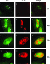
Localization of nucleolin in HCMV-infected cells. HFF cells uninfected (panels 1 to 3) or infected with AD169 (MOI of 3) (panels 4 to 12) were fixed at different time points postinfection (indicated to the right) and stained with antisera recognizing nucleolin (Ncl) and UL44 and secondary antibody conjugated to green (Ncl) or red (UL44) fluorophores. Images were obtained by acquiring and merging sequential optical planes on the z axis. (Left) Cells stained to determine the positions of nucleolin at the indicated time points. (Middle) Cells stained to determine the positions of UL44 at the indicated time points. (Right) Images from the left and middle columns merged. The positions of nucleoli are indicated by arrows.
FIG. 6.
FIG. 6.
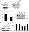
Knockdown of nucleolin with siRNA in HFF cells. (A) Western blotting of HFF cells transfected with siRNA. Proteins from lysates of cells transfected with a pool of siRNA recognizing nucleolin mRNA (lane 2) or control siRNA (lane 1) were assayed by Western blotting using antibodies recognizing nucleolin (Ncl) or β-actin, as indicated to the right. The positions of molecular mass markers (kDa) are indicated to the left. (B) Determination of relative protein levels in cells transfected with siRNA pools. A 2-fold dilution series of protein from lane 1 of A (lanes 1 to 3) was analyzed by Western blotting using antibodies recognizing nucleolin (Ncl) or β-actin, as indicated to the right, compared to undiluted protein from lane 2 of A. The positions of molecular mass markers (kDa) are indicated to the left. (C) Virus production from siRNA-transfected cells. Cells were infected with AD169 at an MOI of 1, and virus was harvested 96 h postinfection. Data points indicate the average virus titers (PFU/ml) from two experiments. Error bars indicate the standard errors of the means of those results. (D) Nucleolin levels from cells transfected with individual nucleolin siRNA. Lysates of cells transfected with control siRNA (lane 1) and each of the 4 individual siRNAs targeting nucleolin (lanes 2 to 5, siRNAs 5 to 8, respectively) were assayed by Western blotting using antibodies recognizing nucleolin (Ncl) or β-actin, as indicated to the right. The positions of molecular mass markers (kDa) are indicated to the left. (E) Determination of relative protein levels in cells transfected with individual siRNAs. A 2-fold dilution series of protein from lane 2 of D (lanes 1 to 3) was analyzed by Western blotting using antibodies recognizing nucleolin (Ncl) compared to undiluted protein from lane 3 of D. The positions of molecular mass markers (kDa) are indicated to the left. (F) Virus production from cells transfected with individual siRNAs. HFF cells transfected with control siRNA or siRNAs 5 to 8 were infected with AD169 at an MOI of 1. Titers of virus (PFU/ml) from these cells were determined in duplicate at 96 h postinfection. Data points indicate the average virus titers (PFU/ml) from titrations of virus performed in duplicate. These data are representative of data from 2 independent experiments.
FIG. 7.
FIG. 7.
Protein synthesis in siRNA-transfected cells. HFF cells were transfected with either nucleolin (Ncl) siRNA or control siRNA and were pulsed with [35S]methionine in the presence or absence of 200 μg/ml cycloheximide (CHX), and cell lysates were prepared. Proteins in each cell lysate were separated on a 10% SDS-polyacrylamide gel, which was stained, dried, and digitally scanned (A) and then exposed to a phosphorimager screen (B). The positions of molecular mass markers (kDa) are indicated to the left.
FIG. 8.
FIG. 8.
Replication of HCMV and reovirus in siRNA-transfected cells. HFF cells transfected with either nucleolin siRNA or control siRNA were infected with HCMV (MOI of 1) or reovirus (Reo) (MOI of 0.1). Virus was harvested at 96 h p.i. for HCMV and at 16 h postinfection for reovirus and titrated onto HFF and L cells, respectively, to determine HCMV and reovirus titers (PFU/ml). The mean titers from three experiments are shown. Error bars represent the standard deviations.
FIG. 9.
FIG. 9.
Levels of viral and cellular proteins during infection of cells containing nucleolin siRNA. (A) Time course of protein levels. HFF cells were transfected with pools of nucleolin siRNA or control siRNA and then infected with AD169 (MOI of 1). Cell lysates were prepared at the indicated time points, and proteins from these lysates were separated on a 10% SDS-polyacrylamide gel. Levels of viral and cellular proteins were assayed by Western blotting using antibodies recognizing immediate-early proteins, UL44, pp28, nucleolin (Ncl), and β-actin, as indicated to the right. (B) Determination of relative protein levels in infected cells at 72 h p.i. A 2-fold dilution series of protein from lane 5 of A (lanes 1 to 3) was analyzed by Western blotting using antibodies recognizing nucleolin (Ncl) and pp28, as indicated to the right, compared to undiluted protein from lane 6 of A. The positions of molecular mass markers (kDa) are indicated to the left.
FIG. 10.
FIG. 10.
Virus production and viral DNA synthesis in HFF cells transfected with siRNA. In three separate experiments, HFF cells were transfected with nucleolin (Ncl) siRNA or control siRNA and then infected with AD169 (MOI of 1). (A) At 96 h postinfection, the virus titer was determined by titrating virus on HFF cells. Virus titer is represented as PFU/ml. (B) Viral DNA synthesis in each experiment was determined by quantitative real-time PCR. The amount of viral DNA assayed is represented as copies of the viral gene UL83 per copy of the cellular gene β-actin.

Similar articles

Cited by

References

    1. Angelov, D., V. A. Bondarenko, S. Almagro, H. Menoni, F. Mongelard, F. Hans, F. Mietton, V. M. Studitsky, A. Hamiche, S. Dimitrov, and P. Bouvet. 2006. Nucleolin is a histone chaperone with FACT-like activity and assists remodeling of nucleosomes. EMBO J. 25:1669-1679. - PMC - PubMed
    1. Appleton, B. A., J. Brooks, A. Loregian, D. J. Filman, D. M. Coen, and J. M. Hogle. 2006. Crystal structure of the cytomegalovirus DNA polymerase subunit UL44 in complex with the C terminus from the catalytic subunit. Differences in structure and function relative to unliganded UL44. J. Biol. Chem. 281:5224-5232. - PubMed
    1. Appleton, B. A., A. Loregian, D. J. Filman, D. M. Coen, and J. M. Hogle. 2004. The cytomegalovirus DNA polymerase subunit UL44 forms a C clamp-shaped dimer. Mol. Cell 15:233-244. - PubMed
    1. Baldick, C. J., Jr., A. Marchini, C. E. Patterson, and T. Shenk. 1997. Human cytomegalovirus tegument protein pp71 (ppUL82) enhances the infectivity of viral DNA and accelerates the infectious cycle. J. Virol. 71:4400-4408. - PMC - PubMed
    1. Bertrand, L., and A. Pearson. 2008. The conserved N-terminal domain of herpes simplex virus 1 UL24 protein is sufficient to induce the spatial redistribution of nucleolin. J. Gen. Virol. 89:1142-1151. - PubMed

Publication types

MeSH terms